
Vol. 93, No. 12, 2003 1553
Bacteriology
Nontoxigenic Strains of Pseudomonas syringae pv. phaseolicola Are
a Main Cause of Halo Blight of Beans in Spain
and Escape Current Detection Methods
Arantza Rico, Ruth López, Carmen Asensio, M. Teresa Aizpún, M. Carmen Asensio-S.-Manzanera, and Jesús Murillo
First, fourth, and sixth authors: Laboratorio de Patología Vegetal, Departamento de Producción Agraria, ETS Ingenieros Agrónomos,
Universidad Pública de Navarra, 31006 Pamplona, Spain; and second, third, and fifth authors: Servicio de Investigación y Tecnología
Agraria, Consejería de Agricultura y Ganadería,
Junta de Castilla y León, 47080 Valladolid, Spain.
Accepted for publication 9 July 2003.
ABSTRACT
Rico, A., López, R., Asensio, C., Aizpún, M. T., Asensio-S.-Manzanera,
M. C., and Murillo, J. 2003. Nontoxigenic strains of Pseudomonas
syringae pv. phaseolicola are a main cause of halo blight of beans in
Spain and escape current detection methods. Phytopathology 93:1553-
1559.
From a collection of 152 pseudomonads isolated from diseased beans
in Spain, 138 (91%) of the strains were identified as Pseudomonas
syringae pv. phaseolicola and the rest as P. syringae pv. syringae. The P.
syringae pv. phaseolicola strains produced typical water-soaked lesions
on bean pods, although 95 of them did not produce phaseolotoxin in
vitro. Ninety-four of these isolates did not produce the expected 0.5-kb
product after polymerase chain reaction (PCR) amplification using
primers specific for open reading frame (ORF) 6 of the phaseolotoxin
(tox) gene cluster and did not contain DNA homologous to ORF 6 in
Southern hybridization experiments. To our knowledge, this is the first
report of the widespread occurrence in the field of strains of P. syringae
pv. phaseolicola lacking the tox cluster, which contrasts sharply with the
general belief that Tox
+
isolates are the only ones with epidemiological
importance. Additionally, the tox
–
isolates were not specifically detected
by a commercial polyclonal antisera in an enzyme-linked immunosorbent
assay. Accordingly, it is possible that the certification of seed lots as free
of the pathogen cannot be reliably done in Spain, or in any other country
where tox
–
strains might occur frequently, using current PCR or sero-
logical protocols. The amplification of three avirulence genes by PCR
allowed us to make predictions of the P. syringae pv. phaseolicola race
structure, as confirmed by plant assays. Six races (races 1, 2, 5, 6, 7, and
9) were identified, with race 7 being the most prevalent (46.1%) followed
by races 6 (21.3%) and 1 (9.0%). All the tox
–
isolates contained gene
avrPphF, typical of races 1, 5, 7, and 9.
The three major bacterial diseases of common bean (Phaseolus
vulgaris L.) are caused by pathovars of Pseudomonas syringae
and Xanthomonas campestris and result in economically impor-
tant losses worldwide (31,41). In particular, P. syringae pv.
phaseolicola, causing halo blight, is probably the most important
bacterial pathogen of bean in Europe, the United States, and many
other countries. Spain is not an exception. Some of these patho-
gens cause field epidemics (1,4,41). P. syringae pv. phaseolicola
was first described in Spain in 1939 and it also appears to be the
main cause of bacterioses of bean, although P. syringae pv.
syringae was reported to be of local importance in certain areas
and bean cultivars (4,7). Although it is still considered a quaran-
tine organism in Spain, X. campestris pv. phaseoli has been re-
peatedly isolated in the field (1,4; C. Asensio, unpublished data)
and works are in progress to determine its importance for bean
production.
Control of halo blight is difficult, and the only practical
methods for its management are the use of pathogen-free seed and
appropriate cultural practices and planting of resistant cultivars.
The presence of extremely low levels of primary inoculum can
initiate severe epidemics under favorable conditions, and in con-
sequence, certification of seed as free of the pathogen requires the
use of highly sensitive and specific methods. Detection and identi-
fication of P. syringae pv. phaseolicola has been done using
different methods that include microbiological assays (15), nucleic
acid hybridization (36), and different serological methods (44,51),
for which there are a number of commercially available anti-
bodies. However, due to its effectiveness and low cost, several
researchers have developed detection assays based on the amplifi-
cation of specific DNA sequences by means of polymerase chain
reaction (PCR) (2,28,37). Further, rapid-cycle, real-time PCR is
currently being evaluated as a routine tool for the diagnosis of P.
syringae pv. phaseolicola in bean seed (38). In all these cases,
specific primers were designed from the available sequence of
DNA coding for phaseolotoxin biosynthesis, which is organized as
a large (>30 kb) gene cluster (tox cluster) (11,54). Phaseolotoxin
is a non-host-specific toxin that induces chlorosis on leaves of
several plant species by inhibition of ornithine carbamoyl trans-
ferase, a critical enzyme in the urea cycle. Toxin production is
very specific to P. syringae pv. phaseolicola, although it also was
described in a single bean isolate of P. syringae pv. syringae and
in P. syringae pv. actinidiae, which causes a canker disease of
kiwifruit (28,43,47). Nontoxigenic (Tox
–
) strains of P. syringae pv.
phaseolicola have been described, and it is known that they are
still pathogenic and occasionally occur in the field (17), although
it is generally believed that Tox
–
strains are of little or no epi-
demiological significance (30,31,37).
Based on their interaction with eight bean differential cultivars,
nine races of P. syringae pv. phaseolicola have been differentiated
involving five pairs of matching resistance-avirulence genes
(Table 1) (45,49). Three of these avirulence (avr) genes (avrPphB,
avrPphE, and avrPphF) were cloned and sequenced (16,42,48).
Although avrPphB and avrPphF are present only in races ex-
pressing the corresponding phenotype, avrPphE is present in all
the examined isolates but is only functional as an avr gene in
races 2, 4, 5, and 7. The geographical distribution of races is not
Corresponding author: J. Murillo; E-mail address: jesus@unavarra.es
Publication no. P-2003-1020-01R
© 2003 The American Phytopathological Society

1554 PHYTOPATHOLOGY
random, and races 3, 4, 5, 8, and 9 are not found, or only rarely,
outside of Africa, whereas races 1, 2, 6, and 7 are distributed
worldwide (45). Race 6, which is compatible with all the differ-
ential cultivars (Table 1), appears to be on average the predomi-
nant race worldwide (19,20,45), but race 8 is by far the most
common in South Africa (5). Therefore, it is necessary to know
beforehand the race structure of the pathogen in a given area, and
to continuously monitor any possible deviations, in order to imple-
ment effective control measures based on the deployment of
vertical resistance.
In the County of Castilla y León, the largest dry bean producing
region in Spain, most of the bean crop is dedicated to high quality
common bean landraces that are, in general, highly susceptible to
bacterioses. Consequently, frequent disease outbreaks are a major
constraint for bean production and cause the disappearance of
valuable local bean cultivars (1). Ongoing breeding programs at
Servicio de Investigación y Tecnología Agraria (directed by C.
Asensio) aim to introduce durable resistance to these local bean
genotypes. In this work, we focused on the identification and
characterization of the local Pseudomonas populations in order to
support the breeding programs and to develop appropriate molecu-
lar tools for the rapid identification of races. Our results demon-
strate the prevalence in Spanish fields of nontoxigenic P. syringae
pv. phaseolicola, which cannot be detected by PCR or enzyme-
linked immunosorbent assay (ELISA) using the currently avail-
able methods. A preliminary report has been published (29).
MATERIALS AND METHODS
Strains and growth conditions. Reference strains of bacteria
and fungi were obtained from international collections and are
listed in Table 2. Unless otherwise indicated, P. syringae isolates
were propagated on King’s medium B (KMB) (18) at 28°C.
Isolation and identification of bacteria from diseased beans.
Bacteria were isolated on KMB following standard methods (21)
from bean leaves or pods with typical symptoms of bacterioses.
From the original isolation plates, only fluorescent colonies were
retained for this work. All isolates were purified by a series of
single colony transfers on KMB and stored at –80°C in KMB plus
20% glycerol before their characterization.
Bacterial isolates were identified essentially as described (21,
39) following the LOPAT scheme (levan production, oxidase ac-
tivity, potato soft rot, arginine dihydrolase activity, and tobacco
hypersensitive response) (21), except that the degradation of po-
tato tissue was not examined and the arginine dihydrolase test was
carried out on petri dishes using anaerobic jars (3). We examined
the utilization of diverse carbohydrates as the sole carbon source,
the degradation of aesculine, degradation of casein on milk-Tween
(MT) medium (8), production of toxins, and pathogenic charac-
teristics. Carbohydrates (
D
-mannitol, inositol,
D
-sorbitol, erythri-
tol,
L
+tartrate,
D
+tartrate, and
L
-lactate) were added to Ayer’s
minimal medium (21) to a final concentration of 0.1% (wt/vol).
Those isolates that did not show significant growth after 1 week
of incubation at 28°C were considered negatives.
Symptomatology on pods and leaves of bean cv. Canadian
Wonder, which is susceptible to all known races of P. syringae pv.
phaseolicola, was examined as described (10,45). Excised pods
were dipped in 70% ethanol, rinsed two times in distilled water,
and inoculated with a toothpick. To confirm the pathogenicity of
the isolates, the symptomatology on leaves of cv. Canadian Won-
der was examined as described below for race identification. At
least two replicate inoculations were made per isolate.
Toxin production. The production of antimetabolite toxins and
phytotoxins was examined following previously published proce-
dures (6,9) with slight modifications (3). For phaseolotoxin, the
isolates were toothpick-inoculated on top of a lawn of Escherichia
coli on solid Ayer’s minimal medium (21) and incubated for
2 days at 22°C. To confirm the identity of the toxins, isolates also
were tested on plates supplemented with 100 µl of a sterile 1%
(wt/vol) solution of the amino acids
L
-ornithine,
L
-citrulline, and
L
-arginine. Isolates were considered to produce phaseolotoxin
when the inhibition of E. coli growth was inverted on plates con-
taining
L
-citrulline and
L
-arginine but not on plates with
L
-
ornithine. The production of syringomycin was assessed by the ca-
pacity of the isolates to inhibit the growth of Geotrichum
candidum F260 on potato dextrose agar plates (9).
Serology. Double-antibody sandwich (DAS)-ELISAs were
performed with a commercial kit (Loewe Biochemica GmbH,
Germany) and following the manufacturer’s instructions. Bacterial
suspensions were prepared on buffered physiological saline
(140 mM NaCl, 2 mM NaH
2
PO
4
, and 15 mM Na
2
HPO
4
) and
adjusted to optical density (OD) at 600 nm of approximately 1
before applying them to antibody-coated plates. In every plate, P.
syringae pv. phaseolicola strain 1449B was used as a positive
control and P. syringae pv. syringae strains B728a and B86-17 as
negative controls. The results were read on a plate reader (Multi-
skan EX; Thermo Electron, Finland) at 405 nm. In order to make
comparisons among plates, we calculated the serological reaction
(SR) for each isolate as described (40), using the formula SR =
[(x – y)/(z – y)] × 100, where x is the average OD for the studied
isolate, z is the average OD for the positive control, and y is the
average OD for the negative controls. Results were considered
negative when the SR was below 50. Strains were tested in at
least two separate experiments with two replicates each.
TABLE 1. Partly validated model to explain observed interactions between races of Pseudomonas syringae pv. phaseolicola and cultivars of the host, bean
Race/avr gene
a
1 2 3 4 5 6 7 8 9
avrPphF
·
·
·
avrPphF
·
avrPphF
·
avrPphF
·
avrPphE
·
avrPphE
avrPphE
·
avrPphE
·
·
·
·
avrPphB
avrPphB
·
·
·
·
·
·
·
·
· 4 ·
·
·
·
Cultivar Resistance
gene
· 5 ·
·
·
·
· 5 5
Canadian Wonder
·
·
·
·
·
+ + + + + + + + +
A52 (ZAA54)
·
·
· 4 ·
+ + + + – + + + +
Tendergreen
·
·
R3
·
·
+ + – – + + + + +
Red Mexican Ul3
R1
·
· 4 ·
– + + + – + – + –
1072
·
R2
·
·
·
+ – + – – + – + +
A53 (ZAA55)
·
·
R3 4 ·
+ + – – – + + + +
A43 (ZAA12)
·
R2
R3
4
5
+ – – – – + – – –
Guatemala 196-B
R1
·
R3 4 ·
– + – – – + – + –
a
(+), susceptible response; (–), resistant response; (·), gene absent; although homologues of avrPphE are present in the nine races (42), it is only functional as
an avirulence gene in the races indicated. Avirulence/resistance matching genes: avrPphF/R1; avrPphE/R2; and avrPphB/R3 (adapted from literature citation 49).

Vol. 93, No. 12, 2003 1555
Molecular genetic techniques. PCR amplifications were per-
formed in a total volume of 25 µl by mixing 20 pmol each primer,
0.15 mM each dNTP, 1
×
PCR reaction buffer, 1.5 mM MgCl
2
,
1 unit of Taq DNA polymerase (Biotaq; Bioline Ltd., London),
and 5 µl of a bacterial cell suspension. Specific primers for the
amplification of the ethylene forming enzyme gene (efe) (34), the
coronafacate ligase gene from the coronatine cluster (22), avrPphB
(42), avrPphE (42), avrPphF (48), open reading frame (ORF) 6
from locus phtE of the phaseolotoxin biosynthesis cluster (primers
P 5.1 and P 3.1) (37,54), and virPphA (14) were as published
previously. Amplifications were carried out using a RoboCycler
Gradient Cycler (Stratagene, La Jolla, CA) and consisted of a
denaturation step of 10 min at 94°C followed by 30 cycles at 94°C
for 1 min, 55°C for 1 min, and 72°C for 1 min, and a final exten-
sion step at 72°C for 10 min.
DNA was isolated from single colonies grown overnight in
KMB with a DNA extraction kit (Puregene; Gentra Systems Inc.,
Minneapolis, MN). Southern transfer was done by a standard
procedure (33), and hybridization of nylon filters was carried out
with a chemiluminescence labeling and detection kit (Roche
Diagnostics, Basel, Switzerland). Appropriate amplicons to be
used as DNA probes were purified from gels and ligated to
pGemT-Easy (Promega, Madison, WI); the identity of the cloned
inserts was routinely confirmed by DNA sequencing. To label the
probe, the fragment was amplified from the clone, purified from a
1% agarose gel, and labeled by random priming.
Sequencing and analysis of gyrB and rpoD. Primer pairs
gyrB-L (5
-AAGTATCCGGCGGCTTG-3) and gyrB-R (5-GTG-
GTCGCGACCTTGTG-3
) and rpoD-L (5-CGCCAAACGTAT-
CGAAGAA-3
) and rpoD-R (5-GCTATTTTCAGGCCGGTTT-
3
) were designed from published sequences of gyrB (EMBL
Accession No. AB016375 [35]) and rpoD (EMBL Accession No.
AB039500 [53]), respectively, from strains of P. syringae pv.
phaseolicola. Genes gyrB and rpoD were amplified as described
previously from the nontoxigenic P. syringae pv. phaseolicola
strains CYL233, CYL275, CYL309, CYL314, CYL325, and
CYL352. Partial sequences of 534 and 572 nucleotides were
determined at MWG-Biotech AG (Ebersberg, Germany) directly
from the purified gyrB and rpoD PCR fragments, respectively,
using the corresponding amplification primers. Sequences were
combined for the same strain and treated as a single 1106 nucleo-
tide sequence, assuming that analysis using longer sequences re-
sults in better resolution and reliability (53). Sequences were
aligned with the corresponding combined sequence fragments of a
range of P. syringae pathovars and other pseudomonads (53) using
ClustalW (46). Phylogenetic trees were constructed with ClustalW
using the neighbor-joining method (32). The nucleotide sequences
were deposited in the nucleotide sequence database as EMBL
Accession Nos. AJ564779 to AJ564784 for gyrB and AJ564785 to
AJ564790 for rpoD.
Race identification of P. syringae pv. phaseolicola strains.
Isolates were inoculated on primary leaves of line 1072 of P.
acutifolious of bean cvs. Canadian Wonder, A52, Tendergreen,
Red Mexican UI3, A53, A43, and Guatemala 196-B as described
previously (45). Infection was scored according to a previously
defined five-point scale (12). At least six replicate plants were
inoculated for each combination of isolate and cultivar.
RESULTS
Isolation and identification of P. syringae from diseased
beans. Sampling was carried out from 1993 to 2001 in com-
mercial fields over the entire bean production zone (4,162 ha in
1998) from Castilla y León County (94,147 km
2
), Spain, mainly
from common bean landraces. From our own experience, these
cultivars are highly susceptible to bacterioses in the field, al-
though it is not known if they display resistance to any particular
race of P. syringae. Bacteria were isolated from leaves and pods
showing necrotic or water-soaked lesions typical of bacterioses,
either surrounded or not by chlorotic haloes, and only fluorescent
isolates were kept for further characterization. The 152 isolates
examined in this work were fluorescent on KMB, levan positive,
oxidase and arginine dihydrolase negative, and elicited a hyper-
sensitive reaction on tobacco (LOPAT group Ia), and were there-
fore considered P. syringae (21).
Fourteen of the isolates (9.2%) displayed a biochemical pattern
typical of P. syringae pv. syringae, produced syringomycins but
not phaseolotoxin, and induced sunken brown lesions upon
inoculation onto pods of bean cv. Canadian Wonder (Table 3).
These isolates were in consequence identified as P. syringae pv.
syringae and were not further characterized.
The remaining 138 isolates (90.8%) were considered P. syringae
pv. phaseolicola because they showed typical metabolic charac-
teristics of this pathovar and their inoculation onto pods of uni-
versally susceptible cv. Canadian Wonder resulted in typical
water-soaked lesions 2 to 3 days after inoculation (Table 3). Addi-
tionally, all of them produced specific amplification bands by
PCR using primers specific for virPphA, shown to be essential for
pathogenicity on beans (13), and for avrPphE. These isolates did
not produce syringomycins and did not produce amplification
bands with primers specific for coronatine genes and for the ethyl-
ene forming enzyme (Table 3), which has been described in P.
syringae pv. glycinea and in kudzu isolates of P. syringae pv.
phaseolicola (52). Assimilation of mannitol is usually negative for
P. syringae pv. phaseolicola, although all the isolates from kudzu
tested positive, as did some isolates from other hosts, including
bean (23,50). However, 72% of the Spanish isolates were able to
utilize mannitol as the sole carbon source (Table 3). Noticeably,
the 94 isolates that lacked the phaseolotoxin gene ORF 6 (tox
–
;
described below) tested positive, whereas 38 of the 44 isolates
containing ORF 6 tested negative as expected.
Phaseolotoxin detection. Unexpectedly, only 43 of the P.
syringae pv. phaseolicola isolates produced phaseolotoxin (Table
3), as determined by the E. coli inhibition bioassay. The remaining
95 isolates (68.8%) did not cause the inhibition of E. coli or, if
they did, it was not specifically reverted by citrulline or arginine.
TABLE 2. Reference bacterial strains and fungi used in this study
Strain
Characteristics
Source or reference
Escherichia coli
CECT831
Sensitive to phaseolotoxin
Colección Española
de Cultivos Tipo,
Spain
Geotrichum candidum
F260
Sensitive to syringomycin
A. de Vicente
Pseudomonas syringae
pv. glycinea
49a/90
Race 4; coronatine pro-
ducer; 1990, Germany
M. Ullrich
4180
Race 4; coronatine pro-
ducer; 1975, New Zealand
(24)
pv. phaseolicola
1281A
Race 1; 1984, UK
(45)
882
Race 2; 1975, USA
(45)
1310A
Race 3; 1984, Tanzania
(45)
1302A
Race 4; 1984, Rwanda
(45)
1375A
Race 5; 1985, Kenya
(45)
1299A
Race 6; 1984, Tanzania
(45)
1449B
Race 7; 1985, Ethiopia
(45)
2656A
Race 8; 1990, Lesotho
(45)
2709A
Race 9; 1990, Malawi
(45)
pv. syringae
B728a Wild
type;
Rif
r
; isolated from
Phaseolus vulgaris, USA
G. W. Sundin
B86-17
Wild type; isolated from
Phaseolus vulgaris, USA
G. W. Sundin
pv. tabaci
CFBP1621
Wild type
C. Manceau

1556 PHYTOPATHOLOGY
Other researchers have described the natural existence of Tox
–
strains of P. syringae pv. phaseolicola (24,37,50) as resulting
either from point mutations or, apparently less frequently, from the
absence of part or all of the tox cluster. However, although they
are still pathogenic and occasionally occur in the field (17), it is
generally believed that Tox
–
strains are of little or no epidemi-
ological significance (30,31,37). This is the basis for the wide-
spread use of DNA sequences from the tox cluster as a target for
the detection, by PCR or DNA hybridization, of P. syringae pv.
phaseolicola in seed lots (2,28,36,37). Accordingly, we examined
if the Tox
–
isolates identified here could be detected by PCR using
primers P 5.1 and P 3.1, which are specific for phaseolotoxin
genes and were designed for a highly sensitive enrichment PCR
assay, BIO-PCR, for seed sample processing (37). All the Tox
+
and one of the Tox
–
isolates produced the expected 0.5-kb band
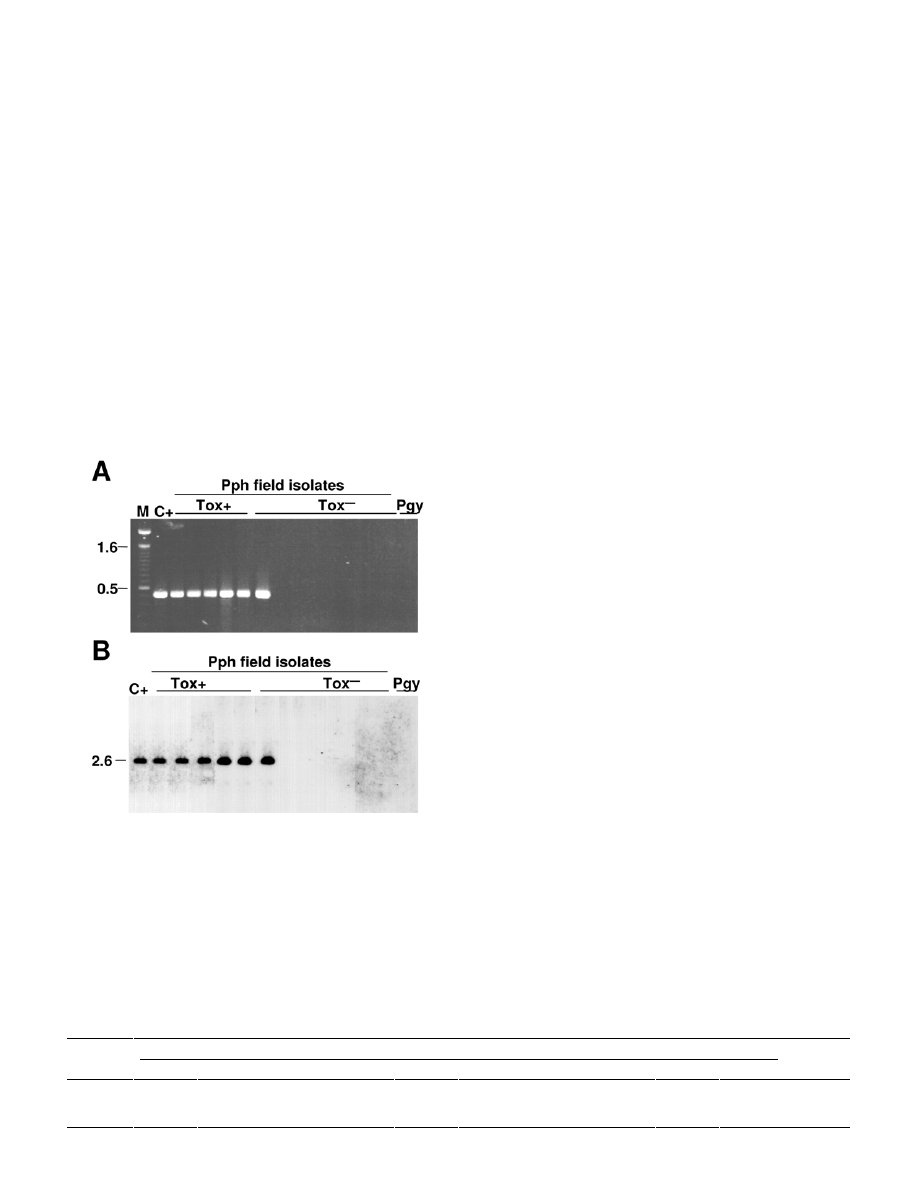
after PCR amplification (Table 3; Fig. 1). The remaining 94 Tox
–
P. syringae pv. phaseolicola isolates, as well as isolates from
pathovars glycinea, syringae, and tabaci, did not produce any
amplification band or occasionally produced some nonspecific
weak bands of higher size than expected. Southern hybridization
experiments of genomic DNA using the P 5.1-P 3.1 amplicon from
strain 1449B as a probe showed that these latter isolates did not
contain sequences homologous to the probe (Fig. 1). This suggests
that the majority of the Tox
–
P. syringae pv. phaseolicola isolates
native to Spain examined here might lack part of or the entire
phaseolotoxin biosynthesis cluster and, in consequence, might
remain undetected after PCR examination of contaminated seed
lots.
Because Tox
–
isolates of P. syringae pv. phaseolicola are only
rarely reported (30,37,50), it is arguable that the Spanish non-
toxigenic isolates lacking tox DNA (tox
–
) might belong to a differ-
ent pathovar. We therefore conducted a phylogenetic analysis by
using partial sequences of the genes for DNA gyrase B subunit
(gyrB) and
s
70
factor (rpoD) from six representative tox
–
isolates.
Both proteins are ubiquitous in bacteria and essential for cell
growth and were used previously for phylogenetic analysis of the
genus Pseudomonas (53). A total of 534 nucleotides for gyrB and
572 nucleotides for rpoD were identical to the corresponding
sequences of four strains each of P. syringae pv. phaseolicola and
P. syringae pv. glycinea (53). The neighbor-joining tree (data not
shown) resulting from the analysis of the combined partial gyrB
and rpoD sequences was essentially identical to the previously
constructed tree (53) and clustered the six tox
–
isolates together
with P. syringae pv. phaseolicola and P. syringae pv. glycinea and
well apart from other P. syringae pathovars.
ELISA. Because most of the Spanish isolates of P. syringae pv.
phaseolicola cannot be detected by the currently available PCR
protocols, we tested the specificity of other available detection
techniques. Isolates were tested by DAS-ELISA using one of the
several commercial antibodies available for the detection of P.
syringae pv. phaseolicola and using two strains of P. syringae pv.
syringae as negative controls. The polyclonal antibody used re-
acted as expected with all the isolates that contained the phaseo-
lotoxin gene cluster, producing clear positive reactions, with SR
mean values ranging between 82 and 113. Conversely, clear nega-
tive reactions were observed for all the tox
–
isolates (mean SR
values between 0.2 and 13) and for two P. syringae pv. glycinea
(mean SR values of 0 and 3). Our results, therefore, indicate that
the Spanish isolates lacking tox DNA cannot be detected using a
commercial ELISA test and suggest that other commercial anti-
bodies also might fail to detect this type of P. syringae pv.
phaseolicola isolate.
Race identification. We wanted to set up a method to allow the
rapid identification of races as well as to assess the diversity of
races in the sampled area. The previous molecular characterization
of P. syringae pv. phaseolicola isolates (7,23) appears to indicate
that races are polyphyletic, which could make the identification of
race-specific molecular markers problematical. We therefore d-
cided to examine the amplification by PCR of avr gene bands
characteristic of races (Table 1). Selected genes were avrPphB,
which is present only in races 3 and 4 (16), avrPphE, producing
an amplification product 104 bp larger in race 8 (42), and
avrPphF, which is carried only by races 1, 5, 7, and 9 (48). From
the 138 isolates examined, 108 isolates (78.3%) contained gene
avrPphF and were tentatively assigned to the group of races 1, 5,
7, and 9. The remaining 30 isolates (21.7%) were tentatively
included in races 2 or 6, because no isolates were found that could
be assigned to races 3, 4, or 8.
The reactions of 89 randomly selected isolates on the bean
differentials resulted in a race assignation (Table 4) that widely
agreed with the PCR analyses: 51 isolates (57.3%) were assigned
to races 1, 5, 7, and 9, while 21 isolates (23.6%) were included in
races 2 and 6. In addition, we found no isolates that corresponded
to races 3, 4, or 8, as was predicted from the PCR results de-
scribed previously. The avr gene content of all these isolates, as
examined by PCR, was as expected for the race in which they
TABLE 3. Characteristics and identification of 152 Pseudomonas syringae isolates from infected beans in Spain
Number of field P. syringae isolates
Reference P. syringae pvs.
pv. phaseolicola
pv. syringae
Characteristics
phaseolicola 1449B syringae
B728a glycinea
49a/90
tabaci CFBP1621 44
94
14
Utilization of
a
D
-Mannitol – + + +
+6/–38
+
+
Inositol –
+
+
+
–
–
+
D
-Sorbitol –
+
+
+
–
–
+
Erythritol –
+
–
–
–
–
+
L
+Tartrate – –
–
+
–
–
–
D
+Tartrate – –
–
+
–
–
–
L
-Lactate –
+
–
–
–
–
+
Esculin hydrolisis
–
+
+
+
–
–
+
Casein hydrolisis
–
+
–
–
–
–
+
Symptoms ws
b
sb
b
HR
b
HR
ws
ws
sb
Production of
Phaseolotoxin +
– – –
+43/–1
–
–
Syringomycins –
+
–
– –
–
+
Phaseolotoxin genes
c
+
–
–
–
+ – –
Coronatine genes
c
–
–
+
– –
–
–
efe gene
c
–
–
+
–
–
–
–
a
Utilization of compounds as the sole carbon source.
b
Symptoms scored 4 days after stab inoculation of pods of the universally susceptible bean cv. Canadian Wonder; ws, water-soaked lesions with occasional
bacterial ooze; sb, sunken brown lesions; and HR, hypersensitive reaction.
c
Examined by polymerase chain reaction with specific primers.
Vol. 93, No. 12, 2003 1557
were classified by the plant assays (Table 1). However, 17 addi-
tional isolates, including 16 tox
–
isolates that contained avrPphF,
produced sets of reactions that were not compatible with any of
the known races. The patterns of these reactions appeared to
warrant their classification in at least five new putative races (data
not shown).
In general, race 7 was the most abundant (46.1%) followed by
races 6 (21.3%) and 1 (9.0%), while only one or two isolates each
were found for races 2, 5, and 9. Noticeably, all the tox
–
isolates
contained gene avrPphF and, in consequence, the majority of the
race 1 and 7 isolates belonged to this group. We did not find any
obvious correlation between the race isolated and the place, date,
or cultivar of isolation.
DISCUSSION
We characterized a collection of 152 fluorescent pseudomonads
isolated between 1993 and 2001 in north-central Spain from le-
sions on field-grown common bean landraces. Similar to many
other bean-growing regions of the world (31,41), P. syringae
pv. phaseolicola was the most abundant bacterial pathogen
found, representing 91% of the total isolates, while the remaining
were P. syringae pv. syringae (Table 3). The majority (68.8%) of
the P. syringae pv. phaseolicola isolates did not produce phaseolo-
toxin (Table 3), which contrasts sharply with the general belief
that Tox
+
isolates are the only ones with epidemiological im-
portance (30,31,37). Additionally, all but one of the nontoxi-
genic isolates lacked DNA homologous to ORF 6 (Table 2), a
putative fatty acid desaturase gene that is essential for the
biosynthesis of phaseolotoxin (54). Identification of the tox
–
as P.
syringae pv. phaseolicola was unambiguously confirmed by
comparison of gyrB and rpoD sequences from six representative
tox
–
isolates to those of other P. syringae pathovars and Pseudo-
monas spp. (53).
The absence of DNA homologous to ORF 6 in the tox
–
strains is
noteworthy, because the primers used in current PCR protocols for
the detection and identification of this pathogen are based on this
DNA sequence (37). In a separate study (J. A. Oguiza, A. Rico, L.
Rivas, L. Sutra, A. Vivian, and J. Murillo, unpublished data), we
showed that strains lacking ORF 6 also lacked DNA homologous
to the genes in the known borders of the tox cluster, argK and
amtA. This suggests that the tox cluster was probably acquired
only by some P. syringae pv. phaseolicola strains and not by
others and indicates the impracticality of using primer sets
directed to other regions of the tox cluster for the detection or the
identification of this pathogen (23,25,28). Furthermore, a com-
mercial kit for the specific detection of P. syringae pv. phaseoli-
cola by DAS-ELISA also failed to detect the Spanish isolates.
Because the kit contains a polyclonal antibody, our results suggest
the existence of significant differences between the isolates
containing the tox cluster and those putatively lacking it and could
imply that other commercial antibodies also might fail to detect
the tox
–
strains. In support of this, a selection of the tox
–
isolates
characterized here did not react with another polyclonal antisera
produced against whole-cell preparations of a Tox
+
P. syringae pv.
phaseolicola strain (F. J. Legorburu and I. Ruiz de Galarreta,
personal communication). In consequence, it is possible that
the certification of seed lots as free of the pathogen cannot be
reliably done in Spain, or in any other country where these
kinds of strains might occur frequently, using current PCR or
serological protocols.
To our knowledge, this is the first report of the widespread
occurrence in the field of nontoxigenic P. syringae pv. phaseoli-
cola strains. Although very rarely, nontoxigenic isolates are re-
ported in the literature (37,50) and were responsible for the occur-
rence of some outbreaks of the so-called “halo-less” halo blight in
Australia (17). However, even though one of these isolates pro-
duced no detectable toxin in the culture medium, the presence in
the medium of a trace of phaseolotoxin was established unequivo-
cally (24), indicating that these isolates putatively contained the
phaseolotoxin biosynthesis cluster. It is possible that the isolates
lacking tox DNA represent the original population of P. syringae
pv. phaseolicola in Spain. In support of this, since 1987 (1) we
have seen a noticeable increase in the number of isolates be-
longing to the primitive race 2, which was later divided into races
2, 6, and 8 (45). This increase was particularly noticeable in areas
where commercial cultivars were planted, suggesting that this
could have contributed to the introduction of these new races. The
increase in the frequency of race 6 isolates could be due to their
capacity to infect a wider range of bean cultivars or to its capacity
to produce phaseolotoxin. However, the predominance of tox
–
isolates is evidence against a role of phaseolotoxin in virulence.
TABLE 4. Race identification of 89 Pseudomonas syringae pv. phaseolicola isolates by pathogenicity tests and distribution of races among isolates containing
or lacking open reading frame (ORF) 6 from the phaseolotoxin gene cluster
Number of isolates per race
ORF
6 1 2 3 4 5 6 7 8 9 ?
Total
Present 1 2 0 0 0 19 6 0 0 1
29
Absent 7 0 0 0 1 0
35 0 1 16
60
Total 8 2 0 0 1 19
41 0 1 17
89
Fig. 1. Detection of a sequence specific for the phaseolotoxin biosynthesis
cluster in Tox
+
and Tox
–
Spanish field isolates of Pseudomonas syringae
pv. phaseolicola (Pph). A, Electrophoretic analysis of polymerase chain
reaction amplification products obtained with primer pair P 5.1-P 3.1,
directed to open reading frame 6 from the phtE locus. C+, P. syringae pv.
phaseolicola 1449B used as a Tox
+
control; Pgy, P. syringae pv. glycinea
49a/90 used as a Tox
–
control; and M, 1-kb marker. B, Southern blot
hybridization of genomic DNA digested with EcoRI. The amplicon
generated from strain 1449B with primers P 5.1-P 3.1 was cloned, purified,
labeled with digoxinenin, and used as a probe. Sizes are indicated to the left
in kilobases.

1558 PHYTOPATHOLOGY
Indeed, nontoxigenic mutants of P. syringae pv. phaseolicola are
still pathogenic and their virulence is comparable to that of the
wild type (26,27), except that there is no formation of chlorotic
haloes; however, there is limited evidence that phaseolotoxin
might contribute to systemic movement of bacteria in planta (26).
Likewise, phaseolotoxin appears to contribute to the formation of
chlorotic halo lesions in kiwifruit canker produced by P. syringae
pv. actinidiae, but not for production of other symptoms or for the
multiplication of the pathogen in planta (43). It is possible how-
ever, that the toxin provides an ecological advantage that explains
its acquisition by two different pathovars. For instance, because
phaseolotoxin inhibits a key metabolic pathway, it is feasible that
it might act by inhibiting the growth of potential competitors.
Nevertheless, it would be necessary to obtain appropriate experi-
mental evidence to support the role, if any, of phaseolotoxin in
virulence.
Race 6 appears to be predominant worldwide (19,20,45); how-
ever, more than 78% of the Spanish isolates contained avrPphF
and only 21% belonged to race 6. An important asymmetry is that
all the tox
–
isolates contained avrPphF, whereas the majority of
the Tox
+
isolates lacked it. The prevalence of avrPphF in the
Spanish population is striking and currently difficult to explain.
This gene is embedded in a plasmid-borne pathogenicity island
(13) and has been shown to increase the virulence of the race 7
isolate 1449B to bean cv. Tendergreen and to different cultivars of
soybean (48). Additionally, avrPphF also restricts the host range
of the bacterium by inciting a resistance reaction in bean cultivars
containing the resistance gene R1, such as Red Mexican and
Guatemala 196-B. It is then possible that avrPphF was acquired
only by certain groups of isolates because it confers a selective
advantage, such as increased virulence to the local bean genotypes
or to other alternative hosts. Alternatively, avrPphF might have
been originally inherited by the ancestor of P. syringae pv.
phaseolicola and has been selectively lost only by certain isolates.
This last possibility is likely taking into account the close
phylogenetic relationships of strains containing or lacking tox
DNA.
The avr gene content of the individual isolates, as determined
by PCR using primer pairs specific for three avr genes, allowed us
to make predictions about the race structure of the native bacterial
population that agreed in general with the results obtained by
plant assays. This PCR method is valuable in that it determines
the existence in the bacterial population of particular avr genes,
which are responsible for specific plant resistance phenotypes that
might be of interest to plant breeders. Of the five genes postulated
to explain the existence of nine races in P. syringae pv. phaseolicola,
only three avr genes were cloned and sequenced (Table 1).
Because we used primers to detect only these last three avirulence
genes, the method only gives a broad idea of the groups of races
present, although it might be used accurately to detect the pres-
ence of races 3, 4, and 8, whose distribution is limited. As more
avr genes are characterized, the method may be refined to pre-
cisely identify each race. In our case, both PCR and plant assays
showed a preponderance of isolates containing avrPphF, which is
characteristic of races 1, 5, 7, and 9, and evidenced the absence of
isolates belonging to races 3, 4, and 8. A source of discrepancy
that lowered the predictive value of the avr-PCR method was the
existence of 19% of isolates, most of which contained avrPphF,
that could not be classified in any of the known races (Table 4).
Other researchers have reported the existence of isolates whose
race could not be established (7,20,23,45). As these results and
ours indicate, it is possible that there are several new, as of yet,
uncharacterized races of P. syringae pv. phaseolicola. This is
further supported (5) by the inconsistencies between reactions
observed after leaf and pod inoculation for certain isolates, but not
for others, suggesting the existence of new avr genes or alleles in
the P. syringae pv. phaseolicola population that could determine
new races.
ACKNOWLEDGMENTS
This work was supported with grant RTA01-005-C2 from the Spanish
Instituto Nacional de Investigación y Tecnología Agraria y Alimentaria
(INIA). A. Rico was funded by a fellowship from the INIA and R. López
was funded by a fellowship from the Consejería de Agricultura y
Ganadería, Junta de Castilla y León. We wish to dedicate this publication
to F. García-Arenal. We thank C. Manceau, G. W. Sundin, J. Taylor, A. de
Vicente, A. Vivian, and M. Ullrich for bacterial and fungal strains, T.
Osinga and T. Williams for critically reading the manuscript and for
helpful suggestions, and S. Fernández for technical assistance.
LITERATURE CITED
1. Asensio, C. 1996. Bacteriosis de judías en Castilla y León: Identi-
ficación de la variación patogénica de Pseudomonas syringae pv.
phaseolicola y factores que influyen en su desarrollo. Estudio de la
herencia de la resistencia a la raza 1 de P.s. pv. phaseolicola. Ph.D.
thesis. Universidad de León, Spain.
2. Audy, P., Braat, C. E., Saindon, G., Huang, H. C., and Laroche, A. 1996.
A rapid and sensitive PCR-based assay for concurrent detection of
bacteria causing common and halo blight in bean seed. Phytopathology
86:361-366.
3. Cazorla, F. M. 1998. Pseudomonas syringae pv. syringae, agente causal
de la necrosis apical del mango. Ecología y caracterización bac-
teriológica y molecular. Ph.D. thesis. Universidad de Málaga, Spain.
4. de Andrés, M. F., García-Arenal, F., López, M. M., and Melgarejo, P.
1998. Patógenos de plantas descritos en España. Ministerio de Agri-
cultura, Pesca y Alimentación, Madrid.
5. Fourie, D. 1998. Characterization of halo blight races on dry beans in
South Africa. Plant Dis. 82:307-310.
6. Gasson, M. J. 1980. Indicator technique for antimetabolic toxin produc-
tion by phytopathogenic species of Pseudomonas. Appl. Environ.
Microbiol. 39:25-29.
7. González, A. J., Landeras, E., and Mendoza, M. C. 2000. Pathovars of
Pseudomonas syringae causing bacterial brown spot and halo blight in
Phaseolus vulgaris L. are distinguishable by ribotyping. Appl. Environ.
Microbiol. 66:850-854.
8. Goszczynska, T., and Serfontein, J. J. 1998. Milk-Tween agar, a semi-
selective medium for isolation and differentiation of Pseudomonas
syringae pv. syringae, Pseudomonas syringae pv. phaseolicola and
Xanthomonas axonopodis pv. phaseoli. J. Microbiol. Methods 32:
65-72.
9. Gross, D. C., and De Vay, S. E. 1977. Production and purification of
syringomycin, a phytotoxin produced by a Pseudomonas syringae.
Physiol. Plant Pathol. 11:13-28.
10. Harper, S., Zewdie, N., Brown, I. R., and Mansfield, J. W. 1987. Histo-
logical, physiological and genetical studies of the responses of leaves
and pods of Phaseolus vulgaris to three races of Pseudomonas syringae
pv. phaseolicola and to Pseudomonas syringae pv. coronafaciens.
Physiol. Mol. Plant Pathol. 31:153-172.
11. Hernández-Guzmán, G., and Alvarez-Morales, A. 2001. Isolation and
characterization of the gene coding for the amidinotransferase involved
in the biosynthesis of phaseolotoxin in Pseudomonas syringae pv.
phaseolicola. Mol. Plant-Microbe Interact. 14:545-554.
12. Innes, N. L., Conway, J., and Taylor, J. D. 1984. Resistance to halo
blight in the Cambridge accession V4604 and V4058 of Phaseolus
beans. Ann. Appl. Biol. 104:307-314.
13. Jackson, R. W., Athanassopoulos, E., Tsiamis, G., Mansfield, J. W.,
Sesma, A., Arnold, D. L., Gibbon, M. J., Murillo, J., Taylor, J. D., and
Vivian, A. 1999. Identification of a pathogenicity island, which contains
genes for virulence and avirulence, on a large native plasmid in the bean
pathogen Pseudomonas syringae pathovar phaseolicola. Proc. Natl.
Acad. Sci. 96:10875-10880.
14. Jackson, R. W., Mansfield, J. W., Ammouneh, H., Dutton, L. C.,
Wharton, B., Ortiz-Barredo, A., Arnold, D. L., Tsiamis, G., Sesma, A.,
Butcher, D., Boch, J., Kim, Y. J., Martin, G. B., Tegli, S., Murillo, J., and
Vivian, A. 2002. Location and activity of members of a family of
virPphA homologues in pathovars of Pseudomonas syringae and P.
savastanoi. Mol. Plant Pathol. 3:205-216.
15. Jansing, H., and Rudolph, K. 1990. A sensitive and quick test for
determination of bean seed infestation by Pseudomonas syringae pv.
phaseolicola. J. Plant Dis. Prot. 97:42-55.
16. Jenner, C., Hitchin, E., Mansfield, J., Walters, K., Betteridge, P.,
Teverson, D., and Taylor, J. 1991. Gene-for-gene interactions between
Pseudomonas syringae pv. phaseolicola and Phaseolus. Mol. Plant-
Microbe Interact. 4:553-562.
17. Johnson, J. C. 1969. “Halo-less” halo blight of French bean in Queens-
land. Queensl. J. Agric. Anim. Sci. 26:293-302.

Vol. 93, No. 12, 2003 1559
18. King, E. O., Ward, N. K., and Raney, D. E. 1954. Two simple media for
the demonstration of pyocyanin and fluorescein. J. Lab. Clin. Med.
44:301-307.
19. Kiryakov, I. 2001. Characterization of Pseudomonas syringae pv. Phaseoli-
cola races in North-Eastern Bulgaria. Bulg. J. Agric. Sci. 7:313-318.
20. Lamppa, R. S., Gross, P. L., and del Río, L. E. 2002. Identification of
races of Pseudomonas syringae pv. phaseolicola present in North
Dakota. (Abstr.) Phytopathology 92(suppl.):S139.
21. Lelliott, R. A., and Stead, D. E. 1987. Methods for the Diagnosis of
Bacterial Diseases of Plants, vol. 2. Blackwell Scientific Publications,
London.
22. Liyanage, H., Palmer, D. A., Ullrich, M., and Bender, C. L. 1995.
Characterization and transcriptional analysis of the gene cluster for
coronafacic acid, the polyketide component of the phytotoxin corona-
tine. Appl. Environ. Microbiol. 61:3843-3848.
23. Marques, A. S. d. A., Corbière, R., Gardan, L., Tourte, C., Manceau, C.,
Taylor, J. D., and Samson, R. 2000. Multiphasic approach for the
identification of the different classification levels of Pseudomonas
savastanoi pv. phaseolicola. Eur. J. Plant Pathol. 106:715-734.
24. Mitchell, R. E. 1978. Halo blight of beans: Toxin production by several
Pseudomonas phaseolicola isolates. Physiol. Plant Pathol. 13:37-49.
25. Molouba, F., Guimier, C., Berthier, C., Guenard, M., Olivier, V., Baril,
C., and Horvais, A. 2001. Detection of bean seed-borne pathogens by
PCR. Acta Hortic. 546:603-607.
26. Patil, S. S., Hayward, A. C., and Emmons, R. 1974. An ultraviolet-
induced non-toxigenic mutant of Pseudomonas phaseolicola of altered
pathogenicity. Phytopathology 64:590-595.
27. Peet, R. C., Lindgren, P. B., Willis, D. K., and Panopoulos, N. J. 1986.
Identification and cloning of genes involved in phaseolotoxin production
by Pseudomonas syringae pv. phaseolicola. J. Bacteriol. 166:1096-1105.
28. Prosen, D., Hatziloukas, E., Schaad, N. W., and Panopoulos, N. J. 1993.
Specific detection of Pseudomonas syringae pv. phaseolicola DNA in
bean seed by polymerase chain reaction-based amplification of a
phaseolotoxin gene region. Phytopathology 83:965-970.
29. Rico, A., López, R., Aizpún, M., Asensio, C., and Murillo, J. 2002.
Strains of P. savastanoi pv. phaseolicola isolated from diseased beans in
Spain cannot be detected using primers directed to the phaseolotoxin
gene cluster. Abstract book of the 6th Int. Conf. Pseudomonas syringae
Pathovars and Related Pathogens.
30. Rudolph, K. W. E. 1995. Pseudomonas syringae pathovars. Pages 47-
138 in: Pathogenesis and Host Specificity in Plant Diseases, vol. 1. U. S.
Singh, R. P. Singh, and K. Kohmoto, eds. Elsevier Science, Oxford, UK.
31. Saettler, A. W. 1991. Diseases caused by bacteria. Pages 29-32 in:
Compendium of Bean Diseases. R. Hall, ed. The American Phyto-
pathological Society, St. Paul, MN.
32. Saitou, N., and Nei, M. 1987. The neighbor-joining method: A new
method for reconstructing phylogenetic trees. Mol. Biol. Evol. 4:406-425.
33. Sambrook, J., Fritsch, E. F., and Maniatis, T. 1989. Molecular Cloning: A
Laboratory Manual. 2nd ed. Cold Spring Harbor Laboratory, Cold
Spring Harbor, NY.
34. Sato, M., Watanabe, K., Yazawa, M., Takikawa, Y., and Nishiyama, K.
1997. Detection of new ethylene-producing bacteria, Pseudomonas
syringae pvs. cannabina and sesami, by PCR amplification of genes for
the ethylene-forming enzyme. Phytopathology 87:1192-1196.
35. Sawada, H., Takeuchi, T., and Matsuda, I. 1997. Comparative analysis of
Pseudomonas syringae pv. actinidiae and pv. phaseolicola based on
phaseolotoxin-resistant ornithine carbamoyltransferase gene (argK) and
16S-23S rRNA intergenic spacer sequences. Appl. Environ. Microbiol.
63:282-288.
36. Schaad, N. W., Azad, H., Peet, R. C., and Panopoulos, N. J. 1989.
Identification of Pseudomonas syringae pv. phaseolicola by a DNA
hybridization probe. Phytopathology 79:903-907.
37. Schaad, N. W., Cheong, S. S., Tamaki, S., Hatziloukas, E., and
Panopoulos, N. J. 1995. A combined biological and enzymatic ampli-
fication (BIO-PCR) technique to detect Pseudomonas syringae pv.
phaseolicola in bean seed extracts. Phytopathology 85:243-248.
38. Schaad, N. W., and Frederick, R. D. 2002. Real-time PCR and its appli-
cation for rapid plant disease diagnostics. Can. J. Plant Pathol. 24:250-
258.
39. Schaad, N. W., Jones, J. B., and Chun, W. (eds.) 2001. Laboratory Guide
for Identification of Plant Pathogenic Bacteria. 3rd ed. The American
Phytopathological Society, St. Paul, MN.
40. Siverio, F., Cambra, M., Gorris, M. T., Corzo, J., and López, M. M.
1993. Lipopolysaccharides as determinants of serological variability in
Pseudomonas corrugata. Appl. Environ. Microbiol. 59:1805-1812.
41. Smith, I. M., Dunez, J., Lelliot, R. A., Phillips, D. H., and Archer, S. A.
1988. European Handbook of Plant Diseases. Blackwell Scientific
Publications, London.
42. Stevens, C., Bennett, M. A., Athanassopoulos, E., Tsiamis, G., Taylor, J.
D., and Mansfield, J. W. 1998. Sequence variations in alleles of the
avirulence gene avrPphE.R2 from Pseudomonas syringae pv. phaseoli-
cola lead to loss of recognition of the AvrPphE protein within bean cells
and a gain in cultivar-specific virulence. Mol. Microbiol. 29:165-177.
43. Tamura, K., Imamura, M., Yoneyama, K., Kohno, Y., Takikawa, Y.,
Yamaguchi, I., and Takahashi, H. 2002. Role of phaseolotoxin produc-
tion by Pseudomonas syringae pv. actinidiae in the formation of halo
lesions of kiwifruit canker disease. Physiol. Mol. Plant Pathol. 60:
207-214.
44. Taylor, J. D. 1970. Bacteriophage and serological methods for the
identification of Pseudomonas phaseolicola (Burkh.) Dowson. Ann.
Appl. Biol. 66:387-395.
45. Taylor, J. D., Teverson, D. M., Allen, D. J., and Pastor-Corrales, M. A.
1996. Identification and origin of races of Pseudomonas syringae pv.
phaseolicola from Africa and other bean growing areas. Plant Pathol.
45:469-478.
46. Thompson, J. D., Gibson, T. J., Plewniak, F., Jeanmougin, F., and
Higgins, D. G. 1997. The ClustalX windows interface: Flexible
strategies for multiple sequence alignment aided by quality analysis
tools. Nucleic Acids Res. 25:4876-4882.
47. Tourte, C., and Manceau, C. 1995. A strain of Pseudomonas syringae
which does not belong to pathovar phaseolicola produces phaseolotoxin.
Eur. J. Plant Pathol. 101:483-490.
48. Tsiamis, G., Mansfield, J. W., Hockenhull, R., Jackson, R. W., Sesma,
A., Athanassopoulos, E., Bennett, M. A., Stevens, C., Vivian, A., Taylor,
J. D., and Murillo, J. 2000. Cultivar specific avirulence and virulence
functions assigned to avrPphF in Pseudomonas syringae pv. phaseoli-
cola, the cause of bean halo-blight disease. EMBO J. 19:3204-3214.
49. Vivian, A., Gibbon, M. J., and Murillo, J. 1997. The molecular genetics
of specificity determinants in plant pathogenic bacteria. Pages 293-328
in: The Gene-for-Gene Relationship in Plant Parasite Interactions. I. R.
Crute, E. B. Holub, and J. J. Burdon, eds. CAB International,
Wallingford, UK.
50. Völksch, B., and Weingart, H. 1997. Comparison of ethylene-producing
Pseudomonas syringae strains isolated from kudzu (Pueraria lobata)
with Pseudomonas syringae pv. phaseolicola and Pseudomonas syringae
pv. glycinea. Eur. J. Plant Pathol. 103:795-802.
51. Vuurde, J. W. L., and van den Bovenkamp, G. W. 1989. Detection of
Pseudomonas syringae pv. phaseolicola. Pages 30-40 in: Detection of
Bacteria in Seed and Other Planting Material. A. W. Saettler, N. W.
Schaad, and D. A. Roth, eds. The American Phytopathological Society,
St. Paul, MN.
52. Weingart, H., and Völksch, B. 1997. Ethylene production by Pseudo-
monas syringae pathovars in vitro and in planta. Appl. Environ.
Microbiol. 63:156-161.
53. Yamamoto, S., Kasai, H., Arnold, D. L., Jackson, R. W., Vivian, A., and
Harayama, S. 2000. Phylogeny of the genus Pseudomonas: Intrageneric
structure reconstructed from the nucleotide sequences of gyrB and rpoD
genes. Microbiology 146:2385-2394.
54. Zhang, Y. X., and Patil, S. S. 1997. The phtE locus in the phaseolotoxin
gene cluster has ORFs with homologies to genes encoding amino acid
transferases, the AraC family of transcriptional factors, and fatty acid
desaturases. Mol. Plant-Microbe Interact. 10:947-960.
Wyszukiwarka
Podobne podstrony:
Fasolotoksyna u Pseudomonas syringae
bez makijazu www prezentacje org
Pseudomonas
miesnie szkieletowe glowy, szyji, brzucha i grzbietu bez ilustr
Bez tytułu 1
wykład z cholestazy (bez zdjęć)
tkanki bez animacji
MIKOLOGIA biol geol 2008 wyklad4 bez ilustracji
koordynacja hormonalna czlowieka bez zdjec
wykład dr Steplewska uklad limfatyczny1 bez zdjęć
MOO wyklad 2 ekstrema bez ograniczen
Jak wgrać BIOS bez stacji dyskietek
1 Wypadki komunikacyjne bez zdjec
więcej podobnych podstron