
Anna Barbara Kłysik MD,
DRCOphth
PAPILLOEDEMA, PAPILLITIS
& PSEUDOPAPILLITS

Definition of Papilloedema;
BILATERAL
swelling of the optic nerve head
causeb by INCREASED INTRACRANIAL
PRESSURE (ICP)
MAY BE ASYMMETRICAL

Definition of Papillitis;
Unilateral or bilateral optic disc swelling
from inflammatory or infectious local
causes.
INFLAMMATION OF THE OPTIC NERVE
HEAD

Causes of Papillitis;
Posterior Uveitis
Toxoplasmosis
CMV retinitis
Thyroid ophthalmopathy etc.
Leukaemia
Anaemna
Policythemia
Macroglobulinaemia

Definition of Pseudopapillitis;
Congenital or developmental
abnormality that mimics
papilloedema/ papillitis on clinical
appearance.

Pseudopapillitis;
Optic disk drusen
Hypermetropia
Low intraocular pressure
(postraumatic, postsurgical or
developmental abnormality)

Pathophysiology of Papilloedema;
Decreased axoplasmic flow in neurits of
the optic nerve causes swelling of the pre-
laminar part
Impaired blood circulation of the optic
nerve head

Causes of increased
intracranial pressure;
Hydrocephalus
Brain Tumor
Meningitis or encephalitis
Brain abscess
Essential (Primary Increased intracranial
pressure)
Intracranial haemorrhage (intracerebral,
subarachnoid etc.)
Cavernous sinus thrombosis
Facial dysistosis

VASCULAR
INFLAMMATORY
INFECTIOUS
METABOLIC
TRAUMATIC
NEOPLASTIC
CONGENITAL / DEVELOPMENTAL
IDIOPATHIC
IATROGENIC
CAUSES OF ANYTHING
;

Causes of increased intracranial
pressure
neolpastic : tumors; glioma, meningioma
Vascular; intracranial bleeding, aneursm
idiopathic : essential intracranial hypertension
infectious : meningitis, encephalitis
Toxic : lead poisonig
chronic vitamin A overdose
metabolic: kidney failure
hiperkapnia, respiratory insuficiency
developmental: Hydrocephalus
dysostoses
Arterio-venous malformations
jatrogenne: brain surgery
tetracyclines
Traumatic; Brain concussion

Symptoms
of increased intracranial pressure :
headache
nusea and vomiting
epileptic fits
general malaise,
fatigue
photopsiae
Smell abnormalities

Signs
of increased intracranial
pressure
- Papilloedema
- VI Nerve Palsy (false localizing sign)
- High BP
- Low Hart Rate
- breathing problems ( end stage; from bulbar
compression)
- impaired pupill reaction to light ( uncal herniation)

Papilloedema is the only
sufficient sign to make the
diagnosis of increased
intracranial pressure

In BRAIN TUMORS development of papilloedema depends
on the type of tumor, localization and the speed of growth
Rapidly growing tumors give papilloedema more often than
slow growing tumors.
In SUBARACHNOID HAEMORRHAGE papilloedema may
develop raidly, over several hours.
If OPTIC ATROPHY preceeds increased intracranial
pressure, papilloedema will NOT develop.

PAPILLOEDEMA IN BRAIN TUMORS;
Frequency depends on age:
Most common in children and young adults.
50 % occours below the age of 20.
20 % occours between age 20 and 40
really very rarely above 70.
More commonly in posterior fossa tumors than in frontal
tumors.

Stages of Papilloedema
Clinical appearance of Papilloedema depends on
how high is the intracranial pressure and how
long it has been going on for.
Stages;
- early
- full blown
- chronic
- optic atrophy
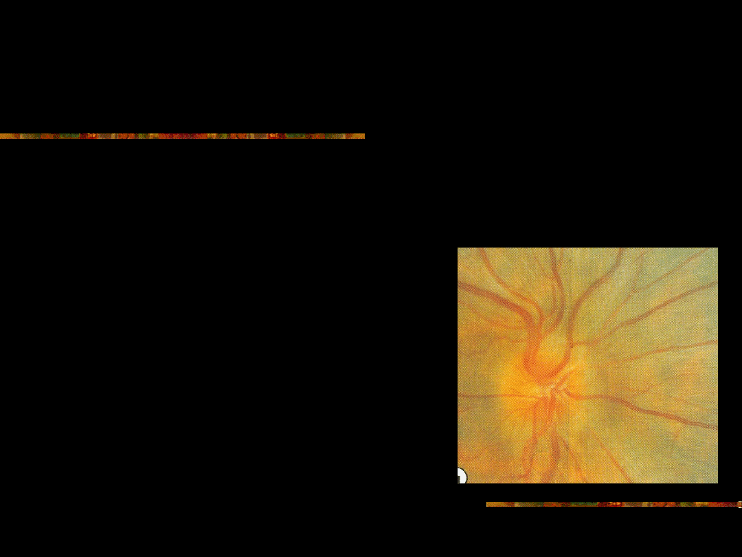
Early Papilloedema:
Blurry nasal margin of the
optic nerve head
Leak on Fluorescein
Angiography.
Dilated veins on the optic
nerve head
lack of venous pulsation
( also in 20% of normal
individuals)
Papillary splinter
haemarrhage
Visual acuity is usually
normal at this stage
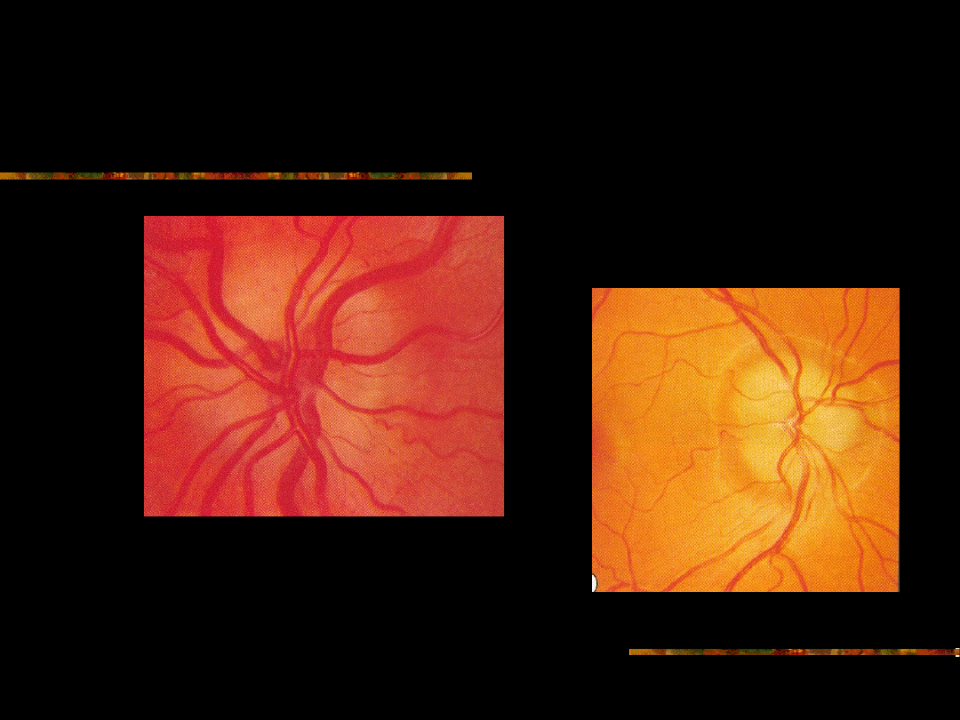
Early Papilloedema

Early Papilloedema
-differential diagnosis
Optic disk drusen
Hypermetropia
Hypertensive retinopathy
Small optic disk
Myelinated nerve fibres
Congenital optic nerve head
abnormalities
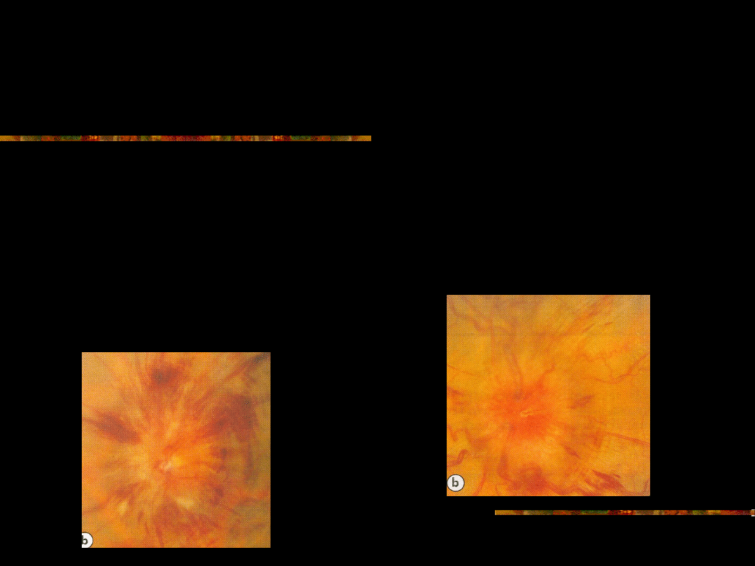
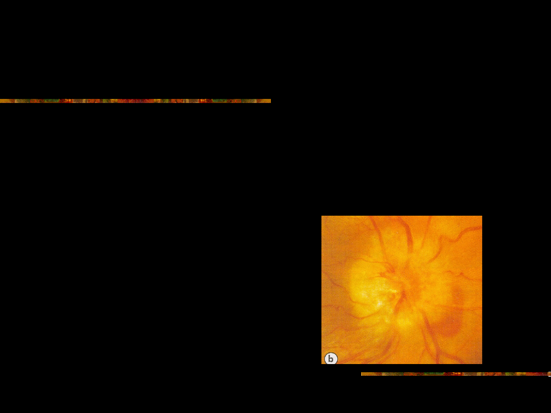
Full blown papilloedema;
Elevated disc, mashroom shape, loss of sharp borders
Wide tortuous disc vessels, flame haemorrhages,cotton
wool spots, hard exudates
Peripapillary retinal folds ( Paton’s folds)
Visual acuity normal or decreased, impaired colour
perception.
Diabetic and hypertensive retinopathy;
Differential diagnosis of fullblown
Papilloedema rerly causes problems.
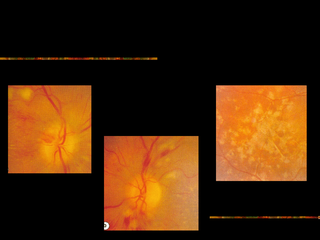
Chronic Papilloedema;
Mashroom shape elevation.
Haemorrhages are diminishing
Drusen-like deposits on the disc
Sometimes macular star
Decreased visual acuity
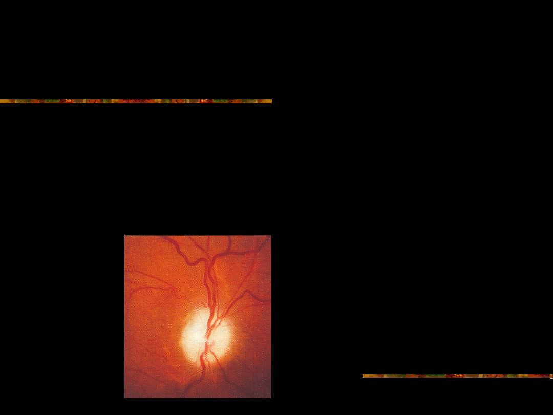
Late; Optic nerve atrophy;
Grey or white optic nerve head with blurry margins, slightly
elevated.
Peripapillary pigmentary changes
VA; no light perception or light perception

Foster-Kenedy’s Syndrome:
Optic atropyh on the side of the tumor and
papilloedema on the contralatelal side.
May be caused by frontal tumor.
Differential diagnosis; new AION on one side and
optic atrophy from old AION on the contralateral
side.

Thank you for your attention.
QUESTIONS?

What is the definition of
Papilloedema?

What are the causes of
Papilloedema?

What are the causes of papilltis?

What are the causes of
pseudopapillitis.
Document Outline
- Slide 1
- Slide 2
- Slide 3
- Slide 4
- Slide 5
- Slide 6
- Slide 7
- Slide 8
- Slide 9
- Slide 10
- Slide 11
- Slide 12
- Slide 13
- Slide 14
- Slide 15
- Slide 16
- Slide 17
- Slide 18
- Slide 19
- Slide 20
- Slide 21
- Slide 22
- Slide 23
- Slide 24
- Slide 25
- Slide 26
- Slide 27
- Slide 28
- Slide 29
Wyszukiwarka
Podobne podstrony:
Human Papillomavirus and Cervical Cancer Knowledge health beliefs and preventive practicies
Human Papillomavirus and Cervical Cancer Knowledge
Pseudomonas and Pseudomonas like Bacteria
08 Polak M A Preventing punching shear failures of reinforced concrete slabs, results of static and
Lulu Papillon FR
tuto papillon brode
Bohm K Papillon
Bague Papillon
collier papillon
Yiruma Papillon (con accordi)
papillon
Brumble, Vine Deloria, Jr , Creationism, and Ethnic Pseudoscience (1998)
ielęgnacja psa rasy Papillon
rosenthal moritz papillons
SZCZEPIONKI PRZECIW HUMAN PAPILLOMAVIRUS (HPV)
Postmodernity and Postmodernism ppt May 2014(3)
Pseudomonas
więcej podobnych podstron