
1
Aluminium in brain tissue in autism
Matthew Mold
a
, Dorcas Umar
b
, Andrew King
c
, Christopher Exley
a*
a
The Birchall Centre, Lennard-Jones Laboratories, Keele University, Staffordshire, ST5 5BG,
United Kingdom.
b
Life Sciences, Keele University, Staffordshire, ST5 5BG, United Kingdom.
c
Department of Clinical Neuropathology, Kings College Hospital, London, SE5 9RS, United
Kingdom.
ABSTRACT
Autism spectrum disorder is a neurodevelopmental disorder of unknown aetiology. It is
suggested to involve both genetic susceptibility and environmental factors including in the
latter environmental toxins. Human exposure to the environmental toxin aluminium has been
linked, if tentatively, to autism spectrum disorder. Herein we have used transversely heated
graphite furnace atomic absorption spectrometry to measure, for the first time, the aluminium
content of brain tissue from donors with a diagnosis of autism. We have also used an
aluminium-selective fluor to identify aluminium in brain tissue using fluorescence
microscopy. The aluminium content of brain tissue in autism was consistently high. The
mean (standard deviation) aluminium content across all 5 individuals for each lobe were
3.82(5.42), 2.30(2.00), 2.79(4.05) and 3.82(5.17) g/g dry wt. for the occipital, frontal,
temporal and parietal lobes respectively. These are some of the highest values for aluminium
in human brain tissue yet recorded and one has to question why, for example, the aluminium
content of the occipital lobe of a 15 year old boy would be 8.74 (11.59) g/g dry wt.?
ACCEPTED MANUSCRIPT

2
Aluminium-selective fluorescence microscopy was used to identify aluminium in brain tissue
in 10 donors. While aluminium was imaged associated with neurones it appeared to be
present intracellularly in microglia-like cells and other inflammatory non-neuronal cells in
the meninges, vasculature, grey and white matter. The pre-eminence of intracellular
aluminium associated with non-neuronal cells was a standout observation in autism brain
tissue and may offer clues as to both the origin of the brain aluminium as well as a putative
role in autism spectrum disorder.
Keywords: Human exposure to aluminium; human brain tissue; autism spectrum disorder;
transversely heated atomic absorption spectrometry; aluminium-selective fluorescence
microscopy
1. Introduction
Autism spectrum disorder (ASD) is a group of neurodevelopmental conditions of unknown
cause. It is highly likely that both genetic [1] and environmental [2] factors are associated
with the onset and progress of ASD while the mechanisms underlying its aetiology are
expected to be multifactorial [3-6]. Human exposure to aluminium has been implicated in
ASD with conclusions being equivocal [7-10]. To-date the majority of studies have used hair
as their indicator of human exposure to aluminium while aluminium in blood and urine have
also been used to a much more limited extent. Paediatric vaccines that include an aluminium
adjuvant are an indirect measure of infant exposure to aluminium and their burgeoning use
has been directly correlated with increasing prevalence of ASD [11]. Animal models of ASD
continue to support a connection with aluminium and to aluminium adjuvants used in human
ACCEPTED MANUSCRIPT

3
vaccinations in particular [12]. Hitherto there are no previous reports of aluminium in brain
tissue from donors who died with a diagnosis of ASD. We have measured aluminium in brain
tissue in autism and identified the location of aluminium in these tissues.
2. Materials and methods
2.1. Measurement of aluminium in brain tissues
Ethical approval was obtained along with tissues from the Oxford Brain Bank (15/SC/0639).
Samples of cortex of approximately 1g frozen weight from temporal, frontal, parietal and
occipital lobes and hippocampus (0.3g only) were obtained from 5 individuals with ADI-R-
confirmed (Autism Diagnostic Interview-Revised) ASD, 4 males and 1 female, aged 15-50
years old (Table 1).
The aluminium content of these tissues was measured by an established and fully validated
method [13] that herein is described only briefly. Thawed tissues were cut using a stainless
steel blade to give individual samples of ca 0.3g (3 sample replicates for each lobe except for
hippocampus where the tissue was used as supplied) wet weight and dried to a constant
weight at 37C. Dried and weighed tissues were digested in a microwave (MARS Xpress
CEM Microwave Technology Ltd.) in a mixture of 1mL 15.8M HNO
3
(Fisher Analytical
Grade) and 1mL 30% w/v H
2
O
2
(BDH Aristar). Digests were clear with no fatty residues and,
upon cooling, were made up to 5mL volume using ultrapure water (cond. <0.067S/cm).
Total aluminium was measured in each sample by transversely heated graphite furnace
atomic absorption spectrometry (TH GFAAS) using matrix-matched standards and an
established analytical programme alongside previously validated quality assurance data [13].
2.2. Fluorescence microscopy
ACCEPTED MANUSCRIPT

4
All chemicals were from Sigma Aldrich (UK) unless otherwise stated. Where available
frontal, parietal, occipital, temporal and hippocampal tissue from 10 donors ( 3 females and 7
males) with a diagnosis of ASD was supplied by the Oxford Brain Bank as three 5μm thick
serial paraffin-embedded brain tissue sections per lobe for each donor (Table S1). Tissue
sections mounted on glass slides were placed in a slide rack and de-waxed and rehydrated via
transfer through 250 mL of the following reagents: 3 min. in Histo-Clear (National
Diagnostics, US), 1 min. in fresh Histo-Clear, 2 min. in 100% v/v ethanol (HPLC grade) and
1 min. in 95, 70, 50 & 30% v/v ethanol followed by rehydration in ultrapure water
(cond.<0.067S/cm) for 35 s. Slides were agitated every 20 s in each solvent and blotted on
tissue paper between transfers to minimise solvent carry-over. Rehydrated brain tissue
sections were carefully outlined with a PAP pen for staining, in order to form a hydrophobic
barrier around the periphery of tissue sections. In between staining, tissue sections were kept
hydrated with ultrapure water and stored in moisture chambers, to prevent sections from
drying out. Staining was staggered to allow for accurate incubation times of brain tissue
sections. We have developed and optimised the fluor lumogallion as a selective stain for
aluminium in cells [14] and human tissues [15]. Lumogallion (4-chloro-3-(2,4-
dihydroxyphenylazo)-2-hydroxybenzene-1-sulphonic acid, TCI Europe N.V. Belgium) was
prepared at ca 1mM via dilution in a 50mM PIPES (1,4-Piperazinediethanesulphonic acid)
buffer, adjusted to pH 7.4 with NaOH. Lumogallion staining was performed via the addition
of 200μL of the staining solution to rehydrated brain tissue sections that were subsequently
incubated at ambient temperature away from light for 45 min. Sections for autofluorescence
analyses were incubated for 45 min in 200μL 50mM PIPES buffer only, pH 7.4. Following
staining, glass slides containing tissue sections were washed six times with 200μL aliquots of
50mM PIPES buffer, pH 7.4, prior to rinsing for 30 s in ultrapure water. Serial sections
numbered 1 and 2 for each lobe were incubated in 50mM PIPES buffer, pH 7.4 or stained
ACCEPTED MANUSCRIPT

5
with 1mM lumogallion in the same buffer, respectively, to ensure consistency across donor
tissues. All tissue sections were subsequently mounted under glass coverslips using the
aqueous mounting medium, Fluoromount™. Slides were stored horizontally for 24 h at 4
o
C
away from light, prior to analysis via fluorescence microscopy.
Stained and mounted human brain tissue sections were analysed via the use of an Olympus
BX50 fluorescence microscope, equipped with a vertical illuminator and BX-FLA reflected
light fluorescence attachment (mercury source). Micrographs were obtained at X 400
magnification by use of a X 40 Plan-Fluorite objective (Olympus, UK). Lumogallion-reactive
aluminium and related autofluorescence micrographs were obtained via use of a U-MNIB3
fluorescence filter cube (excitation: 470 – 495 nm, dichromatic mirror: 505 nm, longpass
emission: 510 nm, Olympus, UK). Light exposure and transmission values were fixed across
respective staining treatment conditions and images were obtained using the CellD software
suite (Olympus, Soft Imaging Solutions, SiS, GmbH). Lumogallion-reactive regions
identified through sequential screening of stained human brain tissue sections were
additionally imaged on autofluorescence serial sections, to assess the contribution of the
fluorophore. The subsequent merging of fluorescence and bright-field channels was achieved
using Photoshop (Adobe Systems Inc. US). When determining intracellular staining the type
of cells stained were estimated by their size and shape in the context of the brain area
sampled and their surrounding cellular environment.
3. Results
3.1. Aluminium content of brain tissues
The aluminium content of all tissues ranged from 0.01 (the limit of quantitation) to 22.11g/g
dry wt. (Table 1). The aluminium content for whole brains (n=4 or 5 depending upon the
ACCEPTED MANUSCRIPT

6
availability of hippocampus tissue) ranged from 1.20 (1.06) g/g dry wt. for the 44 year old
female donor (A1) to 4.77 (4.79) g/g dry wt. for a 33 year old male donor (A5). Previous
measurements of brain aluminium, including our 60 brain study [15], have allowed us to
define loose categories of brain aluminium content beginning with ≤1.00 g/g dry wt. as
pathologically benign (as opposed to ‘normal’). Approximately 40% of tissues (24/59) had
an aluminium content considered as pathologically-concerning (2.00 g/g dry wt.) while
approximately 67% of these tissues had an aluminium content considered as pathologically-
significant (3.00 g/g dry wt.). The brains of all 5 individuals had at least one tissue with a
pathologically-significant content of aluminium. The brains of 4 individuals had at least one
tissue with an aluminium content 5.00g/g dry wt. while 3 of these had at least one tissue
with an aluminium content 10.00g/g dry wt. (Table 1). The mean (SD) aluminium content
across all 5 individuals for each lobe were 3.82(5.42), 2.30(2.00), 2.79(4.05) and 3.82(5.17)
g/g dry wt. for the occipital, frontal, temporal and parietal lobes respectively. There were no
statistically significant differences in aluminium content between any of the 4 lobes.
3.2. Aluminium fluorescence in brain tissues
We examined serial brain sections from 10 individuals (3 females and 7 males) who died
with a diagnosis of ASD and recorded the presence of aluminium in these tissues (Table S1).
Excitation of the complex of aluminium and lumogallion emits characteristic orange
fluorescence that appears increasingly bright yellow at higher fluorescence intensities.
Aluminium, identified as lumogallion-reactive deposits, was recorded in at least one tissue in
all 10 individuals. Autofluorescence of immediately adjacent serial sections confirmed
lumogallion fluorescence as indicative of aluminium. Deposits of aluminium were
significantly more prevalent in males (129 in 7 individuals) than females (21 in 3
ACCEPTED MANUSCRIPT

7
individuals). Aluminium was found in both white (62 deposits) and grey (88 deposits) matter.
In females the majority of aluminium deposits were identified as extracellular (15/21)
whereas in males the opposite was the case with 80 out of 129 deposits being intracellular.
We were only supplied with 3 serial sections of each tissue and so we were not able to do any
staining for general morphology which meant that it was not always possible to determine
which subtype of cell was showing aluminium fluorescence.
Aluminium-loaded mononuclear white blood cells, probably lymphocytes, were identified in
the meninges and possibly in the process of entering brain tissue from the lymphatic system
(Fig.1). Aluminium could be clearly seen inside cells as either discrete punctate deposits or as
bright yellow fluorescence. Aluminium was located in inflammatory cells associated with the
vasculature (Fig. 2). In one case what looks like an aluminium-loaded lymphocyte or
monocyte was noted within a blood vessel lumen surrounded by red blood cells while another
probable lymphocyte showing intense yellow fluorescence was noted in the adventitia (Fig.
2b). Glial cells including microglia-like cells that showed positive aluminium fluorescence
were often observed in brain tissue in the vicinity of aluminium-stained extracellular deposits
(Figs. 3&4). Discrete deposits of aluminium approximately 1m in diameter were clearly
visible in both round and amoeboid glial cell bodies (e.g. Fig. 3b). Intracellular aluminium
was identified in likely neurones and glia-like cells and often in the vicinity of or co-
localised with lipofuscin (Fig. 5). Aluminium-selective fluorescence microscopy was
successful in identifying aluminium in extracellular and intracellular locations in neurones
and non-neuronal cells and across all brain tissues studied (Figs.1-5). The method only
identifies aluminium as evidenced by large areas of brain tissue without any characteristic
aluminium-positive fluorescence (Fig. S1).
ACCEPTED MANUSCRIPT

8
4. Discussion
The aluminium content of brain tissues from donors with a diagnosis of ASD was extremely
high (Table 1). While there was significant inter-tissue, inter-lobe and inter-subject variability
the mean aluminium content for each lobe across all 5 individuals was towards the higher end
of all previous (historical) measurements of brain aluminium content, including iatrogenic
disorders such as dialysis encephalopathy [13,15, 16-19]. All 4 male donors had significantly
higher concentrations of brain aluminium than the single female donor. We recorded some of
the highest values for brain aluminium content ever measured in healthy or diseased tissues in
these male ASD donors including values of 17.10, 18.57 and 22.11 g/g dry wt. (Table 1).
What discriminates these data from other analyses of brain aluminium in other diseases is the
age of the ASD donors. Why, for example would a 15 year old boy have such a high content
of aluminium in their brain tissues? There are no comparative data in the scientific literature,
the closest being similarly high data for a 42 year old male with familial Alzheimer’s disease
(fAD) [19].
Aluminium-selective fluorescence microscopy has provided indications as to the location of
aluminium in these ASD brain tissues (Figs. 1-5). Aluminium was found in both white and
grey matter and in both extra- and intracellular locations. The latter were particularly pre-
eminent in these ASD tissues. Cells that morphologically appeared non-neuronal and heavily
loaded with aluminium were identified associated with the meninges (Fig. 1), the vasculature
(Fig. 2) and within grey and white matter (Figs. 3-5). Some of these cells appeared to be glial
(probably astrocytic) whilst others had elongated nuclei giving the appearance of microglia
[5]. The latter were sometimes seen in the environment of extracellular aluminium
deposition. This implies that aluminium somehow had crossed the blood-brain barrier and
was taken up by a native cell namely the microglial cell. Interestingly, the presence of
ACCEPTED MANUSCRIPT

9
occasional aluminium-laden inflammatory cells in the vasculature and the leptomeninges
opens the possibility of a separate mode of entry of aluminium into the brain i.e.
intracellularly. However, to allow this second scenario to be of significance one would expect
some type of intracerebral insult to occur to allow egress of lymphocytes and monocytes from
the vasculature. The identification herein of non-neuronal cells including inflammatory cells,
glial cells and microglia loaded with aluminium is a standout observation for ASD. For
example, the majority of aluminium deposits identified in brain tissue in fAD were
extracellular and nearly always associated with grey matter [19]. Aluminium is cytotoxic [21]
and its association herein with inflammatory cells in the vasculature, meninges and central
nervous system is unlikely to be benign. Microglia heavily loaded with aluminium while
potentially remaining viable, at least for some time, will inevitably be compromised and
dysfunctional microglia are thought to be involved in the aetiology of ASD [22], for example
in disrupting synaptic pruning [23]. In addition the suggestion from the data herein that
aluminium entry into the brain via immune cells circulating in the blood and lymph is
expedited in ASD might begin to explain the earlier posed question of why there was so
much aluminium in the brain of a 15 year old boy with an ASD.
A limitation of our study is the small number of cases that were available to study and the
limited availability of tissue. Regarding the latter, having access to only 1g of frozen tissue
and just 3 serial sections of fixed tissue per lobe would normally be perceived as a significant
limitation. Certainly if we had not identified any significant deposits of aluminium in such a
small (the average brain weighs between 1500 and 2000g) sample of brain tissue then such a
finding would be equivocal. However, the fact that we found aluminium in every sample of
brain tissue, frozen or fixed, does suggest very strongly that individuals with a diagnosis of
ASD have extraordinarily high levels of aluminium in their brain tissue and that this
ACCEPTED MANUSCRIPT

10
aluminium is pre-eminently associated with non-neuronal cells including microglia and other
inflammatory monocytes.
5. Conclusions
We have made the first measurements of aluminium in brain tissue in ASD and we have
shown that the brain aluminium content is extraordinarily high. We have identified
aluminium in brain tissue as both extracellular and intracellular with the latter involving both
neurones and non-neuronal cells. The presence of aluminium in inflammatory cells in the
meninges, vasculature, grey and white matter is a standout observation and could implicate
aluminium in the aetiology of ASD.
Author contributions
CE designed the study, carried out tissue digests and TH GFAAS. DU carried out tissue
digests and TH GFAAS. AK carried out brain neuropathology on sections prepared by MM.
MM carried out all microscopy and with CE wrote the manuscript. All authors read and
approved the manuscript.
Competing interests
The authors declare that they have no competing interests.
Acknowledgements
ACCEPTED MANUSCRIPT

11
The research is supported by a grant from the Children’s Medical Safety Research Institute
(CMSRI), a not-for-profit research foundation based in Washington DC, USA.
References
1. A. Krishnan, R.Zhang, V. Yao, C.L.Theesfeld, A.K. Wong et al., Genome-wide prediction
and functional characterisation of the genetic basis of autism spectrum disorder, Nature
Neuroscience 19 (2016) 1454-1462.
2. L.A. Sealey, B.W. Hughes, A.N. Sriskanda, J.R. Guest, A.D. Gibson et al., Environmental
factors in the development of autism spectrum disorders, Environ. Int. 88 (2016) 288-298.
3. R. Koyama, Y. Ikegaya, Microglia in the pathogenesis of autism spectrum disorders,
Neurosci. Res. 100 (2015) 1-5.
4. Q. Li, J-M. Zhou, The microbiota-gut-brain axis and its potential therapeutic role in autism
spectrum disorder, Neuroscience 324 (2016) 131-139.
5. C. Kaur, G. Rathnasamy, E-A. Ling, Biology of microglia in the developing brain, J.
Neuropathol Exp. Neurol. 76 (2017) 736-753.
6. M. Varghese, N. Keshav, S. Jacot-Descombes, T. Warda, B. Wicinski et al., Autism
spectrum disorder: neuropathology and animal models, Acta Neuropathol. 134 (2017) 537-
566.
7. H. Yasuda, Y. Yasuda, T. Tsutsui, Estimation of autistic children by metallomics analysis,
Sci. Rep. 3 (2013) 1199.
8. F.E.B. Mohamed, E.A. Zaky, A.B. El-Sayed, R.M. Elhossieny, S.S. Zahra et al.,
Assessment of hair aluminium, lead and mercury in a sample of autistic Egyptian children:
ACCEPTED MANUSCRIPT

12
environmental risk factors of heavy metals in autism, Behavioural Neurol. (2015) Art.
545674.
9. M.H. Rahbar, M. Samms-Vaughn, M.R. Pitcher, J. Bressler, M. Hessabi et al., Role of
metabolic genes in blood aluminium concentrations of Jamaican children with and without
autism spectrum disorder, Int. J. Environ. Res. Public Health 13 (2016) 1095.
10. A.V. Skalny, N.V. Simashkova, T.P. Klyushnik, A.R. Grabeklis, I.V. Radysh et al.,
Analysis of hair trace elements in children with autism spectrum disorders and
communication disorders, Trace Elem. Med. Biol. 177 (2017) 215-223.
11. L. Tomljenovic, C.A. Shaw, Do aluminium vaccine adjuvants contribute to the rising
prevalence of autism?, J. Inorg. Biochem. 105 (2011) 1489-1499.
12. C.A. Shaw, Y. Li, L. Tomljenovic, Administration of aluminium to neonatal mice in
vaccine-relevant amounts is associated with adverse long term neurological outcomes, J.
Inorg. Biochem. 128 (2013) 237-244.
13. E. House, M. Esiri, G. Forster, P. Ince, C. Exley, Aluminium, iron and copper in human
brain tissues donated to the medical research council’s cognitive function and ageing study,
Metallomics 4 (2012) 56-65.
14. M. Mold, H. Eriksson, P. Siesjö, A. Darabi, E. Shardlow, C. Exley, Unequivocal
identification of intracellular aluminium adjuvant in a monocytic THP-1 cell line, Sci. Rep. 4
(2014) 6287.
15. A. Mirza, A. King, C. Troakes, C. Exley, The identification of aluminium in human brain
tissue using lumogallion and fluorescence microscopy, J. Alzh. Dis. 54 (2016) 1333-1338.
ACCEPTED MANUSCRIPT

13
16. C. Exley, M. Esiri, Severe cerebral congophilic angiopathy coincident with increased
brain aluminium in a resident of Camelford, Cornwall, UK, J. Neurol. Neurosurg. Psychiatry
77 (2006) 877-879.
17. C. Exley, E.R. House, Aluminium in the human brain, Monatsh. Chem. 142 (2011) 357-
363.
18. C. Exley, T. Vickers, Elevated brain aluminium and early onset Alzheimer’s disease in an
individual occupationally exposed to aluminium: a case report, J. Med. Case Rep. 8 (2014)
41.
19. A. Mirza, A. King, C. Troakes, C. Exley, Aluminium in brain tissue in familial
Alzheimer’s disease, J. Trace Elem. Med. Biol. 40 (2017) 30-36.
20. R. Shechter, O. Miller, G. Yovel, N. Rosenzweig, A. London et al., Recruitment of
beneficial M2 macrophages to injured spinal cord is orchestrated by remote brain choroid
plexus, Immunity 38 (2013) 555-569.
21. C. Exley, The toxicity of aluminium in humans, Morphologie 100 (2016) 51-55.
22. M.W. Salter, B. Stevens, Microglia emerge as central players in brain disease, Nat. Med.
23 (2017) 1018-1027.
23. U. Neniskyte, C.T. Gross, Errant gardeners: glial-cell-dependent synaptic pruning and
neurodevelopmental disorders, Nat. Rev. Neurosci. 18 (2017) 658-670.
ACCEPTED MANUSCRIPT

14
Figure legends
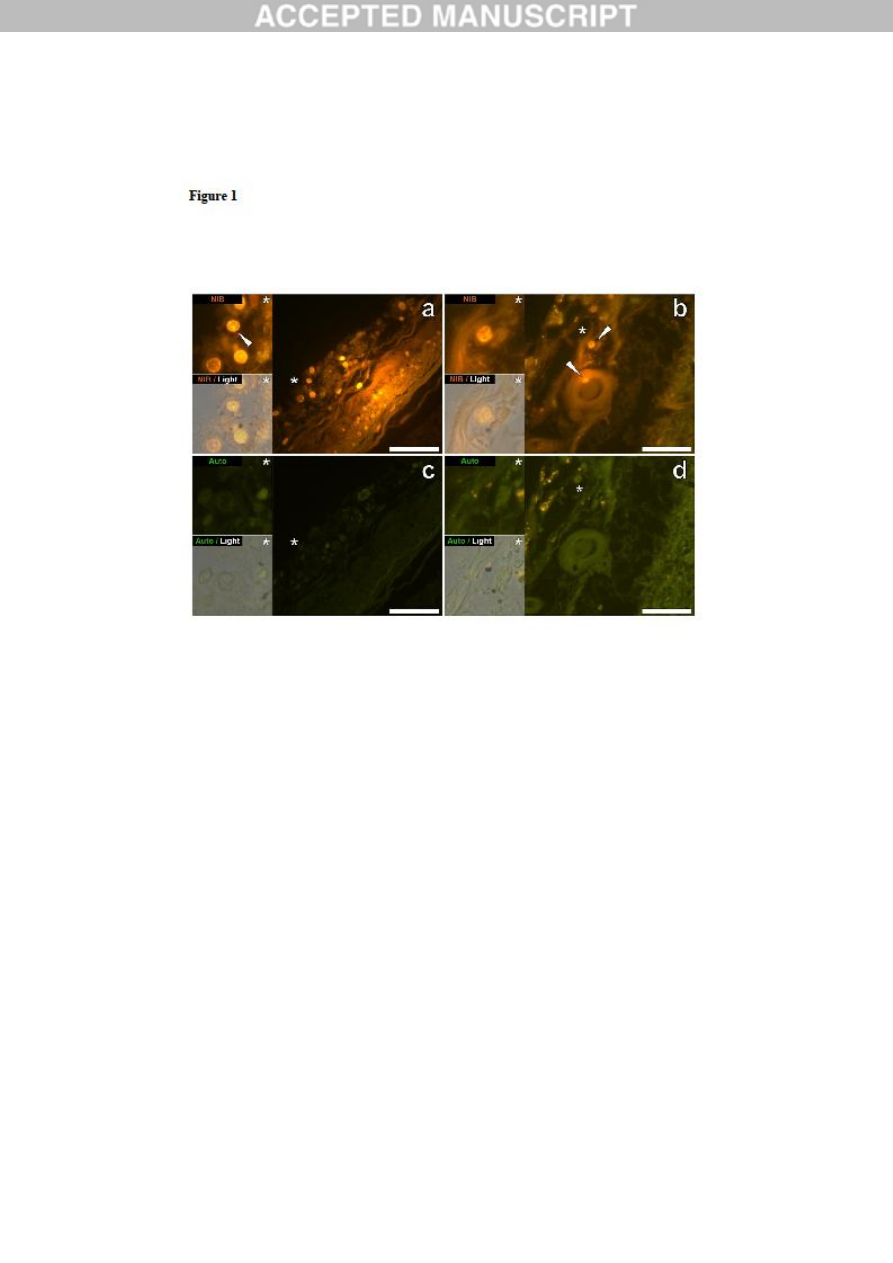
Figure 1. Mononuclear inflammatory cells (probably lymphocytes) in leptomeningeal
membranes in the hippocampus and frontal lobe of a 50-year-old male donor (A2),
diagnosed with autism. Intracellular lumogallion-reactive aluminium was noted via punctate
orange fluorescence emission (white arrows) in the hippocampus (a) and frontal lobe (b). A
green autofluorescence emission was detected in the adjacent non-stained (5μm) serial section
(c & d). Upper and lower panels depict magnified inserts marked by asterisks, of the
fluorescence channel and bright field overlay. Magnification X 400, scale bars: 50μm.
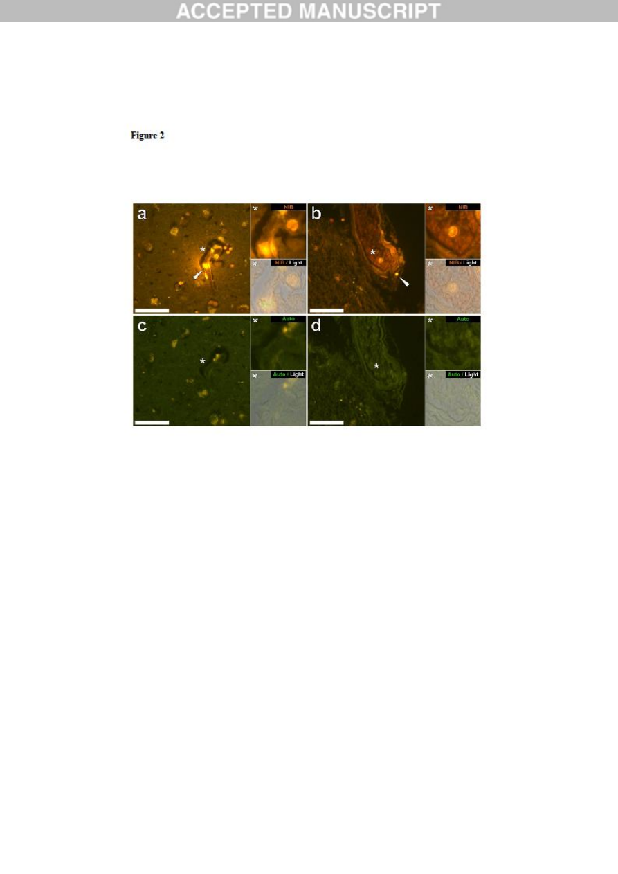
Figure 2. Intracellular lumogallion-reactive aluminium in the vasculature of the
hippocampus of a 50-year-old male donor (A2), diagnosed with autism. Aluminium-loaded
inflammatory cells noted in the hippocampus in the vessel wall (white arrow) (a) and depicting
punctate orange fluorescence in the lumen (b) are highlighted. An inflammatory cell in the
vessel adventitia was also noted (white arrow) (b). Lumogallion-reactive aluminium was
identified via an orange fluorescence emission (a & b) versus a green autofluorescence
emission (c & d) of the adjacent non-stained (5μm) serial section. Upper and lower panels
depict magnified inserts marked by asterisks, of the fluorescence channel and bright field
overlay. Magnification X 400, scale bars: 50μm.
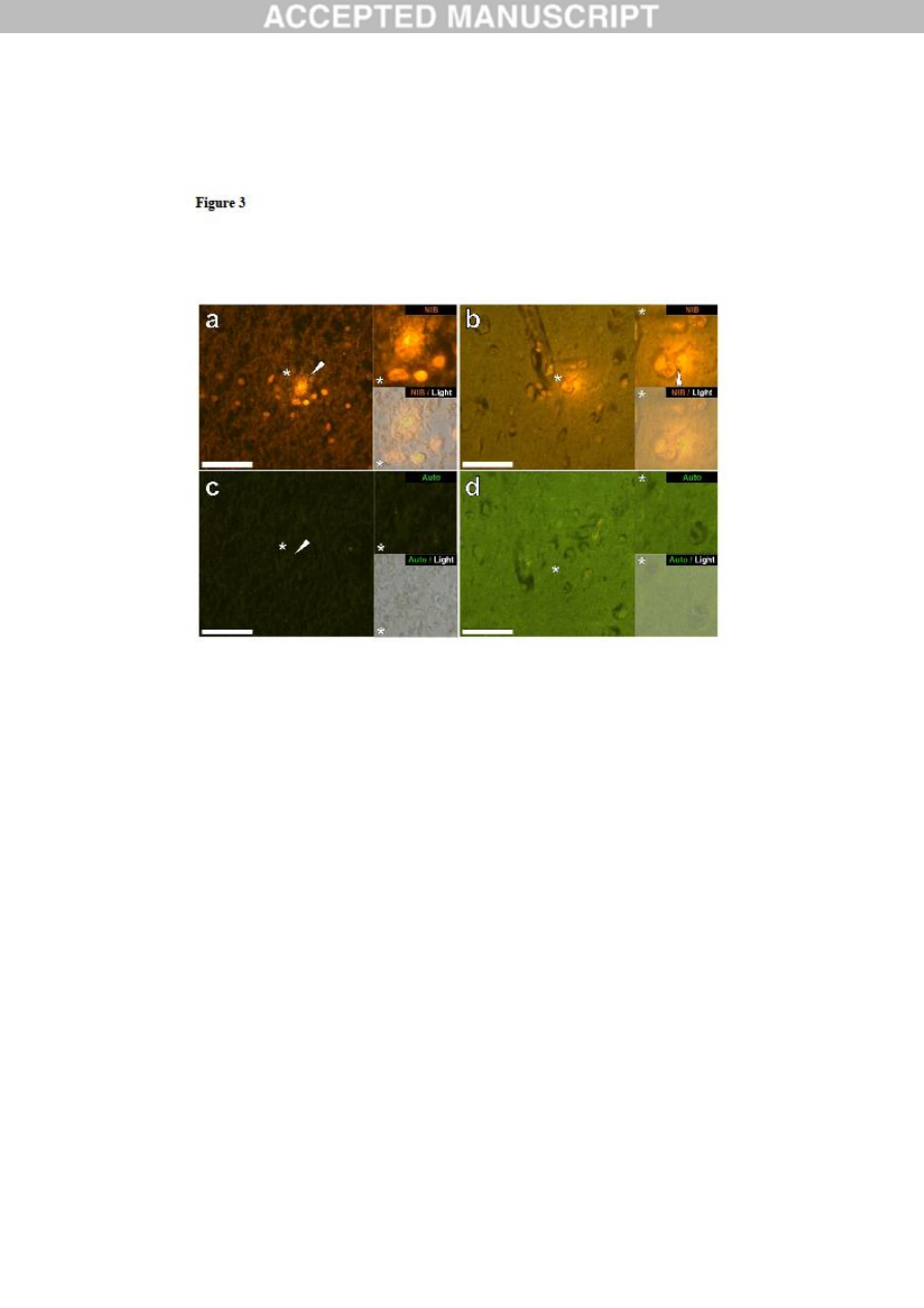
Figure 3. Intracellular aluminium in cells morphologically compatible with glia and
neurones in the hippocampus of a 15-year-old male donor (A4), diagnosed with autism.
Lumogallion reactive cellular aluminium identified within glial-like cells in the hippocampus
(a) and producing a punctate orange fluorescence in glia surrounding a likely neuronal cell
within the parietal lobe (b) are highlighted (white arrows). Lumogallion-reactive aluminium
ACCEPTED MANUSCRIPT

15
was identified via an orange fluorescence emission (a & b) versus a green autofluorescence
emission (c & d) of the subsequent non-stained (5μm) serial section (white arrow/asterisk).
Upper and lower panels depict magnified inserts marked by asterisks, of the fluorescence
channel and bright field overlay. Magnification X 400, scale bars: 50μm.
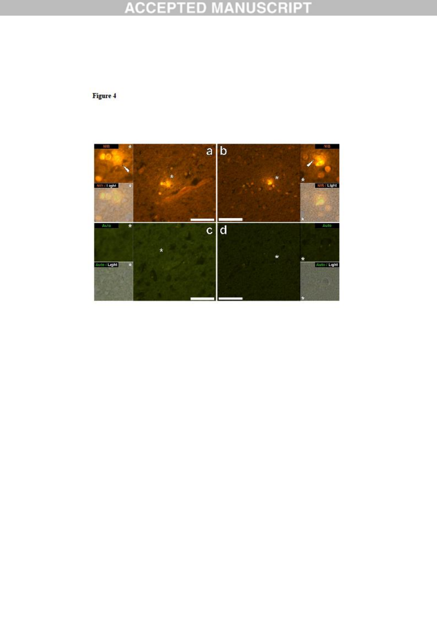
Figure 4. Intracellular aluminium in cells morphologically compatible with microglia
within the parietal and temporal lobes of 29-year-old (A8) and 15-year-old (A4) male
donors, diagnosed with autism. Lumogallion-reactive extracellular aluminium (white arrows)
producing an orange fluorescence emission was noted around likely microglial cells in the
parietal (a) and temporal lobes (b) of donors A8 and A4 respectively. Non-stained adjacent
(5μm) serial sections, produced a weak green autofluorescence emission of the identical area
imaged in white (c) and grey matter (d) of the respective lobes. Upper and lower panels depict
magnified inserts marked by asterisks, of the fluorescence channel and bright field overlay.
Magnification X 400, scale bars: 50μm.
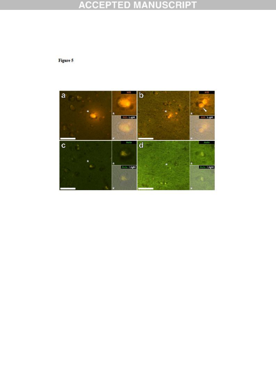
Figure 5. Lumogallion-reactive aluminium in likely neuronal and glial cells in the
temporal lobe and hippocampus of a 14-year-old male donor (A10), diagnosed with
autism. Intraneuronal aluminium in the temporal lobe (a) was identified via an orange
fluorescence emission, co-deposited with lipofuscin as revealed by a yellow fluorescence in
the non-stained autofluorescence serial (5μm) section (c). Intracellular punctate orange
fluorescence (white arrow) was observed in glia in the hippocampus (b) producing a green
autofluorescence emission on the non-stained section (d). Upper and lower panels depict
magnified inserts marked by asterisks, of the fluorescence channel and bright field overlay.
Magnification X 400, scale bars: 50μm.
ACCEPTED MANUSCRIPT
16
ACCEPTED MANUSCRIPT
17
ACCEPTED MANUSCRIPT
18
ACCEPTED MANUSCRIPT
19
ACCEPTED MANUSCRIPT
20
ACCEPTED MANUSCRIPT
21
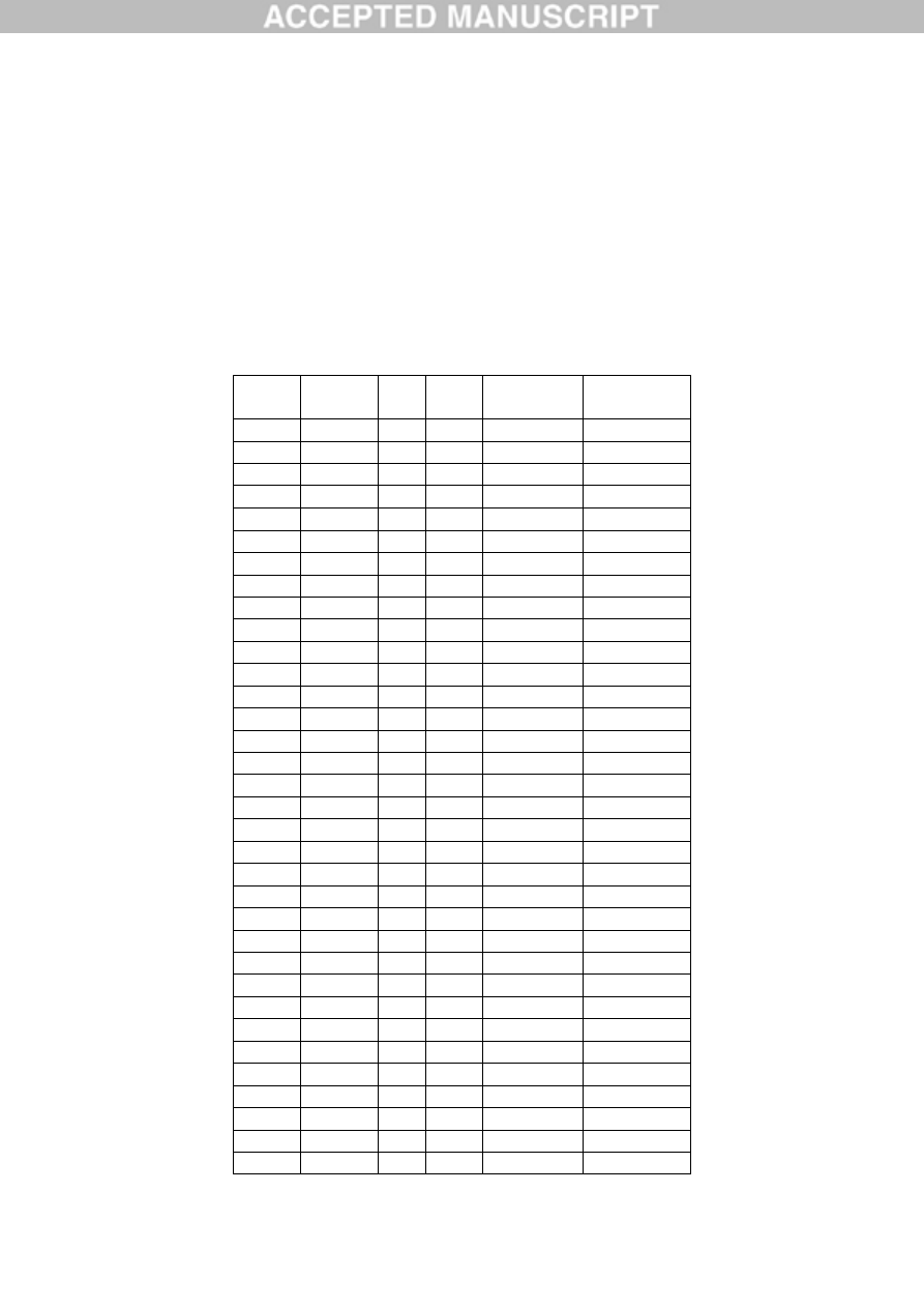
Table 1. Aluminium content of occipital (O), frontal (F), temporal (T) and parietal (P) lobes
and hippocampus (H) of brain tissue from 5 donors with a diagnosis of autism spectrum
disorder.
Donor
ID
Gender Age Lobe Replicate
[Al] g/g
A1
F
44
O
1
0.49
2
4.26
3
0.33
Mean (SD) 1.69 (2.22)
F
1
0.98
2
1.10
3
0.95
Mean (SD) 1.01 (0.08)
T
1
1.13
2
1.16
3
1.12
Mean (SD) 1.14 (0.02)
P
1
0.54
2
1.18
3
NA
Mean (SD) 0.86 (0.45)
All
Mean (SD) 1.20 (1.06)
A2
M
50
O
1
3.73
2
7.87
3
3.49
Mean (SD) 5.03 (2.46)
F
1
0.86
2
0.88
3
1.65
Mean (SD) 1.13 (0.45)
T
1
1.31
2
1.02
ACCEPTED MANUSCRIPT
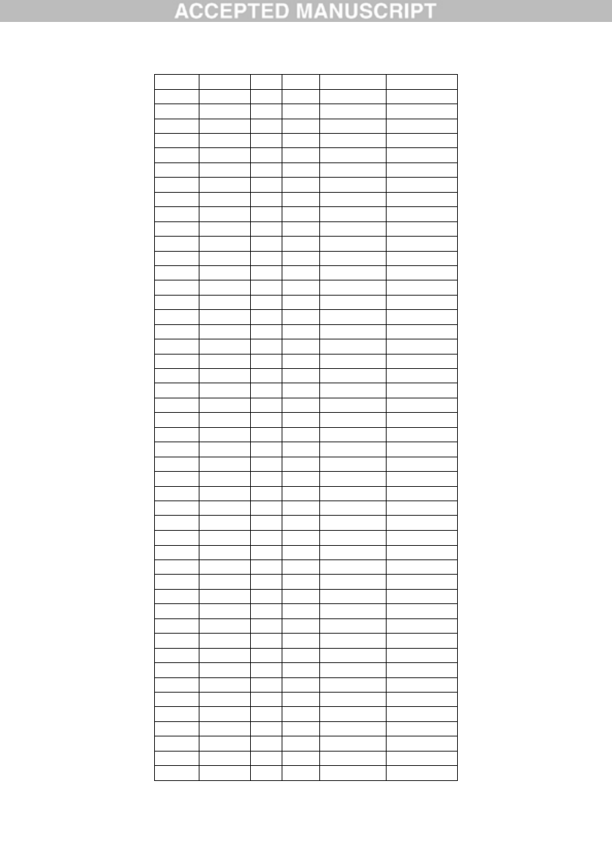
22
3
2.73
Mean (SD) 1.69 (0.92)
P
1
18.57
2
0.01
3
0.64
Mean (SD) 6.41 (10.54)
Hip.
1
1.42
All
Mean (SD) 3.40 (5.00)
A3
M
22
O
1
0.64
2
2.01
3
0.66
Mean (SD) 1.10 (0.79)
F
1
1.72
2
4.14
3
2.73
Mean (SD) 2.86 (1.22)
T
1
1.62
2
4.25
3
2.57
Mean (SD) 2.81 (1.33)
P
1
0.13
2
3.12
3
5.18
Mean (SD) 2.82 (1.81)
All
Mean (SD) 2.40 (1.58)
A4
M
15
O
1
2.44
2
1.66
3
22.11
Mean (SD) 8.74 (11.59)
F
1
1.11
2
3.23
3
1.66
Mean (SD) 2.00 (1.10)
T
1
1.10
2
1.83
3
1.54
Mean (SD) 1.49 (0.37)
P
1
1.38
2
6.71
3
NA
Mean (SD) 4.05 (3.77)
Hip.
1
0.02
ACCEPTED MANUSCRIPT
23
All
Mean (SD) 3.73 (6.02)
A5
M
33
O
1
3.13
2
2.78
3
1.71
Mean (SD) 2.54 (0.74)
F
1
2.97
2
8.27
3
NA
Mean (SD) 5.62 (3.75)
T
1
1.71
2
1.64
3
17.10
Mean (SD) 6.82 (8.91)
P
1
5.53
2
2.89
3
NA
Mean (SD) 4.21 (1.87)
All
Mean (SD) 4.77 (4.79)
ACCEPTED MANUSCRIPT
Wyszukiwarka
Podobne podstrony:
Theory of Mind in normal development and autism
(autyzm) Autismo Gray And White Matter Brain Chemistry In Young Children With Autism
Application of Magnetic Resonance Spectroscopy in the Mental Diseases of Schizophrenia and Autism
Brain,?havior and Media key
The?lance in the World and Man
Brain,?havior and Media
Kundalini Is it Metal in the Meridians and Body by TM Molian (2011)
Topic 13 AHL Plants IB III Lecture 2 Plant Tissues and Organs
Mutations in the CgPDR1 and CgERG11 genes in azole resistant C glabrata
Producing proteins in transgenic plants and animals
Top 5?st Jobs in the US and UK – 15?ition
[41]Hormesis and synergy pathways and mechanisms of quercetin in cancer prevention and management
Flashback to the 1960s LSD in the treatment of autism
A Comparison of the Status of Women in Classical Athens and E
2 Fill in Present Perfect and Present Perfect Continuous
Changes in Brain Function of Depressed Subjects During
The problems in the?scription and classification of vovels
więcej podobnych podstron