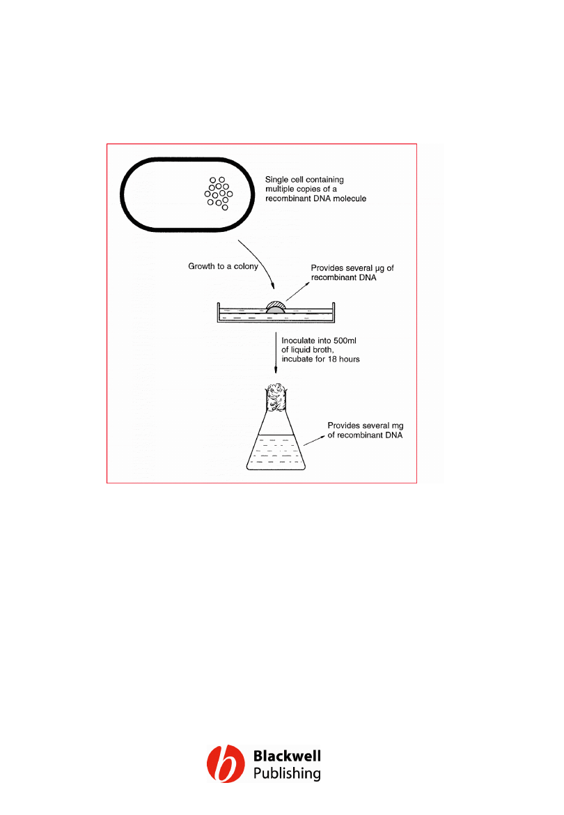
Figure 5.1 Cloning can supply large amounts
of recombinant DNA.
Gene Cloning and DNA Analysis by T.A. Brown. © 2006 T.A.
Brown.
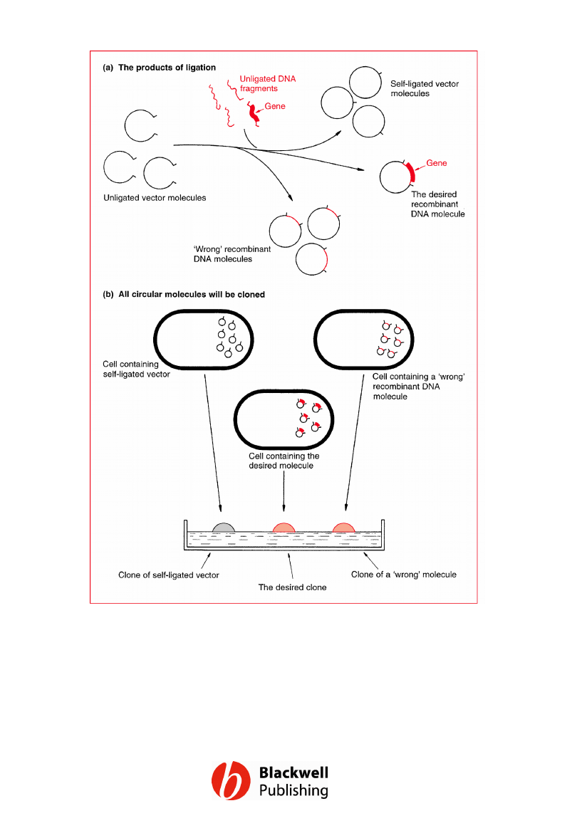
Figure 5.2 Cloning is analogous to
purification. From a mixture of different
molecules, clones containing copies of just
one molecule can be obtained.
Gene Cloning and DNA Analysis by T.A. Brown. © 2006 T.A.
Brown.
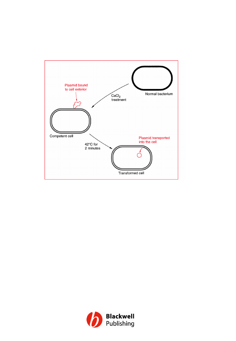
Figure 5.3 The binding and uptake of DNA
by a competent bacterial cell.
Gene Cloning and DNA Analysis by T.A. Brown. © 2006 T.A.
Brown.
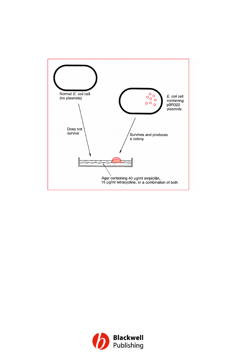
Figure 5.4 Selecting cells that contain
pBR322 plasmids by plating onto agar
medium containing ampicillin and/or
tetracycline.
Gene Cloning and DNA Analysis by T.A. Brown. © 2006 T.A.
Brown.
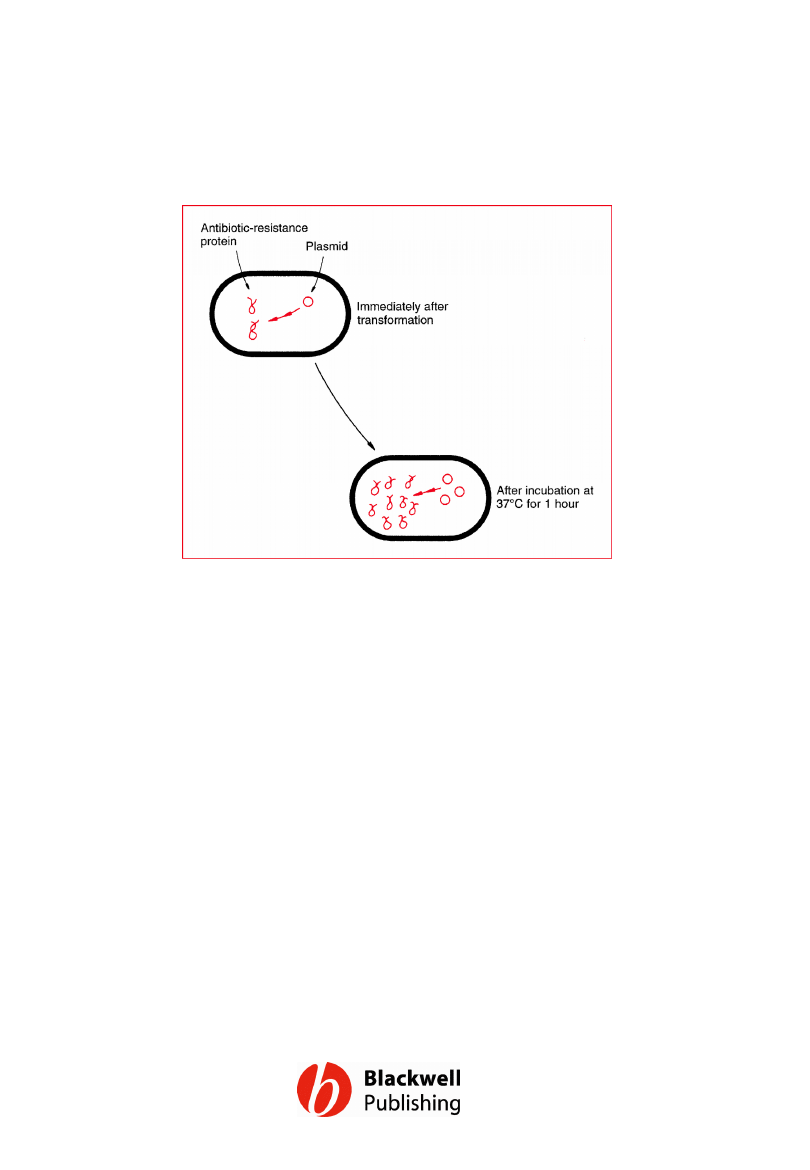
Figure 5.5 Phenotypic expression. Incubation
at 37°C for 1 hour before plating out
improves the survival of the transformants on
selective medium, because the bacteria have
had time to begin synthesis of the antibiotic
resistance enzymes.
Gene Cloning and DNA Analysis by T.A. Brown. © 2006 T.A.
Brown.
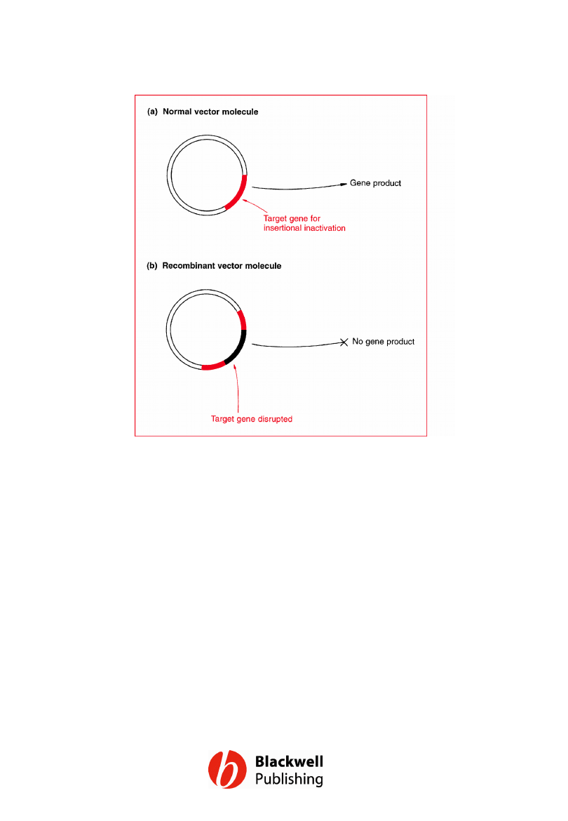
Figure 5.6 Insertional inactivation. (a) The
normal, non-recombinant vector molecule
carries a gene whose product confers a
selectable or identifiable characteristic on the
host cell. (b) This gene is disrupted when new
DNA is inserted into the vector; as a result the
recombinant host does not display the
relevant characteristic.
Gene Cloning and DNA Analysis by T.A. Brown. © 2006 T.A.
Brown.
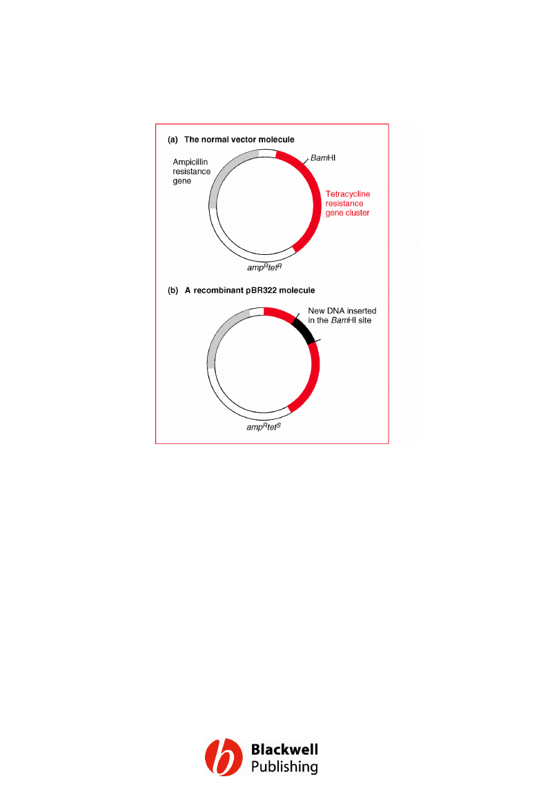
Figure 5.7 The cloning vector pBR322: (a)
the normal vector molecule; (b) a
recombinant molecule containing an extra
piece of DNA inserted into the BamHI site. For
a more detailed map of pBR322 see Figure
6.1.
Gene Cloning and DNA Analysis by T.A. Brown. © 2006 T.A.
Brown.
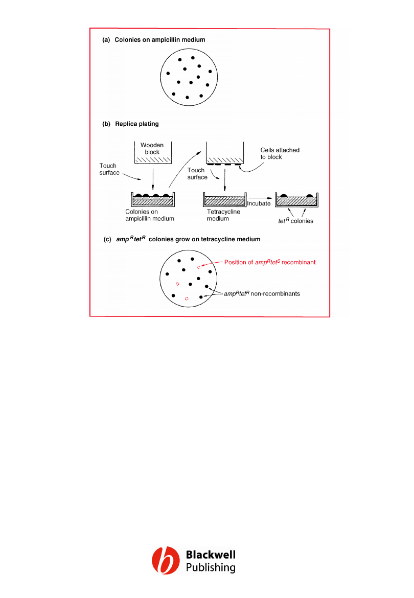
Figure 5.8 Screening for pBR322
recombinants by insertional inactivation of
the tetracycline resistance gene. (a) Cells are
plated onto ampicillin agar: all the
transformants produce colonies. (b) The
colonies are replica plated onto tetracycline
medium. (c) The colonies that grow on
tetracycline medium are ampRtetR and
therefore non-recombinants. Recombinants
(ampRtetS) do not grow, but their position on
the ampicillin plate is now known.
Gene Cloning and DNA Analysis by T.A. Brown. © 2006 T.A.
Brown.
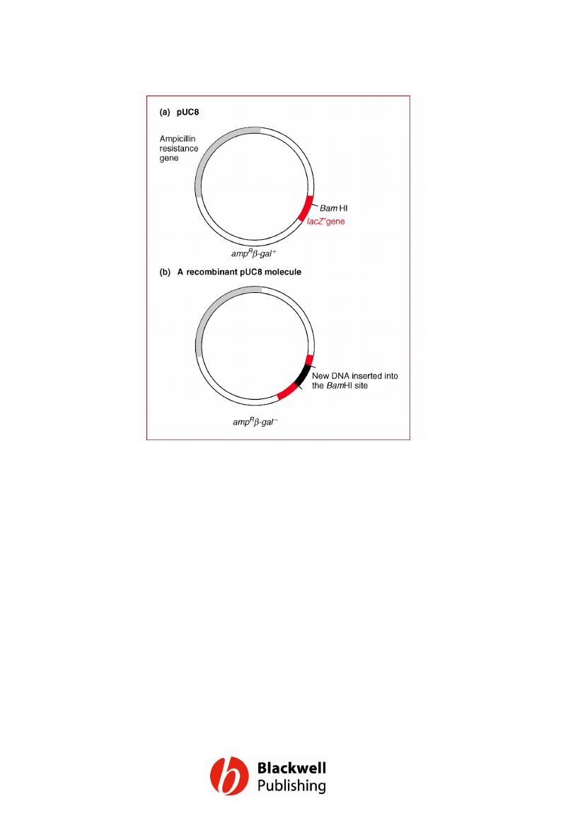
Figure 5.9 The cloning vector pUC8: (a) the
normal vector molecule; (b) a recombinant
molecule containing an extra piece of DNA
inserted into the BamHI site. For more
detailed maps of pUC8 see Figure 6.3.
Gene Cloning and DNA Analysis by T.A. Brown. © 2006 T.A.
Brown.
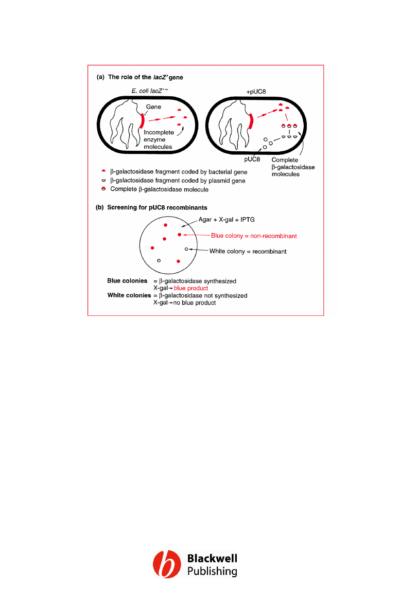
Figure 5.10 The rationale behind insertional
inactivation of the lacZ¢ gene carried by
pUC8. (a) The bacterial and plasmid genes
complement each other to produce a
functional b-galactosidase molecule. (b)
Recombinants are screened by plating onto
agar containing X-gal and IPTG.
Gene Cloning and DNA Analysis by T.A. Brown. © 2006 T.A.
Brown.
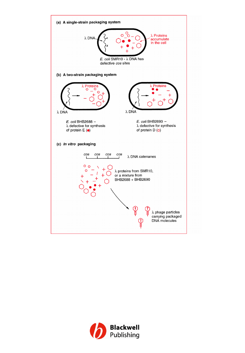
Figure 5.11 In vitro packaging. (a) Synthesis
of l capsid proteins by E. coli strain SMR10,
which carries a l phage that has defective cos
sites. (b) Synthesis of incomplete sets of l
capsid proteins by E. coli strains BHB2688
and BHB2690. (c) The cell lysates provide the
complete set of capsid proteins and can
package l DNA molecules in the test tube.
Gene Cloning and DNA Analysis by T.A. Brown. © 2006 T.A.
Brown.
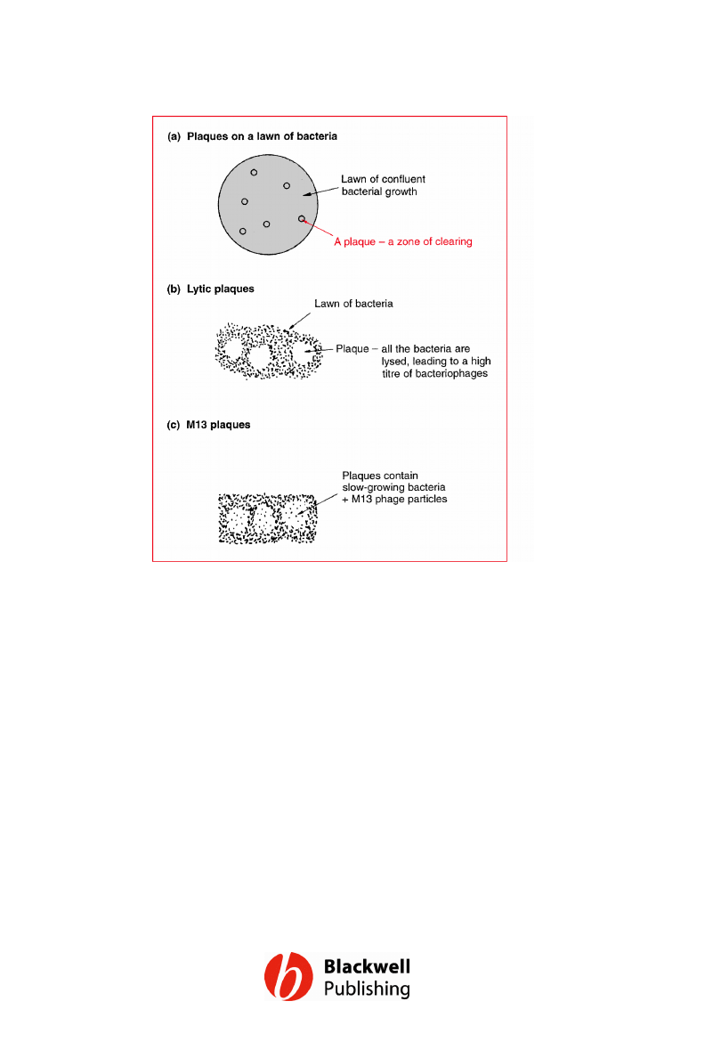
Figure 5.12 Bacteriophage plaques. (a) The
appearance of plaques on a lawn of bacteria.
(b) Plaques produced by a phage that lyses
the host cell (e.g. l in the lytic infection cycle);
the plaques contain lysed cells plus many
phage particles. (c) Plaques produced by
M13; these plaques contain slow-growing
bacteria plus many M13 phage particles.
Gene Cloning and DNA Analysis by T.A. Brown. © 2006 T.A.
Brown.
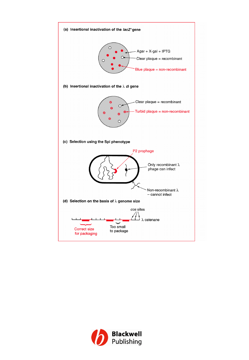
Figure 5.13 Strategies for the selection of
recombinant phage.
Gene Cloning and DNA Analysis by T.A. Brown. © 2006 T.A.
Brown.
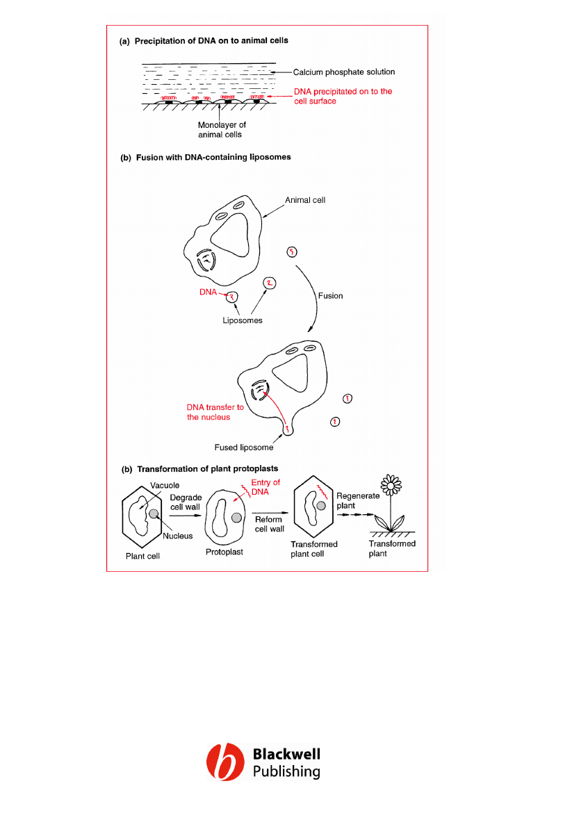
Figure 5.14 Strategies for introducing new
DNA into animal and plant cells: (a)
precipitation of DNA on to animal cells; (b)
introduction of DNA into animal cells by
liposome fusion; (c) transformation of plant
protoplasts.
Gene Cloning and DNA Analysis by T.A. Brown. © 2006 T.A.
Brown.
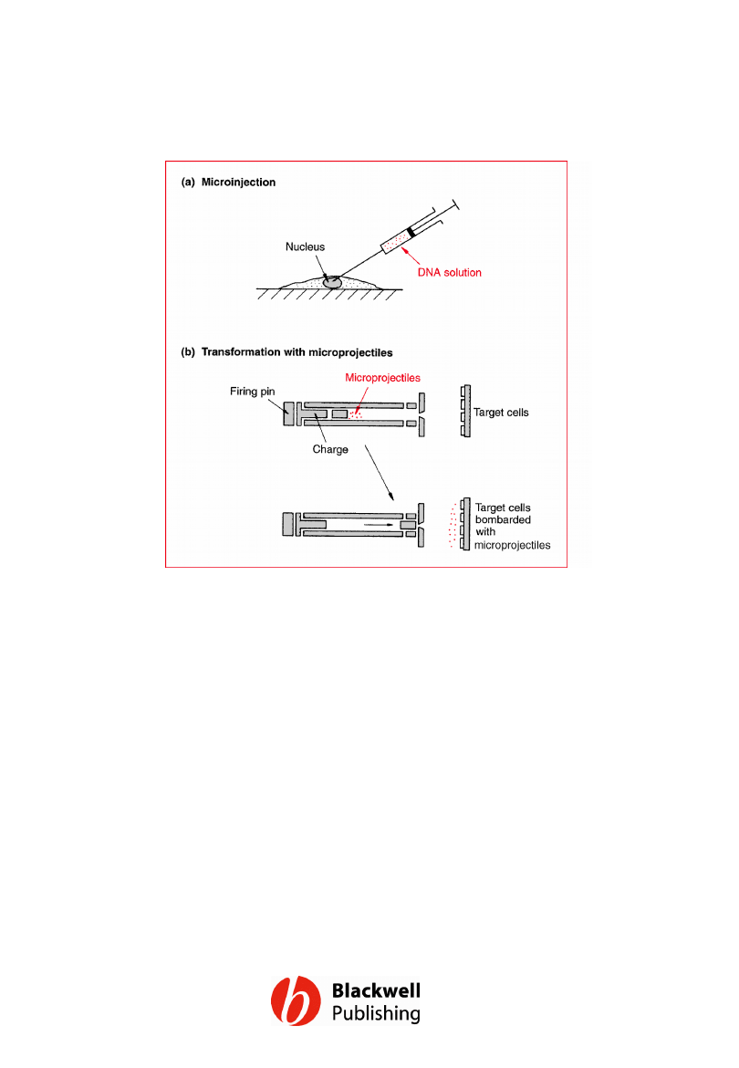
Figure 5.15 Two physical methods for
introducing DNA into cells.
Gene Cloning and DNA Analysis by T.A. Brown. © 2006 T.A.
Brown.
Document Outline
- Figure 5.1 Cloning can supply large amounts of recombinant DNA.
- Figure 5.2 Cloning is analogous to purification. From a mixture of different molecules, clones containing copies of just one molecule can be obtained.
- Figure 5.3 The binding and uptake of DNA by a competent bacterial cell.
- Figure 5.4 Selecting cells that contain pBR322 plasmids by plating onto agar medium containing ampicillin and/or tetracycline.
- Figure 5.5 Phenotypic expression. Incubation at 37°C for 1 hour before plating out improves the survival of the transformants on selective medium, because the bacteria have had time to begin synthesis of the antibiotic resistance enzymes.
- Figure 5.6 Insertional inactivation. (a) The normal, non-recombinant vector molecule carries a gene whose product confers a selectable or identifiable characteristic on the host cell. (b) This gene is disrupted when new DNA is inserted into the vector; as a result the recombinant host does not display the relevant characteristic.
- Figure 5.7 The cloning vector pBR322: (a) the normal vector molecule; (b) a recombinant molecule containing an extra piece of DNA inserted into the BamHI site. For a more detailed map of pBR322 see Figure 6.1.
- Figure 5.8 Screening for pBR322 recombinants by insertional inactivation of the tetracycline resistance gene. (a) Cells are plated onto ampicillin agar: all the transformants produce colonies. (b) The colonies are replica plated onto tetracycline medium. (c) The colonies that grow on tetracycline medium are ampRtetR and therefore non-recombinants. Recombinants (ampRtetS) do not grow, but their position on the ampicillin plate is now known.
- Figure 5.9 The cloning vector pUC8: (a) the normal vector molecule; (b) a recombinant molecule containing an extra piece of DNA inserted into the BamHI site. For more detailed maps of pUC8 see Figure 6.3.
- Figure 5.10 The rationale behind insertional inactivation of the lacZ¢ gene carried by pUC8. (a) The bacterial and plasmid genes complement each other to produce a functional b-galactosidase molecule. (b) Recombinants are screened by plating onto agar containing X-gal and IPTG.
- Figure 5.11 In vitro packaging. (a) Synthesis of l capsid proteins by E. coli strain SMR10, which carries a l phage that has defective cos sites. (b) Synthesis of incomplete sets of l capsid proteins by E. coli strains BHB2688 and BHB2690. (c) The cell lysates provide the complete set of capsid proteins and can package l DNA molecules in the test tube.
- Figure 5.12 Bacteriophage plaques. (a) The appearance of plaques on a lawn of bacteria. (b) Plaques produced by a phage that lyses the host cell (e.g. l in the lytic infection cycle); the plaques contain lysed cells plus many phage particles. (c) Plaques produced by M13; these plaques contain slow-growing bacteria plus many M13 phage particles.
- Figure 5.13 Strategies for the selection of recombinant phage.
- Figure 5.14 Strategies for introducing new DNA into animal and plant cells: (a) precipitation of DNA on to animal cells; (b) introduction of DNA into animal cells by liposome fusion; (c) transformation of plant protoplasts.
- Figure 5.15 Two physical methods for introducing DNA into cells.
Wyszukiwarka
Podobne podstrony:
Figures for chapter 12
Figures for chapter 6
Figures for chapter 14
Figures for chapter 10
Figures for chapter 11
Figures for chapter 8
Figures for chapter 9
Figures for chapter 2
Figures for chapter 16
Figures for chapter 13
Figures for chapter 3
Figures for chapter 7
Figures for chapter 15
Figures for chapter 1
Figures for chapter 12
Figures for chapter 6
Figures for chapter 14
Figures for chapter 10
więcej podobnych podstron