
Polímeros: Ciência e Tecnologia, vol. 19, nº 3, p. 241-247, 2009
241
Autor para correspondência: Odilio B. G. Assis, Embrapa Instrumentação Agropecuária, Rua XV de Novembro 1452, CEP: 13560-970, São Carlos, SP,
Brasil. E-mail: odilio@cnpdia.embrapa.br
!
2
4
)
'
/
$
%
2
%
6
)
3
Ä
/
chitosan depends on its biological origin, molecular weight
and degree of acetylation
[3]
. Since chitosan is soluble
in diluted acid solutions, films can be readily prepared
by casting or dipping, resulting in dense and porous
structure
[4,5]
.
Chitosan film is regarded as biofunctional material, well
tolerated by living tissues, particularly applicable as edible
coatings to prolong shelf-life and preserve quality of fresh
foods
[6]
. In medical field, chitosan films have been tested
as curative wound dressing and as scaffolds for tissue and
bone engineering
[7]
. Additionally the reactive functional
groups present in chitosan (amino group at the C2 position
of each deacetylated unit and hydroxyl groups at the C6
and C3 positions) can be readily subjected to chemical
derivatization allowing the manipulation of mechanical and
solubility properties
[8]
enlarging its biocompatibility.
The Antimicrobial Models of Chitosan
Chitin and chitosan have been investigated as an
antimicrobial material against a wide range of target
organisms like algae, bacteria, yeasts and fungi in
experiments involving in vivo and in vitro interactions
with chitosan in different forms (solutions, films and
composites). Early research describing the antimicrobial
potential of chitin, chitosan, and their derivatives dated from
the 1980-1990s
[9-14]
. Generally, in these studies the chitosan
is considered to be a bacteriocidal (kills the live bacteria or
some fraction therein) or bacteriostatic (hinders the growth
of bacteria but does not imply whether or not bacteria are
killed), often with no distinction between activities. Recent
data in literature has the tendency to characterize chitosan
as bacteriostatic rather than bactericidal
[15]
, although the
exact mechanism is not fully understood and several other
factors may contribute to the antibacterial action
[16]
.
Introduction
Chitin is a polysaccharide of animal origin found
abundantly in nature and characterized by a fibrous
structure. It forms the basis of the main constituent of the
outer skeleton of insects and crustaceans like shrimp, crabs
and lobster
[1]
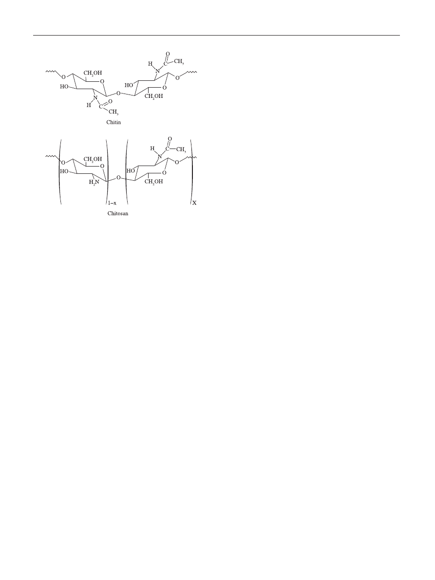
. The chemical structure of chitin is similar to
cellulose, having one hydroxyl group on each monomer
substituted with an acetylamine group (Figure 1). The
extraction of chitin involves an acid removal of calcium
carbonate (demineralization), generally by hot reaction with
HCl, HNO
3
or HCl, etc., followed by a deproteinization
(removal of proteins). This step usually performed by
alkaline treatments (e.g. with NaOH)
[1,2]
. In its extracted
crude form, chitin has a highly ordered crystalline structure,
is translucent, resilient and quite tough. It has, however,
poor solubility and low reactivity.
The chitin structure can be modified by removing the
acetyl groups, which are bond to amine radicals in the
C2 position on the glucan ring, by means of a chemical
hydrolysis in concentrated alkaline solution at elevated
temperature to produce a deacetylated form (Figure 1).
When the fraction of acetylated amine groups is reduced
to 40-35%, the resultant co-polymer, (1
→ 4)-2-amine-2-
deoxy-
β-D-glucan and (1 → 4)-2-acetamide-2-deoxy-β-D-
glucan, is then referred to as chitosan. Chitosan is primarily
characterized by its molecular weight (MW) and the degree
of acetylation (DA). Commercially chitosan is available
with > 85% deacetylated units (DA < 15%), and molecular
weights (MW) between 100 and 1000 kDa. There is no a
specific standard to define MW, but it is accepted that Low
MW < 50 kDa, Medium MW 50 – 150 kDa, and High MW
> 150 kDa.
Chitosan is a weak base and is insoluble in water, but
soluble in dilute aqueous acidic solutions below its pKa
(~6.3), in which it can convert glucosamine units (-NH
2
)
into the soluble protonated form (-NH
+
3
). The solubility of
A Review of the Antimicrobial Activity of Chitosan
Rejane C. Goy, Douglas de Britto, Odilio B. G. Assis
Embrapa Instrumentação Agropecuária, São Carlos/SP
Abstract: Chitosan, a versatile hydrophilic polysaccharide derived from chitin, has a broad antimicrobial spectrum to which
gram-negative, gram-positive bacteria and fungi are highly susceptible. In the current review, three possible and accepted
antimicrobial mechanisms for chitosan are presented and briefly discussed. The activity dependence on polymeric molecular
weight (MW) and degree of acetylation (DA) are described. The chitosan minimum inhibitory concentrations (MIC) are
summarized according to recent data found in the literature. The potential to improve inhibitory growth of bacteria by
using water soluble chitosan derivatives is also discussed. The data indicate that the effectiveness of chitosan varies and is
dependent on species of target microorganisms.
Keywords: Chitosan, polysaccharide, antimicrobial mechanisms.
Goy, R. C. et al. - A Review of the antimicrobial activity of chitosan
242
Polímeros: Ciência e Tecnologia, vol. 19, nº 3, p. 241-247, 2009
worth observing that the amount of polycationic chitosan
available to bind to a charged bacterial surface is apparently
reduced as the concentration of chitosan increases
[15,26]
.
A possible explanation is that in the presence of a larger
number of charged sites, the chains tend to form clusters
by molecules aggregation while they are still in solution
[4]
.
Observations have confirmed that at higher concentrations,
the chitosan tends to form a coating over the bacteria, not
necessary attached to the surface and independently of the
bacteria type
[13]
. In such condition, adjustments on pH could
be decisive for a good solubility and to keep the chains apart
from each other.
Concerning the bacteria surface polarity, the outer
membrane of gram-negative bacteria consists essentially of
lipopolysaccharides containing phosphate and pyrophosphate
groups which render to the surface a density of negative
charges superior to that observed for gram-positive ones
(membrane composed by peptidoglycan associated to
polysaccharides and teichoic acids)
[27]
. This supports the
evidence that the leakage of intracellular material observed
by chitosan in gram-negative is superior to that reported in
gram-positive bacteria
[21-23]
.
The bacterial effectiveness on gram-positive or gram-
negative bacteria is however, somewhat controversial. Some
authors have stated that chitosan generally showed stronger
effects for gram-positive bacteria (e.g. Listeria monocytogenes,
Bacillus megaterium, B. cereus, Staphylococcus aureus,
Lactobacillus plantarum, L. brevis, L. bulgaris, etc.
) than for
gram-negative bacteria (e.g. E. coli, Pseudomonas fluorescens,
Salmonella typhymurium, Vibrio parahaemolyticus
, etc.)
[28-31]
.
Conversely, it has been demonstrated that hydrophilicity in
gram-negative bacteria is significantly higher than in gram-
positive bacteria, making them most sensitive to chitosan
[32]
.
These findings are confirmed by several in vitro experiments
in which gram-negative bacteria appear to be very sensitive
to chitosan, exhibiting increased morphological changes on
treatment when compared to gram-positives
[22,23,33-35]
. The
charge density on the cell surface is a determinant factor to
establish the amount of adsorbed chitosan. More adsorbed
chitosan would evidently result in greater changes in the
structure and in the permeability of the cell membrane.
This would suggest that the antibacterial mode of action is
dependent upon the host microorganism
[24]
.
Another proposed mechanism is the binding of chitosan
with microbial DNA, which leads to the inhibition of the
mRNA and protein synthesis via the penetration of chitosan
into the nuclei of the microorganisms
[10,13,36]
. In this the
chitosan molecules is assumed to be able to pass through the
bacterial cell wall, composed of multilayers of cross-linked
murein, and reach the plasma membrane. Observation by
confocal laser scanning microscopy
[7]
confirmed the presence
of chitosan oligomers (a chain with few number of monomer
units) inside E. coli exposed to chitosan under different
conditions. Raafat et al.
[16]
stated that in spite of been accepted
as a possible mechanism, the probability of it occurring is
Three models have been proposed, the most acceptable
being the interaction between positively charged chitin/
chitosan molecules and negatively charged microbial cell
membranes. In this model the interaction is mediated by the
electrostatic forces between the protonated NH
+
3
groups and
the negative residues
[17]
, presumably by competing with Ca
2+
for electronegative sites on the membrane surface
[18]
.
This electrostatic interaction results in twofold interfe-
rence: i) by promoting changes in the properties of membrane
wall permeability, thus provoke internal osmotic imbalances
and consequently inhibit the growth of microorganisms
[10,12]
,
and ii) by the hydrolysis of the peptidoglycans in the
microorganism wall, leading to the leakage of intracellular
electrolytes such as potassium ions and other low molecular
weight proteinaceous constituents (e.g. proteins, nucleic
acids, glucose, and lactate dehydrogenase)
[9,11,13,19,20]
.
This model was investigated in a recent work by
Raafat et al.
[16]
, who observed under transmission electron
microscope the ultrastructural changes of S. simulans 22 cells
upon exposure to positively charged chitosan. It was possible
to observe and identify chitosan molecules attached on
bacteria cell surfaces. In the interacting sites it was registered
that the cell membrane became locally detached from the
cell wall, giving rise to “vacuole-like” structures underneath
the wall. The detachment generates ions and water efflux,
provoking decreases on the internal bacteria pressure
[16]
.
Visual confirmation of an effective membrane lysis been also
reported on gram-negative and gram-positive bacteria
[21-23]
.
Since such mechanism is based on electrostatic interaction,
it suggests that the greater the number of cationized amines,
the higher will be the antimicrobial activity
[24,25]
. This
suggests that chitosan has higher activity than that found for
chitin and this has been confirmed experimentally
[17,24]
. It is
Figure 1. Schematic representations of the chemical structures of the chitin
and chitosan.
Goy, R. C. et al. - A Review of the antimicrobial activity of chitosan
Polímeros: Ciência e Tecnologia, vol. 19, nº 3, p. 241-247, 2009
243
Influence of the Degree of Acetylation and Molecular
Weight
Several studies have shown that the biological activity of
chitosan depends significantly on its molecular weight (MW)
and degree of acetylation (DA). Both parameters affect the
antimicrobial activity of chitosan independently, though it has
been suggested that the influence of the MW on the antimicrobial
activity is greater then the influence of the DA
[41]
.
To cite recent examples, studies carried out on Bacillus
cereus, E. coli, Staphylococcus aureus, Pseudomonas
aeruginosa, Salmonella enterica, B. subtilis, Listeria
monocytogenes
and Klebsiella pneumoniae
[42-47]
, proved that
for lower chitosan MW (LMW), greater is the observed effect
on the reducing of microorganism growth and multiplication.
The size and conformation appears to be fundamental to
understand the effectiveness of LMW chitosan. The mobility,
attraction and ionic interaction of small chains are easier than of
big ones facilitating the adoption of an extended conformation
and an effective binding to the membrane surface
[1]
.
Similarly but in different intensity, chitosan antimicrobial
effectiveness is improved as the degree of acetylation is
lower
[46,48]
. Studies on chitin and chitosan with different DA
were analyzed against fungi (Aspergillus fumigatus, Aspergillus
parasiticus, Fusarium oxysporum, Candida albicans
);
Gram-positive (Staphylococcus aureus, Staphylococcus
saprophyticus, Bacillus cereus, Listeria monocytogenes)
and
Gram-negative bacteria (Escherichia coli, Samonella
tiphymurium, Pseudomonas aeruginosa, Enterococcus
faecailis, Aeromonas hydrophila, Shigella dysenteriae,
Vibrio cholerae, Vibrio parahaemolyticus).
In all cases the
antimicrobial activity also increased with decreasing DA
[48-50]
.
As already mentioned, the DA is determinant in the
solubility and charge development, where the –NH
2
, –OH
groups in the molecule of chitosan are considered as the
rater low. The prevailing contention is that chitosan acts
essentially as an outer membrane disruptor rather than as a
penetrating material
[16,34]
.
The third mechanism is the chelation of metals, suppression
of spore elements and binding to essential nutrients to
microbial growth
[37,38]
. It is well known that chitosan has
excellent metal-binding capacities where the amine groups
in the chitosan molecules are responsible for the uptake of
metal cations by chelation
[23]
. In general, such mechanism is
more efficient at high pH in where positive ions are bounded
to chitosan, since the amine groups are unprotonated and the
electron pair on the amine nitrogen is available for donation
to metal ions. A model proposed based on the system
chitosan-Cu, relate the pH dependence on the proportion of
available sites for interacting in polysaccharide backbone
[39]
.
At pH < 6 the complexation involves only one NH
2
group and
three hydroxyls or H
2
O molecules, while at pH > 6.7 is likely
to have two NH
2
involved in the complex formation. For
higher pHs, i.e., 7-9, the deprotonation of hydroxyl groups
are considered to occur and the predominant complexation
is ruled by two –NH
2
and two hydroxyl groups dissociated.
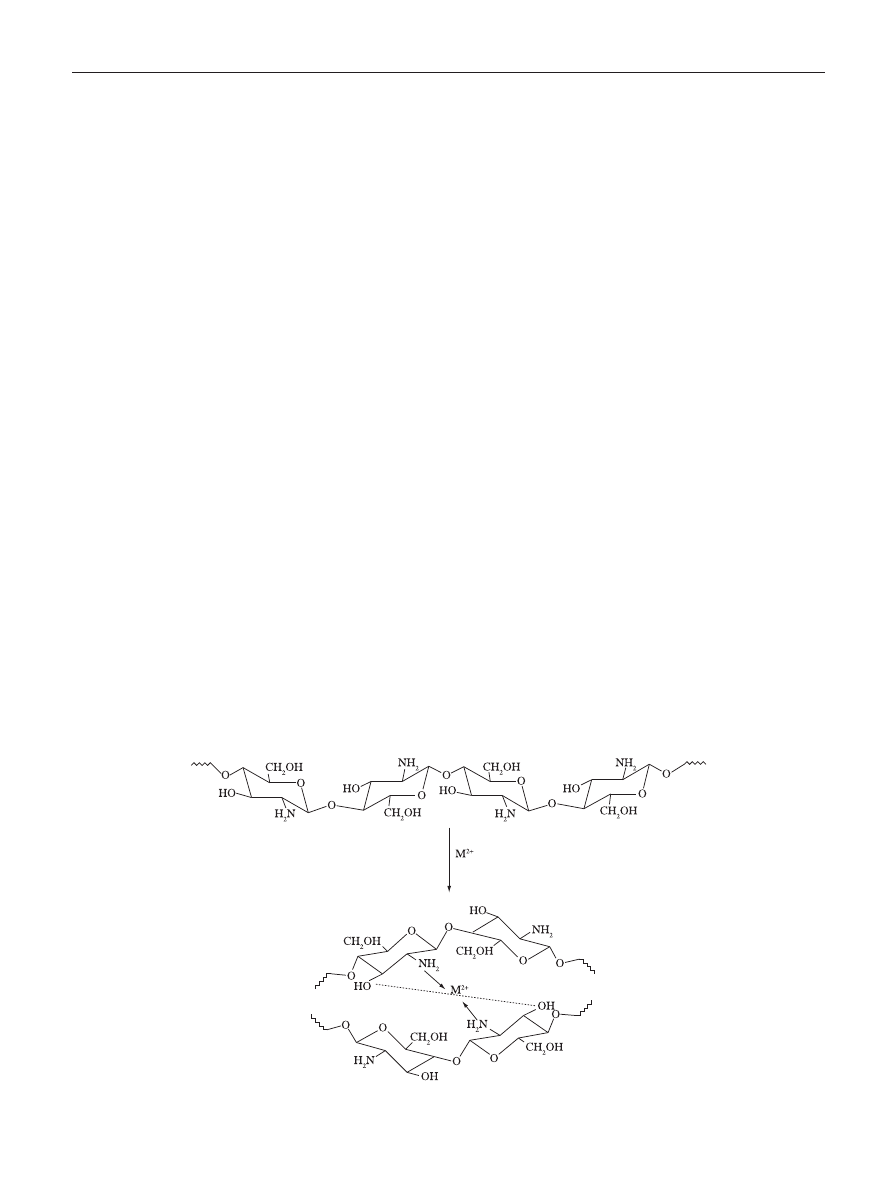
Similarly, in a recent model proposed by Wang et al.
[40]
, the
metal is arranged as an electron acceptor connected to one
or more chitosan chains via –NH
2
and by forming bridges to
hydroxyl groups, as illustrated in Figure 2.
It is unquestionable that chitosan molecules in bacteria
surrounds might complex metals and blockage some essential
nutrients to flow, contributing to cell death
[1]
. Nevertheless, this
is, evidently, not a determinant antimicrobial action since the
sites available for interaction are limited and the complexation
reach saturation in function of metal concentration.
Figure 2. Metal-chitosan complexation model according to Wang et al.
[40]
.

Goy, R. C. et al. - A Review of the antimicrobial activity of chitosan
244
Polímeros: Ciência e Tecnologia, vol. 19, nº 3, p. 241-247, 2009
results from author to author
[58,59]
. MIC however is useful as
a practical indicator of a primary activity against a selected
pathogenic microorganism. In Table 1 is a brief summarization
dominating reactive sites. Hence as the DA is reduced, higher
will be the free amino groups present in chitosan and higher
will be the antimicrobial effect
[48]
.
Antifungal Activity
Similarly to bacteria, the chitosan activity against fungus
is assumed to be fungistatic rather than fungicidal with a
potential to communicate regulatory changes in both the
host and fungus
[16,51]
. Generally chitosan has been reported
as being very effective in inhibiting spore germination, germ
tube elongation and radial growth
[52,53]
. Most of the studies
have been done on yeasts and moulds associated with food
and plant spoilage. For these, in the presence of chitosan,
several biological processes are activated in plant tissue,
where chitinases are induced with action on biotrophic and
necrotrophic mycoparasites, entomopathogenic fungi and
vesicular arbuscular mycorrhizal fungi
[53]
.
The antifungic mechanism of chitosan involves cell
wall morphogenesis with chitosan molecules interfering
directly with fungal growth, similarly to the effects observed
in bacteria cells
[52]
. Microscopic observation reported that
chitosan oligomers diffuse inside hyphae interfering on the
enzymes activity responsible for the fungus growth
[54]
. The
intensity of degradation action of chitosan on fungal cell
walls is also dependant upon the concentration, DA and
local pH
[55]
. Studies conducted in nutrient agar on cultures
of R. solani and S. rolfsii reveled that the percentage of
fungus germination decreased with increasing the chitosan
concentration in the medium. Generally the primary observed
influence is on the length of the lag phase. As the inhibition
process takes place, the medium shifted toward alkalinity
which reduces the effectiveness of the chitosan
[55]
.
Inhibition rate in order of 80% against plant fungus such
as Phomopsis asparagi and as high as 95% against Fusarium
oxysporum, Cucumernum owen, Rhizoctonia solani
and
Fusarium oxysporum
have been, however, known to occur
with low chitosan concentration (20-150 mg.L
-1
)
[56]
.
Sensitivity of Microorganism Strains to Chitosan
Chitosan has several advantages over regular type
of disinfectants owing to its broad spectrum of activity.
Chitosan has been observed to act more quickly on fungi
than on bacteria
[57]
, and activity against typhoid organisms
are comparable to the standard antibiotics used in clinical
practice
[26,33,57]
. As discussed this antimicrobial activity has a
strong dependence on MW and DA characteristics and also
varied according microorganism strains.
There are many studies about the minimum inhibitory
concentration (MIC) for chitin, chitosan, their derivatives or
combination, with different results for different microorganism.
MIC is defined as the lowest concentration of an antimicrobial
that will inhibit the visible growth of a microorganism after
overnight incubation. It is dependent of many factors and the
non-standardized procedures make difficult to compare MIC
Table 1. Minimum inhibitory concentration (MIC) for chitosan against
several microorganisms (concentration normalized to ppm).
Sensible organisms
MIC (ppm)
Reference
Gram negative
Escherichia coli
20
100
468
650
1000
[7]
[50]
[60]
[49]
[31,61,62]
Xanthomonas campestris
500
[63]
Salmonella enterica
2000
3000
[64]
[65]
Samonella tiphymurium
>1000
1500
2000
[31]
[50]
[61]
Pseudomonas aeruginosa
>200
1700
[50]
[61]
Aeromonas hydrophila
1000
[50]
Shigella dysenteriae
>200
[50]
Vibrio cholerae
200
[50]
Vibrio parahaemolyticus
150
1000
[50]
[31]
Pseudomonas fluorescens
250
500
~1000
[19]
[7]
[31]
Enterobacter aerogenes
250
[19]
Gram positive
Bacillus cereus
<1000
1000
[31]
[7,50]
Bacillus megaterium
800
[44]
Staphylococcus aureus
20
100
>800
700
>1250
[7]
[19]
[44]
[61]
[49]
Listeria monocytogenes
150
250
800
[50]
[19]
[31]
Candida lambica
250
[19]
Lactobacillus plantarum
<1000
2000
[44]
[64]
Lactobacillus brevis
1000
[31]
Lactobacillus bulgaricus
>1000
[31]
Fungi
Aspergillus fumigatus
>2000
[50]
Aspergillus parasiticus
>2000
[50]
Fusarium oxysporum
100
[7]
Botrytis cinerea
10
[7]
Byssochlamys
spp.
1000-5000
[38]
Candida albicans
500
600
>1250
[50]
[61]
[49]
Drechstera sorokiana
10
[7]
Microsporum canis
1100
[61]
Trichophyton mentagrophytes
2200
[61]
Goy, R. C. et al. - A Review of the antimicrobial activity of chitosan
Polímeros: Ciência e Tecnologia, vol. 19, nº 3, p. 241-247, 2009
245
of recent works showing some MIC values found for chitosan
against several microorganisms.
Water Soluble Chitosan
Although chitin and chitosan have been confirmed as
attractive biomacromolecules with relevant antimicrobial
properties, applications are somewhat limited due to both
being water-insoluble. Water soluble chitosan derivatives
can be obtained by the introduction of permanent positive
charges in the polymer chains, resulting in a cationic
polyelectrolyte characteristic independently of the pH of the
aqueous medium. This can be accomplished for instance by
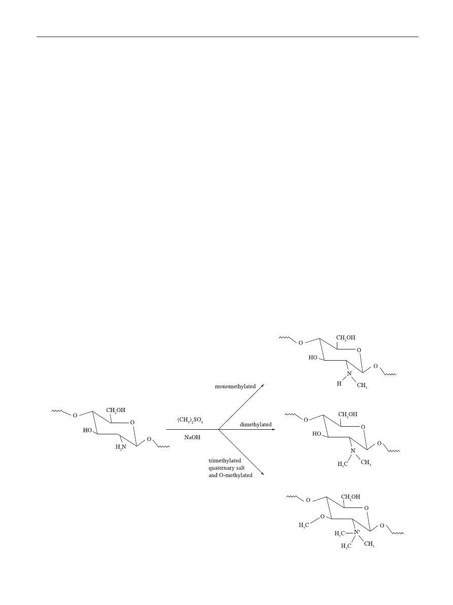
the quaternization of the nitrogen atoms of the amino groups.
To attain this, an extensive methylation of chitosan is required
that is carried out in suspension of dimethylsulfate, NaOH
and NaCl resulting in N,N,N-trimethylchitosan (Figure 3)
[66]
.
The synthesis of chitosan derivatives takes place by grafting
methyl functionality onto chitosan amino groups at the C-2
position
[67]
.
Studies with quaternary salts of chitosan reveled that
the antimicrobial activity against bacteria is higher than
that of chitosan
[68]
. Jia et al.
[69]
, reported that the activity of
N-propyl-N,N-dimethyl chitosan against E. coli is 20 times
higher then that of chitosan, indicating that the derivatives
with cationic charge exhibit particularly high activity.
An important feature of the chitosan derivatives is the
evidence that the alkyl moiety also plays an important role
in the antimicrobial activity
[69]
. According to Xie et al.
[70]
,
at neutral pH, the degree of protonation of NH
2
is very low,
so the repulsion of NH
3
+
is weak. Under such condition the
intermolecular and intramolecular hydrogen bonds result
in a formation of hydrophobic micro-area in the polymer
chain rendering hydrophobic and hydrophilic parts, favoring
the structural affinity between the bacteria cell wall and the
derivative
[26,70]
.
It would be expected that antimicrobial activity would
increase as the content of the alkyl moiety increases, as
confirmed by Rabea et al.
[71]
, who found that the antibacterial
activity had improved with an increasing on the chain
length of the alkyl substituent. This better performance was
attributed to the contribution of the hydrophobic portions of
the derivatives.
Besides the quaternary salts of chitosan, other aqua-
soluble derivatives such as hydroxypropyl and carboxymethyl
chitosans exist. Hydroxypropyl chitosan derivatives with
high degree of substitution (DS) are water insoluble, but after
graftization with maleic acid they become soluble in neutral
pH with antibacterial activity higher than that of the parent
chitosan
[70]
. Studies with this kind of derivative are very
important to help understand the mechanism of microbial-
antimicrobial agent interaction. For example, it has been
concluded that for neutral or alkaline media, the cationic
nature of chitosan can no longer explain its antibacterial
activity
[70]
. In this case, the strong coordination capability
of NH
2
groups in chitosan chain might be one possible
mechanism.
Figure 3. Schematic representation of the reaction leading to the quaternization of the amino groups of chitosan and resulting in N,N,N-trimethylchitosan
[66]
.

Goy, R. C. et al. - A Review of the antimicrobial activity of chitosan
246
Polímeros: Ciência e Tecnologia, vol. 19, nº 3, p. 241-247, 2009
6. Assis, O. B. G. & Pessoa, J. D. C. - Braz. J. Food Technol.,
7, p.17-22 (2004).
7. Liu, X. F.; Guan, Y. L.; Yang, D. Z.; Li, Z. & Yao, K. D.
- J. Appl. Polymer Sci., 79, p. 1324-1335 (2001).
8. Britto, D.; Campana Filho, S. P. & Assis, O. B. G. -
Polimeros: Ciência & Tecnologia, 15, p.129-132
(2005).
9. Chen, C. S.; Liau, W. Y. & Tsai, G. J. - J. Food Prot., 61,
p.1124-1128 (1998).
10. Hadwiger, L. A.; Kendra, D. G.; Fristensky, B. W. &
Wagoner, W. - Chitosan both activated genes in plants
and inhibits RNA synthesis in fungi
, in: “Chitin in nature
and technology”. Muzzarelli, R. A. A., Jeuniaux, C. &
Gooday, G. W. (Eds.), Plenum, New York (1981).
11. Papineau, A. M.; Hoover, D. G.; Knorr, D. & Farkas, D.
F. - Food Biotechnol., 5, p.45-57 (1991).
12. Shahidi, F.; Arachchi, J. & Jeon, Y. J. - Trends Food Sci.
Technol., 10, p.37-51 (1999).
13. Sudarshan, N. R.; Hoover, D. G. & Knorr, D. - Food
Biotechnol., 6, p.257-272 (1992).
14. Young, D. H.; Kohle, H. & Kauss, H. - Plant Physiol., 70,
p.1449-1454 (1982).
15. Coma, V.; Martial-Gros, A.; Garreau, S.; Copinet,
A.; Salin, F. & Deschamps, A. - J. Food Sci.,
67, p.1162-1169 (2002).
16. Raafat, D.; von Bargen, K.; Haas, A. & Sahl, H. G. -
Appl. Environ. Microbiol., 74, p.3764-3773 (2008).
17. Tsai, G. J. & Su, W. H. - J. Food. Prot., 62, p.239-243
(1999).
18. Young, D. H. & Kauss, H. - Plant Physiol., 73, p.698-702
(1983).
19. Devlieghere, F.; Vermeulen. A. & Debevere. J. - Food
Microbiol., 21, p.703-714 (2004).
20. Fang, S. W.; Li, C. F. & Shih, D. Y. C. - J. Food Prot., 57,
p.136-140 (1994).
21. Chung, Y. C. & Chen, C. Y. - Bioresource Technol., 99,
p.2806-2814 (2008).
22. Eaton, P.; Fernandes, J. C.; Pereira, E.; Pintado, M. E.
& Malcata, F. X. - Ultramicroscopy, 108, p.1128-1134
(2008).
23. Helander, I. M.; Nurmiaho-Lassila, E. L.; Ahvenainen,
R.; Rhoades, J. & Roller, S. - Int. J. Food Microbiol.,
30, p.235-44 (2001).
24. Másson, M.; Holappa, J.; Hjálmarsdóttir, M.; Rúnarsson,
Ö. V.; Nevalainen, T. & Järvinen, T. - Carbohyd.
Polym., 74, p.566-571 (2008).
25. Yalpani, M.; Johnson F. & Robinson L. E. - Antimicrobial
activity of some chitosan derivatives, in: Brine, C. J.;
Sandford, P. A. & Zikakis, J. P. (Eds), “Advances in
Chitin and Chitosan”, Elsevier, London, p.543-548
(1002).
26. Je, J. Y. & Kim, S. K.- J. Am. Chem. Soc., 128, p.4532
(2006).
27. Prescott, L. M.; Harley, J. P. & Klein, D. A. -
“Microbiology”, McGraw-Hill Co., New York (2002).
The study with carboxymethyl chitosan realized by Sun
et al.
[72]
is also very interesting since its derivative had both
negative and positive substituint groups. They demonstrate
that antimicrobial activities of carboxymethyl chitosan are
affected either by the DS of quaternary group or by the MW,
while no clear effect of the DS of carboxymethyl group
was observed. A further and important conclusion was that
when the derivative is complexed with calcium hydroxide as
pulp-cap, it has better ability in inducing reparative dentine
formation when compared to calcium hydroxide itself.
Conclusions
Chitosan is a versatile material with proved antimicrobial
activity. Three antibacterial mechanisms have been
proposed: i) the ionic surface interaction resulting in wall
cell leakage; ii) the inhibition of the mRNA and protein
synthesis via the penetration of chitosan into the nuclei of the
microorganisms; and iii) the formation of an external barrier,
chelating metals and provoking the suppression of essential
nutrients to microbial growth. It is likely that all events occur
simultaneously but at different intensities. The molecular
weight (MW) and the degree of acetylation (DA) are also
important factors in determining such activity. In general the
lower the MW and the DA, the higher will be the effectiveness
on reducing microorganism growth and multiplication. A
study of previous work from the literature has not lead to
any conclusive data as to whether the chitosan has higher
activity on gram-positive or on gram-negative bacteria. On
both species chitosan seems to act differently, though in both
cases satisfactorily. Water soluble derivatives, which can be
attained by chemical introduction of CH
3
in the main chain,
enhancing the chitosan applicability in a large pH range and
also improve the antimicrobial activity, opening up a broad
range of possibilities.
Acknowledgement
The authors would like to express their gratitude to
Embrapa and FAPESP
References
1. Kumar, A. B. V.; Varadaraj, M. C.; Gowda, L. R. &
Tharanathan, R. N. - Biochem. J., 391, p.167-175
(2005).
2. Percot, A.; Viton, C. & Domard, A. -. Biomacromolecules,
4, p.12-18 (2003).
3. Shepherd, R.; Reader, S. & Falshaw, A. - Glycoconjugate
J., 14, p.535-542 (1997).
4. Assis, O. B. G.; Bernardes Filho, R.; Viera, D. C. &
Campana Filho, S. P. - Int. J. Polymer. Mater., 51,
p.633-638 (2002).
5. Assis, O. B. G. & Silva, V. L. (2003). Polímeros: Ciência
& Tecnologia, 13, p.223-228 (2003).

Goy, R. C. et al. - A Review of the antimicrobial activity of chitosan
Polímeros: Ciência e Tecnologia, vol. 19, nº 3, p. 241-247, 2009
247
28. Coma, V.; Deschamps, A. & Martial-Gros, A. - J. Food
Sci., 68, p.2788-2792 (2003).
29. Dutta, P. K.; Tripath, S.; Mehrotra, G. K. & Dutta, J. -
Food Chem., 114, p.1173-1182 (2009).
30. Jeon, Y. J.; Park, P. J. & Kim, S. K. - Carbohyd. Polym.,
44, p.71-76 (2001).
31. No, H. K.; Park, N. Y.; Lee, S. H. & Meyers, S. P. - Int. J.
Food Microbiol., 74, p.65-72 (2002).
32. Chung, Y. C.; Su, Y. P.; Chen, C. C.; Jia, G.; Wang, H. L.;
Wu, J. C. G. & Lin, J. G. - Acta Pharmacol. Sinica, 25,
p.932-936 (2004).
33. Chen, Y. M.; Chung, Y. C.; Wang, L. W.; Chen, K. T. &
Li, S Y. - J. Environ. Sci. Health A, 37, p.1379-1390
(2002).
34. Eldin, M. S. M.; Soliman, E. A.; Hashem, A. I. & Tamer,
T. M. - Trends Biomater. Artif. Organs., 22, p.121-133
(2008).
35. Simunek, J.; Tishchenko, G.; Hodrová, B. & Bartonová,
H. - Folia Mocrobiol., 51, p.306-308 (2006).
36. Sebti, I.; Martial-Gros, A.; Carnet-Pantiez, A.; Grelier,
S. & Coma, V. - J. Food Sci., 70, p.M100 - M104
(2005).
37. Cuero, R. G.; Osuji, G. & Washington, A. - Biotechnol.
Lett., 13, p.441-444 (1991).
38. Roller, S. & Covill, N. - Int. J. Food Microbiol., 47,
p.67-77 (1999).
39. Guibal, E. - Sep. Purif. Techn., 38, p.43-74 (2004).
40. Wang, X.; Du, Y.; Fan, L.; Liu, H. & Hu, Y. - Polymer
Bulletin, 55, p.105-113 (2005).
41. Sekiguchi, S.; Miura, Y.; Kaneko, H.; Nishimura, S. I.;
Nishi, N.; Iwase, M. & Tokura, S. - Molecular weight
dependency of antimicrobial activity by chitosan
oligomers
, in: Nishinari, K. & Doi, E. (Eds), “Food
Hydrocolloids: Structures, Properties and Functions”,
Plenum Press, New York (1994).
42. Jing, Y. J.; Hao, Y. J.; Qu, H.; Shan, Y.; Li, D. S. & Du, R.
Q. - Acta Biologica Hungarica, 58, p.75-86 (2007).
43. Jung, B. O.; Chung, S. J. & Lee, G. W. - J. Chitin
Chitosan, 7, p.231-236 (2002).
44. Omura, Y.; Shigemoto, M.; Akiyama, T.; Saimoto,
H.; Shigemasa, Y.; Nakamura, I. & Tsuchido, T. -
Biocontrol. Sci., 8, p.25-30 (2003).
45. Tikhonov, V. E.; Stepnova, E. A.; Babak, V. G.; Yamskov,
I. A.; Palma-Guerrero, J.; Jansson, H. B.; Lopez-Llorca,
L. V.; Salinas, J.; Gerasimenko, D. V.; Avdienko, I. D.
& Varlamov, V. P. - Carbohyd. Polym., 64, p.66-72
(2006).
46. Tsai, G. J.; Zhang, S. L. & Shieh, P. L. - J. Food Prot.,
67, p.396-398 (2004).
47. Zivanovic, S.; Basurto, C. C.; Chi, S.; Davidson, P. M. &
Weiss, J. - J. Food Prot., 67, p.952-959 (2004).
48. Andres, Y.; Giraud, L.; Gerente, C. & Le Cloirec, P. -
Environ. Technol., 28, p.1357-1363 (2007).
49. Hongpattarakere, T. & Riyaphan, O. - Songklanakarin J.
Sci. Technol.. 30, p.1-9 (2008).
50. Tsai, G. J.; Su, W. H.; Chen, H. C. & Pan, C. L. - Fisheries
Sci., 68, p.170-177 (2002).
51. Assis, O. B. G. - “Avaliação, por análise de imagens,
da ação fungistática de coberturas de quitosana em
maçãs minimamente processadas
”, Monografia de
Especialização, Universidade Federal de Lavras, Brasil
(2008).
52. El Ghaouth, A.; Arul, J.; Grenier, J. & Asselin, A. -
Phytopathology, 82, p.398-402 (1992).
53. Sashai, A. S. & Manocha, M. S. - FEMS Microbiol. Rev.,
11, p.317-338 (1993).
54. Eweis, M.; Elkholy, S. S. & Elsabee, M. Z. - Int. J.
Biological Macrom., 38, p.1-8 (2006).
55. Stössel, O. & Leuba, J. L. - J. Phytopathol., 111, p.82-90
(1984).
56. Zhang, M.; Tan, T.; Yuan, H. & Rui, C. - J. Bioactive
Compatible Polym., 18, p.391-400 (2003).
57. Cuero, R. G. - EXS, 87, p.315-33 (1999).
58. Andrews, J. M. - Chemotherapy, 48, p.5-16 (2001).
59. Whithear, K. G.; Bowtell, D. D.; Ghiocas, E. & Hughes,
K. L. - Avian Dis., 27, p.937-949 (1983).
60. Du, W. L.; Xu, Y. L.;. Xu, Z. R. & Fan, C. L. -
Nanotechnology, 19, p.1-5 (2008).
61. Balicka-Ramisz, A.; Wojtasz-Pajak, B.; Pilarczyk, A. &
Ramisz, L. L. - Antibacterial and antifungal activity of
chitosan
, in: 12
th
ISAH Congress on Animal Hygiene,
Warsaw, p. 406 (2005).
62. Fernandes, J. C.; Tavaria, F. K.; Soares, J. C.; Ramos, Ó.
S.; Monteiro, M. J.; Pintado, M. E. & Malcata, F. X. -
Food Microbiol., 25, p.922-928 (2008).
63. Kurita, K. - Mar. Biotechnol., 8, p.203-226 (2006).
64. Barzegar, H.; Karbassi, A.; Jamalian, J. & Aminlari, M.
- J. Sci. Technol. Agric. Natur. Resour., 12, p.371-375
(2008).
65. Roller, S. & Covill, N. - J. Food Prot., 63, p.202-209
(2000).
66. Britto, D. & Assis, O. B. G. - Carbohyd. Polym., 69,
p.305-310 (2007).
67. Britto, D.; Forato, L. A. & Assis, O. B. G. - Carbohyd.
Polym., 74, p.86-91 (2008).
68. Sadeghi, A. M. M.; Amini, M.; Avadi, M. R.; Siedi, F.;
Rafiee-Tehrani, M. & Junginger, H. E. - J. Bioactive
Compatible Polym., 23, p.262-275 (2008).
69. Jia, Z.; Shen, D. & Xu W. - Carbohyd. Research., 333,
p.1-6 (2001).
70. Xie, W.; Xu, P., Wang, W. & Liu, Q. - Carbohyd. Polym.,
50, p.35-40 (2002).
71. Rabea, E. I.; Badawy, M. E. T.; Stevens, C.; Smagghe, G.
& Steurbaut, W. - Biomacromolecules, 4. p.1457-1465
(2003).
72. Sun, L.; Du, Y.; Fan, L.; Chen, X. & Yang. J. - Polymer,
47, p.1796-1804 (2006).
Enviado: 27/04/09
Reenviado: 06/07/09
Aceito: 15/07/09
Wyszukiwarka
Podobne podstrony:
Antioxidant and antimicrobial activity of extracts
Book Review of The Color Purple
A Review of The Outsiders Club Screened on?C 2 in October
Short review of the book entitled E for?stasy
Book Review of The Burning Man
Differential Heat Capacity Calorimeter for Polymer Transition Studies The review of scientific inst
animals of the farm activities
Review of Richard Milton The Facts of Life, Shat
Review of Blueprints, Solving the Mystery of Evolu
A review of the epidemiological evidence on tea, flavanoids, and lung cancer
A Review of B Alan Wallace’s ‘The Taboo of Subjectivity’
Harvard Business Review Harnessing the Science of Persuasion
Philosophy Of Mind Minds,Machines,And Mathematics A Review Of Shadows Of The Mind By Roger Penrose
Review of The New Economic Sociology
więcej podobnych podstron