
Control of respiratory functions.
Sleep apnea syndrome
Dariusz Nowak
Appropriate rate of alveolar ventilation and lung
perfusion
maintain the relative stable oxygen pressure (PO
2
),
carbon dioxide pressure (PCO
2
) in the arterial blood
PO
2
85-100 mm Hg
PCO
2
35-45 mm Hg
receptors (chemoreceptors)
nerves
respiratory center
effectors

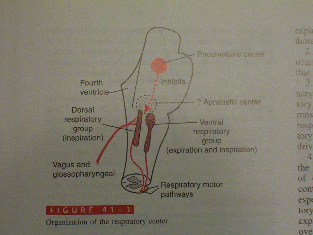
Respiratory center
Localization bilateral , in medulla oblongata and
pons.
Dorsal respiratory group
Ventral respiratory group
Pneumotaxic center
Apneustic center
Chemosensitive area
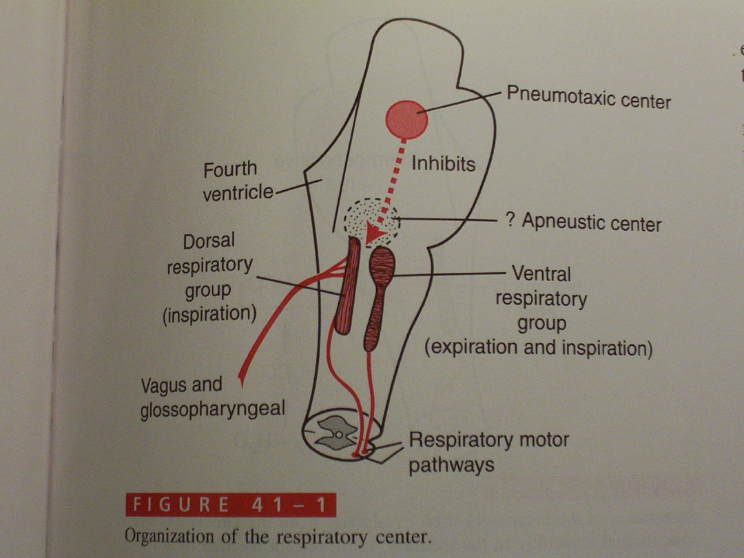

Respiratory center – dorsal
respiratory group
• Sensory terminal of vagus and glossopharyngeal nerves
Signals from peripheral chemoreceptors
baroreceptors
various type receptors in the lung
• Generates basal rhythm of respiration
2 second inspiratory signal
3 second breake (no inspiratory signal) – passive expiration
Control of inspiratory signal:
A/ rate of signal increase (velocity and strenght of one
breathe)
B/ point at which signal is ceased – frequency of respiration

Respiratory center – pneumotaxic center
• Limits the duration of inspiration
• Increases the respiratory rate
Controls the „switch-off” time-point of
inspiratory signal
Respiratory rate 40 breaths/min or 3
breath/min
Strong or weak pneumotaxic signal.

Respiratory center- ventral respiratory
group
• Inactive during normal quiet respiration
• Active when high levels of pulmoary
ventilation are required (e.g. during exercise)
• Contributes to both heavy inspiration and
expiration (impulses to abdominal muscles)

Respiratory center – apneustic center
• Sends signals to dorsal respiratory group
• Prevents or retards the „switch-off” the
inspiratory signal
• Controls the intensity of inspiration , allows
full inspiration

Additional way controlling
respiration
Hering-Breuer inflation reflex
• Strech receptors (located in muscle wall of bronchi and
bronchioles)
• Vagus nerve
• Dorsal respiratory group
Lungs become overstreached – hyperinflation
Signals act on dorsal respiratory group to „switch-of” inspiratory
signal
Reflex stops further inspiration and increases respiratory rate
(like the pneumotaxic center)
• In humans prevents excessive lung inflation ( reflex is
activated when TV > 1.5 l)

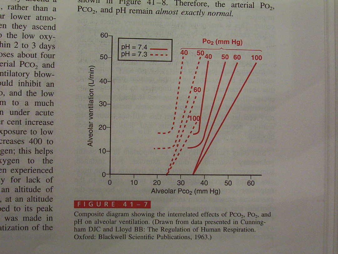
Control of ventilation
We know how the respiratory rate
and the intensity (quiet , deep) of
inspiration and expiration is
controlled
What is the mechanism controlling
current respiratory pattern to
match the ventilatory (metabolic
needs) of our body ?

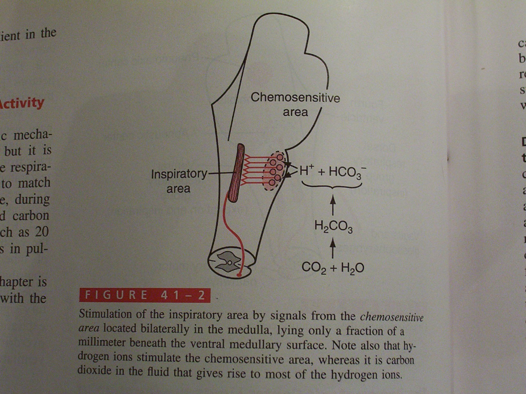
Chemoreceptors
• Central chemoreceptors – chemosensitive area (in medulla)
• Peripheral chemoreceptors located in carotid and aortic bodies
• [H
+
] – directly stimulates respiratory center via central receptors
• PCO
2
also directly stimulates respiratory center
• PO
2
acts only via peripheral chemoeceptors
Changes in blood PCO
2
or [H
+
]
↓
Chemosensitive area
↓
other parts of respiratory center
↓
increased lung ventilation

chemoreceptors
• [H
+
] does not cross blood-brain barrier
• [H
+
] is the main stimulator ot the chemosensitive
area
• CO
2
rapidly diffuses through b-b barrier
What is the role of CO
2
?
Low buffering capacity and low protein concentration
in the fluid surrounding chemosensitive area
Under normal conditions (healthy subject) [H
+
] and
CO
2
contribute to 80% of impulses responsible for
respiratory drive
20 % - oxygen (PO
2
)
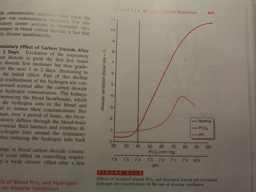

Respiratory center adaptation to
increased PCO
2
Prolonged increase in blood PCO
2
• Stimulation is highest during first few hours after
increase in PCO
2
• Then it decreases to about 20% of the initial (maximal)
value ( after 1 or 2 days)
Why ?
Due to buffering function of the kidneys
• Increase in blood bicarbonate levels
• Diffusion of bicarbonate through blood cerebrospinal fluid
barrier
• Binding of H
+
by bicarbonates in the close neighborhood
of the chemosensitive area

What is a consequence of
adaptation to increased pCO
2
?
Patient with severe chronic respiratory
insufficiency (e.g. With pulmonary emphysema
or COPD)
Has hypercapnia and hypoxemia
Decreased pO
2
is the major factor stimulating
respiratory center
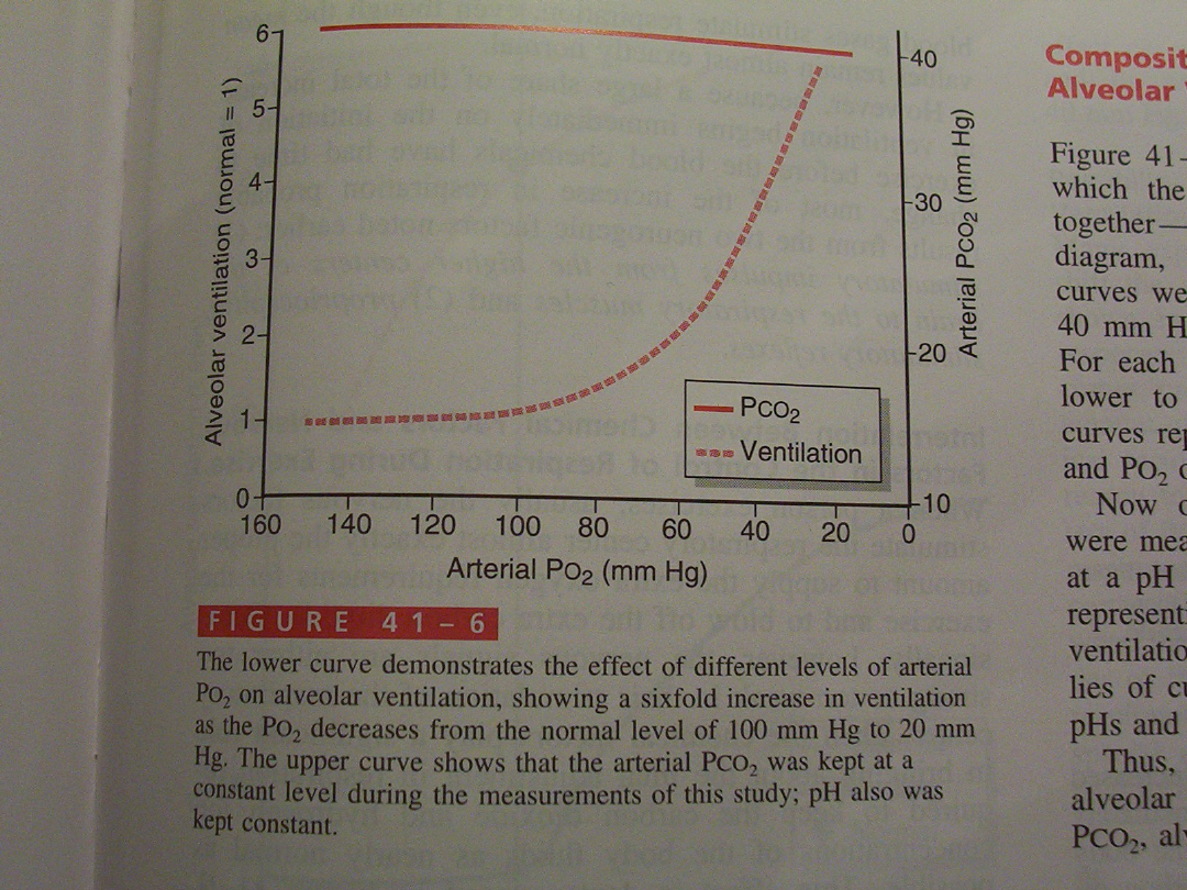
How can we treat patient with oxygen ?
Low flows
Low O
2
concentrations

Why O
2
is not important as CO
2
in
stimulation of respiratory centre under
normal conditions ?
• Hemoglobin saturation is almost total in PO
2
range 60 – 100 mmHg
• Changes of PO
2
from 60 to 100 mmHg have no
significant influence on O
2
content in the
blood and O
2
delivery to tissues.
Thus CO
2
is the most important variable
responsible for respiratory drive regulation
under normal conditions.

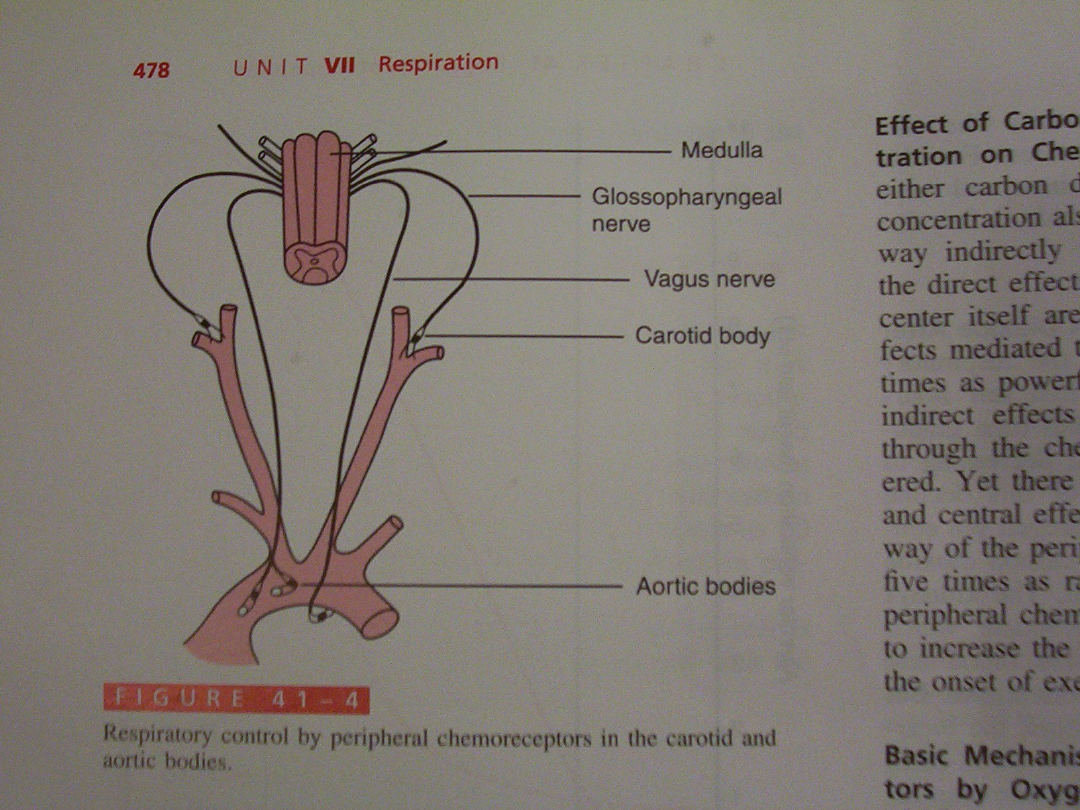
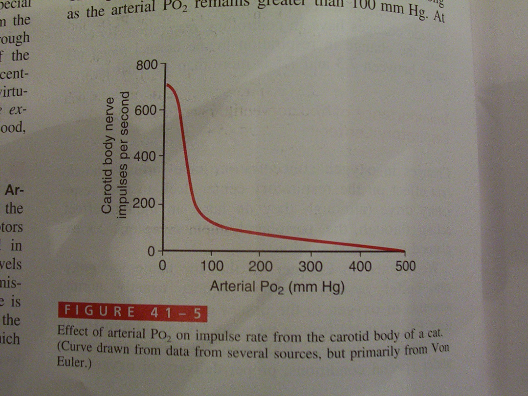
Peripheral chemoreceptor
system
localization:
Aortic bodies – along the aorta arch
Carotid bodies – bifurcation of the common carotid artery
• Very high blood flow through these bodies
• They are exposed continously to arterial blood O
2
(PAO
2
)
• Respond to changes in PAO
2
( decrease) and to a
lesser extent to PCO
2
and [H
+
]
• Hihgly senstive to PAO
2
in the range 60 to 30 mmHg
• Why ??

Peripheral chemoreceptors
• PCO
2
– mediated stimulation via peripheral
receptors is 7-8 times lower than that via
chemosensitive area
• But the onset of stimulation occurs 5 times
rapidly
• Increases rapidity of ventilatory response to
CO
2
when we start exercise

Regulation of respiration during
exercise
• Mean arterial PO
2
, PCO
2
and pH remain almost
normal during exercise
What stimulates intense ventilation during exercise ?
• Brain ; motor impulses to muscles are collaterally
transmitted to neurons in respiratory center
• movement of arms and legs ; excited
proprioreceptors (in joints and muscles) transmit
impulses to respiratory centre
• Hypoxia in the muscles during exercise – afferent
nerve signals to respiratory centre ?
• Changes of PCO
2
and PO
2
between expiration and
inspiration ?

Voluntary control of
respiration
• Talking
• Singing
• Eating
• Defecation
Voluntary hypoventilation or hyperventilation
• Not mediated through respiratory center in the
medulla
• Pathway: cortex , spinal tract ,spinal neurons ,
muscles

Depressant of the respiratory center
• Morphine
• Pentobarbital
• Sleep
Alveolar hypoventilation – chemosensitive areas
reveal decreased response to CO
2
Upper airways muscles relaxation – increase in
the airflow resistance
Document Outline
- Slide 1
- Slide 2
- Slide 3
- Slide 4
- Slide 5
- Slide 6
- Slide 7
- Slide 8
- Slide 9
- Slide 10
- Slide 11
- Slide 12
- Slide 13
- Slide 14
- Slide 15
- Slide 16
- Slide 17
- Slide 18
- Slide 19
- Slide 20
- Slide 21
- Slide 22
- Slide 23
- Slide 24
- Slide 25
- Slide 26
Wyszukiwarka
Podobne podstrony:
06 Control of respiratory funct Nieznany
Ebsco Gross The cognitive control of emotio
Control of Redundant Robot Manipulators R V Patel and F Shadpey
epigenetic control of plant dev Nieznany
Causes and control of filamentous growth in aerobic granular sludge sequencing batch reactors
Nonlinear Control of a Conrinuously Variable Transmission (CVT) for Hybrid Vehicle Powertrains
Control of a 4 leg Inverter for Standalone Photovoltaic Systems
The Hormonal Control of Sexual?velopment
Holysz, Jedraszak, Szarycz THE CONTROL OF THE SIMULATION
Ebsco Gross The cognitive control of emotio
Control of Redundant Robot Manipulators R V Patel and F Shadpey
Control of a 4 leg Inverter for Standalone Photovoltaic Systems
Microwave irradiation of hazelnuts for the control of aflatoxin producing Aspergillus parasiticus
The Discrete Time Control of a Three Phase 4 Wire PWM Inverter with Variable DC Link Voltage and Bat
Variable Speed Control Of Wind Turbines Using Nonlinear And Adaptive Algorithms
the struggle over control of kievdimnik
więcej podobnych podstron