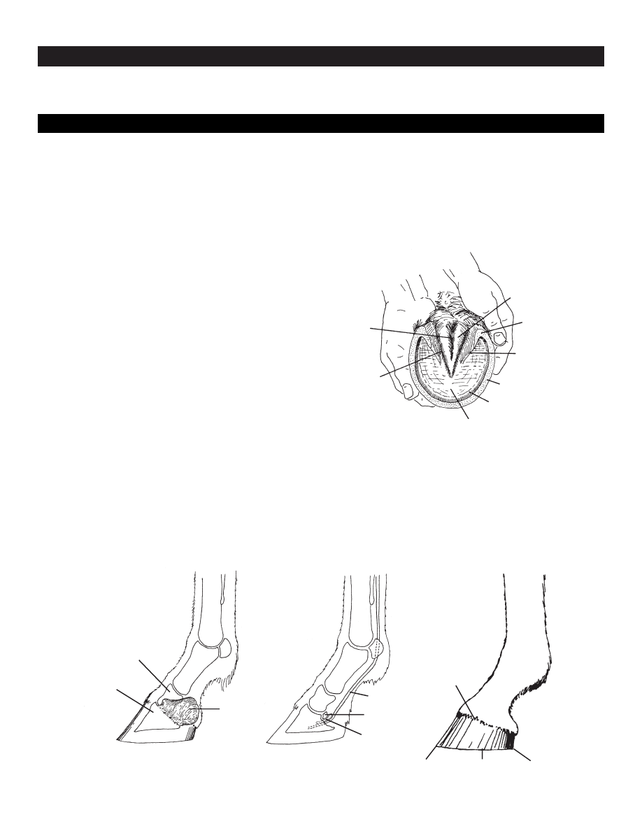
A horse’s hoof is composed of the wall, sole and
frog. The wall is simply that part of the hoof that is visi-
ble when the horse is standing. It covers the front and
sides of the third phalanx, or coffin bone. The wall is
made up of the toe (front), quarters (sides) and heel.
When the foot is lifted off the ground, the sole and
frog are visible, as well as the bars of the wall and the
collateral grooves (see Figure 1).
The wall of the hoof is composed of a horny material
that is produced continuously and must be worn off or
trimmed off. The hoof wall does not contain blood
vessels or nerves. In the front feet, the wall is thickest at
the toe; in the hind feet the hoof wall is of a more
uniform thickness. The wall, bars and frog are the
weight-bearing structures of the foot. Normally the sole
does not contact the ground.
Inside the hoof, lateral cartilages extend back and up
from the inner and outer sides of the third phalanx
(Figure 2a). These cartilages are flexible, but as the horse
ages, they are usually ossified and replaced by bone.
Between the second and third phalanges and above the
deep flexor muscle tendon is a small bone called the
navicular bone (Figure 2b). The navicular bone and its
associated bursa — a fluid-filled sac that reduces friction
between the tendon and the bone — are involved in
navicular disease, which is a common cause of lameness.
The digital cushion is a mass of flexible material that
contributes to the formation of the heels (see Figure 3).
This structure is one of the primary shock absorbers of
the foot.
As weight is placed on the hoof, pressure is trans-
mitted through the phalanges to the wall and onto the
$.25
G 2740
Printed with soy ink on recycled paper
MU Guide
AGRICULTURAL
PUBLISHED BY MU EXTENSION, UNIVERSITY OF MISSOURI-COLUMBIA
muextension.missouri.edu/xplor/
Horses
Functional Anatomy of the Horse Foot
Robert C. McClure
Department of Veterinary Anatomy, College of Veterinary Medicine
Central
groove
of frog
Collateral
groove
Sole
White line
Wall
Bar of wall
Angle of wall
Frog
Figure 1. The wall, bars and frog are the weight-bearing struc-
tures of the foot.
3rd phalanx
(coffin bone)
Lateral
cartilage
Tendon of
deep digital flexor
Navicular bone
Navicular bursa
Coronet
Toe
Quarter
Heel
2nd phalanx
Figure 2. Internal and external structure of the horse foot.
a
b
c
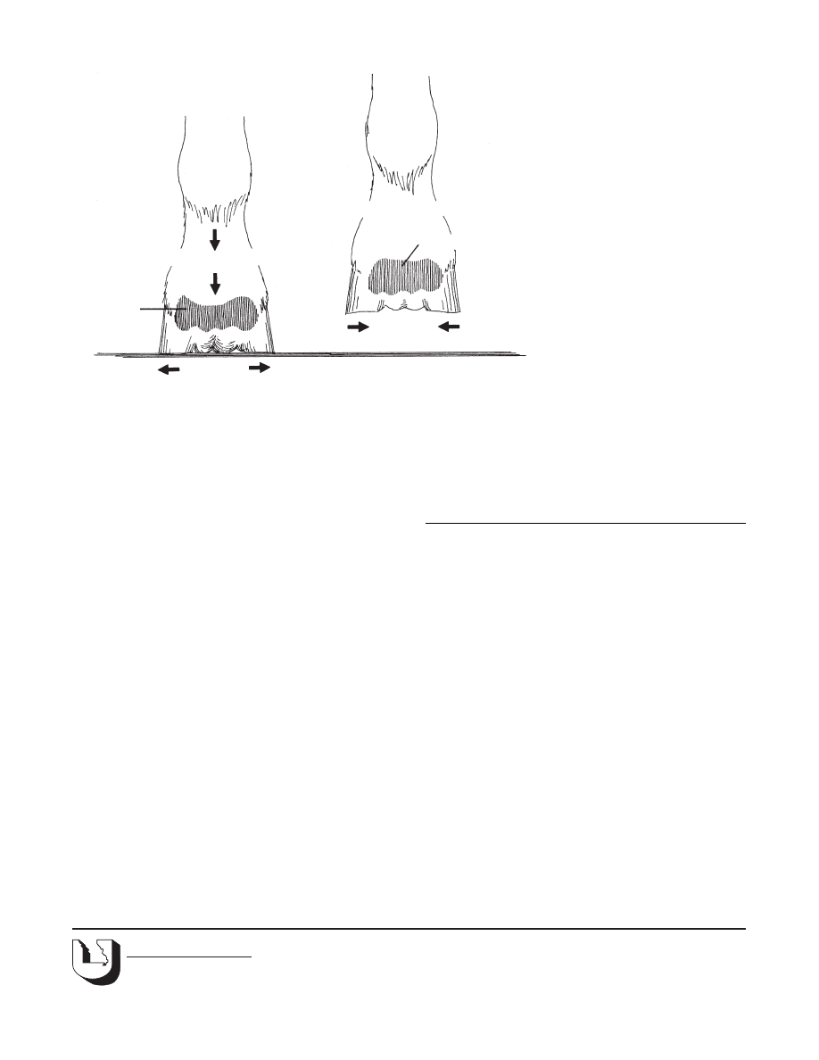
digital cushion and frog. The frog, a highly elastic
wedge-shaped mass, normally makes contact with the
ground first. The frog presses up on the digital cushion,
which flattens and is forced outward against the lateral
cartilages. The frog also is flattened and tends to push
the bars of the wall apart (Figure 3). When the foot is
lifted, the frog and other flexible structures of the foot
return to their original position.
When the foot is placed on the ground, blood is
forced from the foot to the leg by the increase in pressure
and by the change in shape of the
digital cushion and the frog. The
pressure and the change in shape
compress the veins in the foot.
When the foot is lifted, the com-
pression is relieved and blood
flows into the veins again. In this
way, the movement of these struc-
tures in the hoof acts as a pump.
Exercise increases the blood circu-
lation in the foot and favors good
hoof growth. Lack of exercise,
dryness of the horny wall, and
poor nutrition inhibit hoof growth.
Normally, the hoof wall grows
at the rate of about three-eighths
inch per month. New layers of
hoof wall are produced continu-
ously from just below an area
called the coronet at the junction of
the skin and the hoof wall (see
Figure 2c).
The hoof wall is covered with
material that prevents evaporation of moisture. When
this material is deficient, the hoof wall becomes dry and
excessive flaking and cracking may occur. A good hoof
paint aids in preventing excessive drying.
This publication was originally written jointly by Robert C.
McClure, Gerald R. Kirk and Phillip D. Garrett. Kirk and Garrett
are former faculty members in the Department of Veterinary
Anatomy, College of Veterinary Medicine. Illlustrations are by
Phillip D. Garrett.
Page 2
G 2740
Reviewed and reprinted 10/99/5M
■ Issued in furtherance of Cooperative Extension Work Acts of May 8 and June 30, 1914, in cooperation with the United States Department of
Agriculture. Ronald J. Turner, Director, Cooperative Extension, University of Missouri and Lincoln University, Columbia, MO 65211. ■ University
Outreach and Extension does not discriminate on the basis of race, color, national origin, sex, religion, age, disability or status as a Vietnam
era veteran in employment or programs. ■ If you have special needs as addressed by the Americans with Disabilities Act and need this publication
in an alternative format, write ADA Officer, Extension and Agricultural Information, 1-98 Agriculture Building, Columbia, MO 65211, or call
(573) 882-7216. Reasonable efforts will be made to accommodate your special needs.
OUTREACH & EXTENSION
UNIVERSITY OF MISSOURI
COLUMBIA
Weight of horse
Foot is lifted
Digital cushion expands
Walls of hoof contract
Walls of hoof
expand slightly
Digital cushion
is flattened
Figure 3. Flexible structures in the horse’s hoof expand and contract with each step as weight
is transferred from one foot to another.
Wyszukiwarka
Podobne podstrony:
Goel, Dolan The Functional anatomy of H segregating cognitive and affective components
Kinesio® Taping in Stroke Improving Functional Use of the Upper Extremity in Hemiplegia
Anatomy of the Linux System
SHSBC 284 ANATOMY OF THE GPM
On the functional validity of the worm killing worm
Niven, Larry The Flight of the Horse
Wójcik, Marcin; Tobiasz Lis, Paulina Functional Potential of the Novosibirsk Urban Region in Russia
Fussell An Anatomy of the Classes
Kinesio® Taping in Stroke Improving Functional Use of the Upper Extremity in Hemiplegia
Anatomy Based Modeling of the Human Musculature
A systematic review and meta analysis of the effect of an ankle foot orthosis on gait biomechanics a
anatomy of horse, kopyta konia
Functions of the Nervous System
Functions of the Department of Defense
Soliwoda, Katarzyna i inni The influence of the chain length and the functional group steric access
Jane Elliot End of the Trail 2 The Devil in Dead Horse
The Three Investigators 26 The Mystery of the Headless Horse us
więcej podobnych podstron