
http://www.autism.com/ari/mercurylong.html
Autism:
A Unique Type of
Mercury Poisoning
Sallie Bernard*
Albert Enayati, B.S., Ch.E., M.S.M.E.**
Teresa Binstock
Heidi Roger
Lyn Redwood, R.N., M.S.N., C.R.N.P.
Woody McGinnis, M.D.
*Contact: sbernard@nac.net
**Contact: (201) 444-7306
njcan@aol.com
Copyright (c) 2000 by ARC Research
14 Commerce Drive
Cranford, NJ 07016
April 3, 2000
Revision of April 21, 2000
ABSTRACT
Autism is a syndrome characterized by impairments in social relatedness, language and communication,
a need for routine and sameness, abnormal movements, and sensory dysfunction. Mercury (Hg) is a
toxic metal that can exist as a pure element or in a variety of inorganic and organic forms and can cause
immune, sensory, neurological, motor, and behavioral dysfunctions similar to traits defining or
associated with autism. Thimerosal, a preservative frequently added to childhood vaccines, has become
a major source of Hg in human infants and toddlers. According to the FDA and the American Academy
of Pediatricians, fully vaccinated children now receive, within their first two years, Hg levels that exceed
safety limits established by the FDA and other supervisory agencies. A thorough review of medical
literature and U.S. government data indicates (i) that many and perhaps most cases of idiopathic autism,
in which an extended period of developmental normalcy is followed by an emergence of symptoms, are
induced by early exposure to Hg; (ii) that this type of autism represents a unique form of Hg poisoning
(HgP); (iii) that excessive Hg exposure from thimerosal in vaccine injections is an etiological
mechanism for causing the traits of autism; (iv) that certain genetic and non-genetic factors establish a
predisposition whereby thimerosal's adverse effects occur only in some children; and (v) that vaccinal
Hg in thimerosal is causing a heretofore unrecognized mercurial syndrome.
Page 1 of 62
Autism: A Unique Type of Mercury Poisoning
2/5/2004
http://www.vaccinationnews.com/DailyNews/July2001/AutismUniqueMercPoison.htm

SYNOPSIS
A review of medical literature indicates that the characteristics of autism and of mercury poisoning
(HgP) are strikingly similar. Traits defining or associated with both disorders are summarized in Table A
immediately following the Table of Contents and are discussed and cited in the body of this document.
The parallels between the two diseases are so thorough as to suggest, based on total Hg injected into
U.S. children, that many cases of autism are a form of mercury poisoning.
For these children, the exposure route is childhood vaccines, most of which contain thimerosal, a
preservative which is 49.6% ethylmercury by weight. The amount of mercury a typical child under two
years receives from vaccinations equates to 237.5 micrograms, or 3.53 x 10¹⁷ molecules
(353,000,000,000,000,000 molecules). Most such vaccinal Hg may not be excreted and instead migrates
to the brain.
The total amount injected into infants and toddlers (i) is known to exceed Federal safety standards, (ii) is
officially considered to be a “low” level; whereby (iii) only a small percentage of exposed individuals
exhibit symptoms of toxicity. In fact, children who develop Hg-related autism are likely to have had a
predisposition derived from genetic and non-genetic factors.
Importantly, the timings of vaccinal Hg-exposure and its latency period coincide with the emergence of
autistic-symptoms in specific children. Moreover, excessive mercury has been detected in urine, hair,
and blood samples from autistic children; and parental reports, though limited at this date, indicate
significant improvement in symptoms subsequent to heavy-metal chelation therapy.
The HgP phenotype is diverse and depends upon a number of factors - including type of Hg, route of
entry into the body, rate and level of dose, individual genotype, and the age and immune status of the
patient. Historically, variation among these factors has caused slightly different manifestations of
mercurialism; Mad Hatter’s disease, Minamata disease, acrodynia, and industrial exposures provide
examples.
The pathology arising from the mercury-related variables involved in autism - intermittent bolus doses
of ethylmercury injected into susceptible infants and toddlers - is heretofore undescribed in medical
literature. Therefore, in accord with existing HgP data and HgP’s ability to induce virtually all the traits
defining or associated with autism spectrum disorders, we hypothesize that many and perhaps most
cases of autism represent a unique form of mercury poisoning.
This conclusion and its supporting data have important implications for the affected population of
autistic individuals and their families, for other unexplained disorders with symptoms similar to those of
heavy metal intoxication, for vaccine content, and for childhood vaccination programs. Due to its high
potential for neurotoxicity, thimerosal should be removed immediately from all vaccine products
designated for infants and toddlers.
Table of Contents
ABSTRACT & SYNOPSIS
TABLE
OF
CONTENTS
AUTISM-MERCURIALISM COMPARISONS
Page 2 of 62
Autism: A Unique Type of Mercury Poisoning
2/5/2004
http://www.vaccinationnews.com/DailyNews/July2001/AutismUniqueMercPoison.htm

INTRODUCTION
Autism
Mercury
Diagnosing
Mercury
Poisoning
in Autism
I. SYMPTOM COMPARISON
a. Affect/Psychological Presentation
b. Language & Hearing
c. Sensory Perception
d. Movement/Motor Function
e. Cognition/Mental Function
f. Behaviors
g. Vision
h. Physical Presentations
j. Gastrointestinal Function
II. COMPARISON OF BIOLOGICAL ABNORMALITIES
a. Biochemistry
b. Immune System
c. CNS Structure
d. Neurons & Neurochemicals
e. EEG Activity/Epilepsy
III. MECHANISMS, SOURCES & EPIDEMIOLOGY OF EXPOSURE
a. Exposure Mechanism
b. Population Susceptibility
c. Sex Ratio
d. Exposure Levels & Autism Prevalence
e. Genetic Factors
f. Course
of
Disease
g. Thimerosal Interaction with Vaccines
IV. DETECTION OF MERCURY IN AUTISTIC CHILDREN
Case Studies
Discussion
DISCUSSION
Diagnostic Criteria Are Met
Unique
Form
Would be Expected, Implicates Vaccinal Thimerosal
Historical Precedent Exists
Barriers Preventing Earlier Discovery Are Removed
MEDICAL & SOCIETAL IMPLICATIONS
Affected Population
Other Disorders
Vaccination Programs
REFERENCES
Page 3 of 62
Autism: A Unique Type of Mercury Poisoning
2/5/2004
http://www.vaccinationnews.com/DailyNews/July2001/AutismUniqueMercPoison.htm

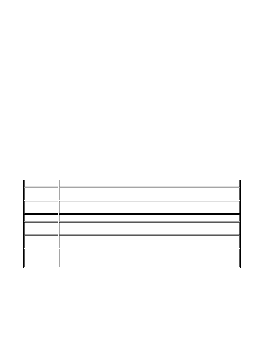
Table A:
Summary Comparison of Characteristics
of Autism & Mercury Poisoning
Mercury Poisoning
Autism
Psychiatric Disturbances
Social deficits, shyness, social withdrawal
Social deficits, social withdrawal, shyness
Depression, mood swings; mask face
Depressive traits, mood swings; flat affect
Anxiety
Anxiety
Schizoid tendencies, OCD traits
Schizophrenic & OCD traits; repetitiveness
Lacks eye contact, hesitant to engage others
Lack of eye contact, avoids conversation
Irrational fears
Irrational fears
Irritability, aggression, temper tantrums
Irritability, aggression, temper tantrums
Impaired face recognition
Impaired face recognition
Speech, Language & Hearing Deficits
Loss of speech, failure to develop speech
Delayed language, failure to develop speech
Dysarthria; articulation problems
Dysarthria; articulation problems
Speech comprehension deficits
Speech comprehension deficits
Verbalizing & word retrieval problems
Echolalia; word use & pragmatic errors
Sound sensitivity
Sound sensitivity
Hearing loss; deafness in very high doses
Mild to profound hearing loss
Poor performance on language IQ tests
Poor performance on verbal IQ tests
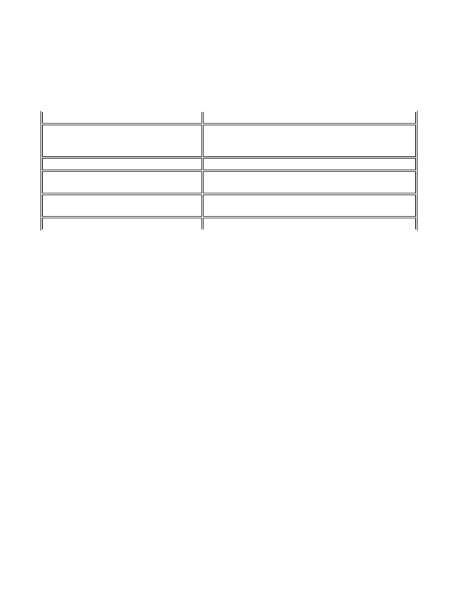
Sensory Abnormalities</TD< tr>
Abnormal sensation in mouth & extremities
Abnormal sensation in mouth & extremities
Sound sensitivity
Sound sensitivity
Abnormal touch sensations; touch aversion
Abnormal touch sensations; touch aversion
Vestibular abnormalities
Vestibular abnormalities
Motor Disorders
Involuntary jerking movements - arm flapping, ankle
jerks, myoclonal jerks, choreiform movements,
circling, rocking
Stereotyped movements - arm flapping,
jumping, circling, spinning, rocking;
myoclonal jerks; choreiform movements
Deficits in eye-hand coordination; limb apraxia;
intention tremors
Poor eye-hand coordination; limb apraxia;
problems with intentional movements
Gait impairment; ataxia - from incoordination &
clumsiness to inability to walk, stand, or sit; loss of
motor control
Abnormal gait and posture, clumsiness and
incoordination; difficulties sitting, lying,
crawling, and walking
Difficulty in chewing or swallowing
Difficulty chewing or swallowing
Unusual postures; toe walking
Unusual postures; toe walking
Cognitive Impairments
Borderline intelligence, mental retardation - some
Borderline intelligence, mental retardation -
Page 4 of 62
Autism: A Unique Type of Mercury Poisoning
2/5/2004
http://www.vaccinationnews.com/DailyNews/July2001/AutismUniqueMercPoison.htm

cases reversible
sometimes "recovered"
Poor concentration, attention, response inhibition
Poor concentration, attention, shifting attention
Uneven performance on IQ subtests
Uneven performance on IQ subtests
Verbal IQ higher than performance IQ
Verbal IQ higher than performance IQ
Poor short term, verbal, & auditory memory
Poor short term, auditory & verbal memory
Poor visual and perceptual motor skills, impairment in
simple reaction time
Poor visual and perceptual motor skills, lower
performance on timed tests
Difficulty carrying out complex commands
Difficulty carrying out multiple commands
Word-comprehension difficulties
Word-comprehension difficulties
Deficits in understanding abstract ideas & symbolism;
degeneration of higher mental powers
Deficits in abstract thinking & symbolism,
understanding other’s mental states,
sequencing, planning & organizing
Unusual Behaviors
Stereotyped sniffing (rats)
Stereotyped, repetitive behaviors
ADHD traits
ADHD traits
Agitation, unprovoked crying, grimacing, staring
spells
Agitation, unprovoked crying, grimacing,
staring spells
Sleep difficulties
Sleep difficulties
Eating disorders, feeding problems
Eating disorders, feeding problems
Self injurious behavior, e.g. head banging
Self injurious behavior, e.g. head banging
Visual Impairments
Poor eye contact, impaired visual fixation
Poor eye contact, problems in joint attention
“Visual impairments,” blindness, near-sightedness,
decreased visual acuity
“Visual impairments”; inaccurate/slow
saccades; decreased rod functioning
Light sensitivity, photophobia
Over-sensitivity to light
Blurred or hazy vision
Blurred vision
Constricted visual fields
Not described
Physical Disturbances
Increase in cerebral palsy; hyper- or hypo-tonia;
abnormal reflexes; decreased muscle strength,
especially upper body; incontinence; problems
chewing, swallowing, salivating
Increase in cerebral palsy; hyper- or
hypotonia; decreased muscle strength,
especially upper body; incontinence; problems
chewing and swallowing
Rashes, dermatitis/dry skin, itching; burning
Rashes, dermatitis, eczema, itching
Autonomic disturbance: excessive sweating, poor
circulation, elevated heart rate
Autonomic disturbance: unusual sweating,
poor circulation, elevated heart rate
Gastro-intestinal Disturbances</TD< tr>
Gastroenteritis, diarrhea; abdominal pain, constipation,
“colitis”
Diarrhea, constipation, gaseousness,
abdominal discomfort, colitis
Anorexia, weight loss, nausea, poor appetite
Anorexia; feeding problems/vomiting
Page 5 of 62
Autism: A Unique Type of Mercury Poisoning
2/5/2004
http://www.vaccinationnews.com/DailyNews/July2001/AutismUniqueMercPoison.htm

Lesions of ileum & colon; increased gut permeability Leaky gut syndrome
Inhibits dipeptidyl peptidase IV, which cleaves
casomorphin
Inadequate endopeptidase enzymes needed for
breakdown of casein & gluten
Abnormal Biochemistry
Binds -SH groups; blocks sulfate transporter in
intestines, kidneys
Low sulfate levels
Has special affinity for purines & pyrimidines
Purine & pyrimidine metabolism errors lead to
autistic features
Reduces availability of glutathione, needed in neurons,
cells & liver to detoxify heavy metals
Low levels of glutathione; decreased ability of
liver to detoxify heavy metals
Causes significant reduction in glutathione peroxidase
and glutathione reductase
Abnormal glutathione peroxidase activities in
erythrocytes
Disrupts mitochondrial activities, especially in brain Mitochondrial dysfunction, especially in brain
Immune Dysfunction
Sensitivity due to allergic or autoimmune reactions;
sensitive individuals more likely to have allergies,
asthma, autoimmune-like symptoms, especially
rheumatoid-like ones
More likely to have allergies and asthma;
familial presence of autoimmune diseases,
especially rheumatoid arthritis; IgA
deficiencies
Can produce an immune response in CNS
On-going immune response in CNS
Causes brain/MBP autoantibodies
Brain/MBP autoantibodies present
Causes overproduction of Th2 subset; kills/inhibits
lymphocytes, T-cells, and monocytes; decreases NK
T-cell activity; induces or suppresses IFNg & IL-2
Skewed immune-cell subset in the Th2
direction; decreased responses to T-cell
mitogens; reduced NK T-cell function;
increased IFNg & IL-12
CNS Structural Pathology
Selectively targets brain areas unable to detoxify or
reduce Hg-induced oxidative stress
Specific areas of brain pathology; many
functions spared
Damage to Purkinje and granular cells
Damage to Purkinje and granular cells
Accummulates in amygdala and hippocampus
Pathology in amygdala and hippocampus
Causes abnormal neuronal cytoarchitecture; disrupts
neuronal migration & cell division; reduces NCAMs
Neuronal disorganization; increased neuronal
cell replication, increased glial cells; depressed
expression of NCAMs
Progressive microcephaly
Progressive microcephaly and macrocephaly
Brain stem defects in some cases
Brain stem defects in some cases
Abnormalities in Neuro-chemistry
Prevents presynaptic serotonin release & inhibits
serotonin transport; causes calcium disruptions
Decreased serotonin synthesis in children;
abnormal calcium metabolism
Alters dopamine systems; peroxidine deficiency in rats
resembles mercurialism in humans
Possibly high or low dopamine levels; positive
response to peroxidine (lowers dopamine
levels)
Elevates epinephrine & norepinephrine levels by
blocking enzyme that degrades epinephrine
Elevated norepinephrine and epinephrine
Page 6 of 62
Autism: A Unique Type of Mercury Poisoning
2/5/2004
http://www.vaccinationnews.com/DailyNews/July2001/AutismUniqueMercPoison.htm

INTRODUCTION
Autism
Autism, or Autistic Spectrum Disorder (ASD), is considered a neurodevelopmental syndrome, emerging
early in life and exhibiting a constellation of seemingly unrelated features and a wide variation in
symptom expression and level of severity by individual (Filipek et al, 1999; Bailey et al, 1996). The
diagnostic criteria for autism are qualitative impairments in social relatedness, deficits in verbal and
nonverbal communication, and the presence of repetitive and restricted behaviors or interests (APA,
1994). As will be cited below, other traits associated with autism are movement disorder, sensory
dysfunction, and cognitive impairments as well as gastrointestinal difficulties and immune abnormalities
(Gillberg & Coleman, 1992; Warren et al, 1990; Horvath et al, 1999). Onset must occur before age 36
months (APA, 1994); although in some instances deficits are apparent at birth, in the great majority of
cases there are at least several months of normal development followed by clear regression or failure to
progress normally (Gillberg & Coleman, 1992; Filipek et al, 1999; Bailey et al, 1996). Formerly
regarded as a rare disease, autism is now said to affect one in 500 children (Bristol et al, 1996), with
some estimates suggesting one in 100 for a broader phenotype often labeled as the "autism-spectrum" of
disorders and which includes both higher and lower functioning individuals (Arvidsson et al, 1997;
Wing, 1996).
Autism and autistic symptoms can arise from a number of known disorders, most notably tuberous
sclerosis, Rhett syndrome, Landau-Kleffner syndrome, Fragile X, Phenylketonuria, purine autism, and
other purine metabolic diseases such as PRPP synthetase defects and 5'-nucleotidase superactivity. The
etiology and pathogenesis of the vast majority of autism cases - 70% - 90% (Gillberg and Coleman,
Elevates glutamate
Elevated glutamate and aspartate
Leads to cortical acetylcholine deficiency; increases
muscarinic receptor density in hippocampus &
cerebellum
Cortical acetylcholine deficiency; reduced
muscarinic receptor binding in hippocampus
Causes demyelinating neuropathy
Demyelination in brain
EEG Abnormalities / Epilepsy
Causes abnormal EEGs, epileptiform activity
Abnormal EEGs, epileptiform activity
Causes seizures, convulsions
Seizures; epilepsy
Causes subtle, low amplitude seizure activity
Subtle, low amplitude seizure activities
Population Characteristics
Effects more males than females
Male:female ratio estimated at 4:1
At low doses, only affects those geneticially
susceptible
High heritability - concordance for MZ twins
is 90%
First added to childhood vaccines in 1930s
First "discovered" among children born in
1930s
Exposure levels steadily increased since 1930s with
rate of vaccination, number of vaccines
Prevalence of autism has steadily increased
from 1 in 2000 (pre1970) to 1 in 500 (early
1990s), higher in 2000.
Exposure occurs at 0 - 15 months; clinical silent stage
means symptom emergence delayed; symptoms
emerge gradually, starting with movement & sensation
Symptoms emerge from 4 months to 2 years
old; symptoms emerge gradually, starting with
movement & sensation
Page 7 of 62
Autism: A Unique Type of Mercury Poisoning
2/5/2004
http://www.vaccinationnews.com/DailyNews/July2001/AutismUniqueMercPoison.htm
1992; Bailey et al, 1996) - remain unexplained, however, despite ASD being "one of the most
extensively studied disorders in child psychiatry today" (Malhotra and Gupta, 1999). Nevertheless, there
is general agreement that most cases of autism arise "from the interaction of an early environmental
insult and a genetic predisposition" (Trottier et al, 1999; Bristol et al, 1996).
Mercury
A heavy metal, mercury (Hg) is widely considered one of the most toxic substances on earth (Clarkson,
1997). Instances of Hg poisoning or "mercurialism" have been described since Roman times. The Mad
Hatter in Alice in Wonderland was a victim of occupational exposure to mercury vapor, referred to as
"Mad Hatter's Disease." Further human data has been derived from instances of widespread poisonings
during the 20th Century. These misfortunes include an outbreak in Minamata, Japan, caused by
consumption of contaminated fish and resulting in "Minamata Disease;" outbreaks in Iraq, Guatemala
and Russia due to ingestion of contaminated seed grains; and, in the first half of the century, poisoning
of infants and toddlers by mercury in teething powders, leading to acrodynia or Pink Disease. Besides
these epidemics, numerous instances of individual or small group cases of Hg intoxication and
subsequent phenotype are described in the literature.
The constellation of mercury-induced symptoms varies enormously from individual to individual. The
diversity of disease manifestations derives from a number of interacting variables which are summarized
in Table I. The variables which affect phenotype include an individual's age, the total dosage, dose rate,
duration of exposure, type of mercury, routes of exposure such as inhaled, subcutaneous, oral, or
intramuscular, and, most importantly, by individual sensitivity arising from immune and genetic factors
(Dales, 1972; Koos and Longo, 1976; Matheson et al, 1980; Eto et al, 1999; Feldman, 1982; Warkany
and Hubbard, 1953).
Table I: Summary of Mercury Exposure Variables
Leading to Diverse & Non-Specific Symptomatology
While these variations in exposure, individual status, and genotype give rise to a diverse clinical
phenotype, there are nevertheless obvious commonalities across all mercury-caused disorders. Thus, for
example, victims will almost always develop a movement disorder, but in some individuals this may
manifest as mere clumsiness, while others will develop severe involuntary jerking movements.
Likewise, psychological disturbances are usually present, but in some individuals these might manifest
as anxiety while in others it might present as aggression or irritability.
Variable
Level of Variable
Exposure
Amount
Ranges from high doses, leading to death or near death with severe impairments, to
low "safe" doses, leading to subtle neurological and other physical impairments
Duration of
exposure
One time vs. multiple times over the course of weeks, months, or years
Dose rate
Bolus dose, daily dose
Individual
sensitivity
A function of (a) the age at which exposure occurs, that is, prenatal, infant, child,
adolescent, or adult, (b) genetically determined reactivity to mercury, and (c) gender
Common types
of mercury
The organic alkyl forms - methylmercury and ethylmercury; and inorganic forms -
metallic mercury, elemental (liquid) mercury, and ionic mercury/mercuric salt
Primary routes
of exposure
Inhalation of mercury vapors, orally through the intestinal tract, subcutaneous and
intramuscular injections, topically through ear drops, teething powders, skin creams
and ointments, and intravenously during medical treatments
Page 8 of 62
Autism: A Unique Type of Mercury Poisoning
2/5/2004
http://www.vaccinationnews.com/DailyNews/July2001/AutismUniqueMercPoison.htm

Diagnosing Mercury Poisoning in Autism
Mercury poisoning can be difficult to diagnose and is often interpreted by clinicians as a psychiatric
disorder, especially if exposure is not suspected (Diner and Brenner, 1998; Frackelton and Christensen,
1998). The difficulty in diagnosis derives primarily from two notable characteristics of this heavy metal.
First, there can be a long latent period between time of exposure and onset of overt symptoms, so that
the connection between the two events is often overlooked. The latency period is discussed in more
detail below. Second, the diverse manifestations of the disease make it difficult for the clinician to find a
precise match of his particular patient's symptoms with those described in other case reports (Adams et
al, 1983, Kark et al, 1971; Florentine and Sanfilippo, 1991; Matheson et al, 1980; Frackelton and
Christensen, 1998; Warkany & Hubbard, 1953).
Due to the difficulty of diagnosing mercurialism based on presentation of non-specific symptoms alone,
clinicians have come to rely on the following criteria (Warkany & Hubbard, 1953; Vroom and Greer,
1972).
1. Observation of impairments in many but not all of the following domains: (a)
movement/motor disorder, (b) sensory abnormalities, (c) psychological and behavioral
disturbances, (d) neurological and cognitive deficits, (e) impairments in language, hearing, and
vision, and (f) miscellaneous physical presentations such as rashes or unusual reflexes (Adams et
al, 1983; Snyder, 1972; Vroom & Greer, 1972).
2. Known exposure to Hg (a) at a level that has been documenting as causing impairment in
similar individuals under similar circumstances, and (b) at approximately the same time as the
symptoms emerge, with allowances given for the latency period (Ross et al, 1977; Amin-Zaki et
al, 1978). It should be noted that the dose which is considered "toxic" vs. "safe" is unresolved
among toxicologists; some researchers feel that any amount of exposure is "unsafe" (see EPA,
1997, pp.6-47 to 6-59, for dose discussion).
3. Detectable levels of mercury in urine, blood, or hair (Florentine and Sanfilippo, 1991;
Frackelton and Christensen, 1998; EPA, 1997, p.ES-2). Importantly, because mercury can clear
from biologic samples before the patient feels symptoms or is tested, the lack of detectable
mercury is not cause for ruling out mercury poisoning; and conversely, detectable levels have
been observed in unaffected individuals (Adams et al, 1983; Warkany & Hubbard, 1953;
Cloarec, 1995).
4. Improvement in symptoms after chelation. While many patients' symptoms resolve with
chelation, some clearly poisoned individuals do not improve. Other exposed subjects have also
been known to improve without intervention (Vroom & Greer, 1972; Warkany & Hubbard,
1953).
Thus, none of these criteria is sufficient on its own for a certain diagnosis. Rather, observed effects
within two or three domains are generally required. This paper, which reviews and compares the
extensive literature available on both ASD and mercury, provides citations documenting that, based on
these four diagnostic criteria, many if not most cases of autism meet the requirements for mercury
poisoning. In fact, this review and its citations (i) delineate a single mechanism for inducing all of the
primary domains of impairment and biological abnormalities in autism, including its genetic component,
prevalence levels, and sex ratios; and (ii) identify that mechanism as arising from the "environmental
insult" of early childhood exposure to mercury. Furthermore, the route of exposure is thimerosal, which
is 50% ethylmercury by weight and which is a preservative used in many childhood vaccines.
Page 9 of 62
Autism: A Unique Type of Mercury Poisoning
2/5/2004
http://www.vaccinationnews.com/DailyNews/July2001/AutismUniqueMercPoison.htm

We are not suggesting that the previous reports of mercurialism described in the literature are in fact
cases of autism; rather, we claim that autism represents its own unique form of Hg poisoning, just like
acrodynia, Minamata disease, and Mad Hatter's disease represent distinct yet closely related
presentations of mercurialism. A unique expression would be expected in cases of autism, given that the
effects of repeated vaccinal administration of ethylmercury to infants and toddlers have never been
described before in mercury-related literature. We maintain that the diverse phenotype that is autism
matches the diverse phenotype that is mercurialism to a far greater degree that could reasonably be
expected to occur by chance. Given the known exposure to mercury via vaccination of autistic children
and the presence of mercury found in biologic samples from a number of autistic subjects, also
described here, we are confident that our claim is substantiated. Our paper discusses some important
medical and societal ramifications of this conclusion.
I. SYMPTOM COMPARISON
The overt symptoms of ASD and mercury poisoning, described in the literature and presented here, are
strikingly similar. Summary tables have been provided after each section to aid in symptom
comparisons.
a. Affect/Psychological Presentation
Since its initial description in 1943 by Leo Kanner, a psychiatrist, autism has been defined primarily as a
psychiatric condition. One of the three requirements for diagnosis is a severe deficit in social
interactions (APA, 1994). Self and parental reports describe children and adults who prefer to be alone
and who will withdraw to their rooms if given the chance (MAAP, 1996-1999). Even high functioning
autistics tend to be aloof, have poor social skills, are unable to make friends, and find conversation
difficult (Tonge et al, 1999; Capps et al, 1998). Face recognition and what psychologists call "theory of
mind" are impaired (Klin et al, 1999, Baron-Cohen et al, 1993). Poor eye contact or gaze avoidance is
present in most cases, especially in infancy and childhood (Bernabei et al, 1998).
The second psychobehavioral diagnostic characteristic of autism is the presence of repetitive,
stereotyped activities and the need for sameness (APA, 1994). Traits in this domain strongly resemble
obsessive-compulsive tendencies in both thought and behavior (Lewis, 1996; Gillberg & Coleman,
1992, p.27), especially as the individual becomes more high functioning (Roux et al, 1998): "it [is] very
difficult.to distinguish between obsessive ideation and the bizarre preoccupations so commonly seen in
autistic individuals" (Howlin, 2000). Serotonin uptake inhibitors known to be effective for OCD also
reduce repetitive behaviors in some autistic patients (Lewis, 1996). Most autistic subjects - 84% in one
study - show high levels of anxiety and meet diagnostic criteria for anxiety disorder (Muris et al, 1998).
ASD has been linked to depression, based on symptoms, familial history of depression and the positive
response to SSRIs among many autistics (Clarke et al, 1999; DeLong, 1999; Piven and Palmer, 1999).
One subset of autistics has been described as "passive", with flat affect, "absence of facial expression,"
lack of initiative, and diminished outward emotional reactions. Some autistics have a strong family
history of manic depression and mood swings, and, among those who are verbal, psychotic talk is
frequently observed (Plioplys, 1989). Autism is also said to strongly resemble childhood schizophrenia.
In the past it was often misdiagnosed as such (Gillberg & Coleman, 1992, p.100), and there are a
number of instances of dual ASD-schizophrenia diagnoses in the literature (Clarke et al, 1999).
Furthermore, irrational fears, aggressive behaviors, and severe temper tantrums are common (Muris et
al, 1998; McDougle et al, 1994), as are chronic hyperarousal and irritability (Jaselskis et al, 1992).
"Inexplicable changes of mood can occur, with giggling and laughing or crying for no apparent
reason" (Wing & Attwood, 1987).
Page 10 of 62
Autism: A Unique Type of Mercury Poisoning
2/5/2004
http://www.vaccinationnews.com/DailyNews/July2001/AutismUniqueMercPoison.htm

Mercury poisoning, when undetected, is often initially diagnosed as a psychiatric disorder in both
children and adults (Fagala and Wigg, 1992). Common psychiatric symptoms are (a) depression,
including "lack of interest" and "mental confusion;" (b) "extreme shyness," indifference to others, active
avoidance of others or "a desire to be alone"; (c) irritability in adults and tantrums in children; and (d)
anxiety and fearfulness. Neurosis, including schizoid and obsessive-compulsive traits, has been reported
in a number of cases (Fagala and Wigg, 1992; Kark et al, 1971; O'Carroll et al, 1995; Florentine and
Sanfilippo, 1991; Amin-Zaki, 1974 and 1979; Matheson et al, 1980; Joselow et al, 1972; Smith, 1972;
Lowell, 1996; Tuthill, 1899; Clarkson, 1997; Camerino et al, 1981; Grandjean et al, 1997; Piikivi et al,
1984; Rice, 1996; Vroom & Greer, 1972; Adams et al, 1973; Hua et al, 1996).
Juvenile monkeys prenatally exposed to mercury exhibit decreased social play and increased passive
behavior (Gunderson et al, 1986, 1988), as well as impaired face recognition (Rice, 1996). Humans
exposed to mercury vapor also perform poorly on face recognition tests and may present with a "mask
face" (Vroom & Greer, 1972); emotional instability can occur in children and adults exposed to Hg. For
instance, Iraqi children poisoned by methylmercury had a tendency "to cry, laugh, or smile without
obvious provocation" (Amin-Zaki et al, 1974 & 1979), like the autistic group described by Wing and
Attwood (1987).
Table II: Summary of Psychiatric Disturbances
Found in Autism & Mercury Poisoning
Since traditionally autism has been characterized and studied by researchers primarily in psychiatric
terms, providing case studies illustrating the psychiatric aspects of ASD and of mercurialism are
necessary in establishing the similarities of the two disorders on this critical domain. Also included is a
Mercury Poisoning
Autism
Extreme shyness, social withdrawal, feeling overly
sensitive, introversion
Social deficits, social withdrawal, self
reports of extreme shyness, aloofness
Mood swings; flat affect; mask face; laughing or crying
without provocation; episodes of hysteria
Mood swings; flat affect in some; no facial
expression; laughing or crying without
reason
Anxiety; nervousness; tremulousness; somatization of
anxious feelings
Anxiety, nervousness; anxiety disorder
Schizoid tendencies, neurosis, obsessive-compulsive
traits, repetitive dreams
Schizophrenic traits; OCD traits; repetitive
behaviors and thoughts
Lack of eye contact; being less talkative; hesitancy to
engage others
Lack of eye contact, gaze avoidance; avoids
conversation
Depression, lack of interest in life, lassitude, fatigue,
apathy; feelings of hopelessness; melancholy
Association with depression; lack of
initiative, diminished outward emotions
On the one hand, less overtly active, unwilling to go
outside or be with others; on the other hand, increased
restlessness
Tendency to withdraw, especially to own
rooms, prefer to be alone; hyperactivity
Irrational fears
Irrational fears
Irritability, anger, and aggression; in children this may
manifest as frequent and severe temper tantrums
Irritability and aggression; severe temper
tantrums in children
Psychotic episodes; hallucinations, hearing voices;
paranoid thoughts
Psychotic talk, paranoid thoughts
Impaired face recognition
Impaired face recognition
Page 11 of 62
Autism: A Unique Type of Mercury Poisoning
2/5/2004
http://www.vaccinationnews.com/DailyNews/July2001/AutismUniqueMercPoison.htm

comparison of "Lenny," an autistic adult described by Rhea Paul (1987), and the Mad Hatter from Alice
in Wonderland, considered to be an accurate portrayal of victims of the disease. Of particular relevance
in all these cases are social withdrawal and deficits in social communication, traits (i) always prominent
in autism and (ii) clearly associated with mercurialism.
Case Studies: Autism
"I am 18 years old. My parents found out I was autistic when I was 18 months old. My parents
said I banged my head a lot when I got frustrated when I was young. Head banging motions help
me deal with nervousness. I also take 2 medications to help me cope with stress. I have very few
friends. It is also somewhat painful for me to look people in the eye. This sometimes makes
people think I am not paying attention" (The MAAP, Vol. II, 1997).
"I have a high-functioning autistic eight-year-old boy. My mistake was putting him in the second
grade with a teacher who was determined to 'socialize' him. After three months, the anxiety
proved to be too great for him. He spent a lot of time crying, withdrawing to his room, becoming
compulsive and belligerent. In another era, he would have been seen as having a 'nervous
breakdown'" (The MAAP, Vol. II, 1997).
"I am writing regarding our 25 year old son who was diagnosed only a few months ago as having
Asperger's Syndrome. All his life he displayed the 'classic' symptoms of Asperger's (lack of
social skills, disorganization, anxiety, etc.). A few months ago, he became clinically depressed,
phobic about being around people for fear of more rejection or being laughed at. He now has
obsessive thoughts that our home is electronically 'bugged' and all his actions are being observed
and belittled" (The MAAP, Vol. II, 1997).
"Several people have asked me what it's like to have Asperger's Syndrome. Today, I still prefer
to work on my computer or with electronics rather than socialize. I've never been able to tolerate
any kind of physical contact or intimacy. I like wrestling and rough-housing, but I hate being
caressed or held." (The MAAP, Vol. II, 1997).
"My son Brian is a 6-year-old with high functioning autism. Our main problem now is his
rigidity and obsessive/compulsive behaviors. He gets extremely upset when activities don't go as
he thinks they should. He first gets mad, screaming and yelling, then begins to obsessively talk
about how he can remedy the situation, then often begins to cry uncontrollably. These tantrums
can go on for hours" (The MAAP, Vol. IV, 1996).
"[I'm] age 12r. I have Autism/PDD. I don't really know any real social skills, though my brother
Isaiah says I am a social outcast. I do have trouble making new friends because I get real shy and
nervous" (The MAAP, Vol. IV, 1997).
"I am the mother of three autistic boys. Nate was considered very shy. Poor eye contact but very
smart and doing well in school. Nate was also diagnosed with Hypotonia of the face (which
answered all the mumbling he did wasn't just shyness) and extremities" (The MAAP, Vol. III,
1999)
"I spent many hours sitting in the trees or under the bed or in a dark closet. I had a loud flat
voice. Socialization has always been beyond me" (The MAAP, Vol. II, 1998).
"I sit in my room a prisoner to my autism. Mom and sis doing their loving best to get me out. I
wanted to get out - really get out. I wanted to love, to feel, to connect. But, I couldn't. I was
Page 12 of 62
Autism: A Unique Type of Mercury Poisoning
2/5/2004
http://www.vaccinationnews.com/DailyNews/July2001/AutismUniqueMercPoison.htm

stuck. I was slowly dying. There were days I truly wanted to end it all. If any days were good, I
didn't deserve it. I shouldn't be happy. Autism teaches you that - because it's a life sentence" (The
MAAP, Vol. VI, 1996).
Case Studies: Mercury Poisoning
A 12 year old girl with recent mercury vapor poisoning was initially diagnosed as having a
psychiatric disturbance. Her behavior was more normal when she was unaware of being
watched. She became upset when people were around, was reluctant to speak when others were
present, spoke in a soft, mumbling voice, lacked eye contact, had a flat affect, was sometimes
tearful, experienced auditory hallucinations of voices laughing at her, wished to stay alone in her
room with the lights off and her head covered, and had frequent temper tantrums (Fagala and
Wigg, 1992).
Sufferers of Mad Hatter's disease, arising from prolonged mercury vapor exposure, were known
to suffer from depression, lassitude, acute anxiety, and irrational fears. They also became
nervous, timid, and shy. They blushed readily, were embarrassed in social situations, objected to
being watched, and sought to avoid people. They felt a constant impulse to return home. They
were easily upset, and were prone to agitation, irritability, anger, and aggressive behavior
(O'Carroll et al, 1995).
A survey on an Internet site of adult acrodynia victims, which compared the symptoms of adults
who suffered from acrodynia as children with controls, reported the following symptoms as seen
to a greater degree in acrodynia sufferers than in controls: dislikes being touched or hugged, is a
loner, lacks self confidence, feels nervousness and has a racing heart, has depression and suicidal
feelings (Farnesworth, 1997). One acrodynia victim described his own situation: "not having
learnt normal social skills I spent a lot of my time alone.Gradually by age 11 or so, I was
becoming 'normal'.But, I have never overcome the headache problem, irritability, shyness with
real people, not wanting to be touched, depression, fear of doctors, great anxiety." (Neville's
Recollection, Pink Disease site)
A doctor from the 19th century described several cases of mercury poisoning from dental
amalgams: "There is mental excitability as well as mental depression; perplexing events cause
the highest degree of excitement, ordinary conversation sometimes causes complete confusion,
headache, palpitation, intense solicitude, and anxiety, without reason for it. Such are some of the
symptoms attending these cases." As an example he cites the case of a young woman who "had
come to be melancholic and to withdraw herself from her family and friends, seeking the
seclusion of her room -- refusing to go out or to associate with others, or even with the members
of her own household." (Tuthill, 1899)
Nearly a century later, initial questioning of a 28 year old woman, subsequently found to have
mercury vapor poisoning, "elicited the fact that she had become increasingly withdrawn from
social activities and had felt most uncomfortable when with strangers. She also felt that her
friends had turned against her. She had a repetitive disturbing dream of electric fire around the
frames of the windows in her bedroom." (Ross et al, 1977)
Lenny and The Mad Hatter
(a) Rigid literal interpretation of word meaning; word meaning and pragmatic errors which
interfere with social communication
Lenny -
Page 13 of 62
Autism: A Unique Type of Mercury Poisoning
2/5/2004
http://www.vaccinationnews.com/DailyNews/July2001/AutismUniqueMercPoison.htm

"He was very literal minded, and words spoken to him became matters of immutable fact. For
example, he was trying on new shoes. His mother asked him if they slipped up and down. He
said they didn't, and when asked again if he were sure, he replied, 'No, they don't slip up and
down; they slip down and then they slip up.' "
The Mad Hatter -
"Take some more tea," the March Hare said to Alice, very earnestly.
"I've had nothing yet," Alice replied in an offended tone: "so I ca'n't take more."
"You mean you ca'n't take less," said the Hatter: "It's very easy to take more than nothing."
(b) Social deficits, inability to interpret social rules, leading to perceived rude behavior
Lenny -
"Although he tried working in his father's business for a time, his immaturity, self-centered
behavior, and lack of social judgment required his return to a sheltered setting."
The Mad Hatter - "Your hair wants cutting," said the Hatter. He had been looking at Alice for
some time with great curiosity, and this was his first speech.
"You should learn not to make personal remarks," Alice said with some severity: "it's very rude."
The Hatter opened his eyes wide upon hearing this; but all he said was "Why is a raven like a
writing desk?"
(c) Inability to engage in meaningful social conversation; poor conversational interpretation
skills; perseverative thoughts
Lenny - "During one interview he engaged in a 20 minute monologue about a broken washing
mashine. The interviewer momentarily dozed off. Upon rousing, the interviewer exclaimed, 'Oh,
Lenny, I'm sorry!' 'It's all right,' Lenny replied calmly, 'the washing machine got fixed."
The Mad Hatter (who talks obsessively/perseveratively about Time for a good portion of the
chapter) -
"What a funny watch!" she remarked. "It tells the day of the month, and doesn't tell what o'clock
it is!"
"Why should it?" muttered the Hatter. "Does your watch tell you what year it is?"
"Of course not, " Alice replied very readily: "but that's because it stays the same year for such a
long time altogether."
"Which is just the case with mine," said the Hatter.
Alice felt dreadfully puzzled. The Hatter's remark seemed to her to have no sort of meaning in it,
and yet it was certainly plain English.
b. Language and Hearing
The third diagnostic criterion for autism is a qualitative impairment in communication (APA, 1994), and
such impairment is a primary feature of mercury poisoning.
Delayed language onset is often among the first overt signs of ASD (Eisenmajer et al, 1998).
Historically, half of those with classic autism failed to develop meaningful speech (Gillberg & Coleman,
1992; Prizant, 1996); and oral-motor deficits (e.g. chewing, swallowing) are often present (Filipek et al,
1999). When speech develops, there may be "specific neuromotor speech disorders," including verbal
dyspraxia, a dysfunction in the ability to plan the coordinated movements to produce intelligible
sequences of speech sounds, or dysarthria, a weakness or lack of control of the oral musculature"
Page 14 of 62
Autism: A Unique Type of Mercury Poisoning
2/5/2004
http://www.vaccinationnews.com/DailyNews/July2001/AutismUniqueMercPoison.htm

leading to articulation problems (Filipek et al, 1999). Echolalic speech and pronoun reversals are
typically found in younger children. Many ASD subjects show poorer performance on tests of verbal IQ
relative to performance IQ (Dawson, 1996; Filipek at al, 1999). Higher functioning individuals, such as
those with Asperger's Syndrome, may have language fluency but still exhibit semantic (word meaning)
and pragmatic (use of language to communicate) errors (Filipek et al, 1999).
Auditory impairment is also common. Two separate studies, for example, both found that 24% of
autistic subjects have a hearing deficit (Gillberg & Coleman, 1992). More recently Rosenhall et al
(1999) have diagnosed hearing loss ranging from mild to profound, as well as hyperacusis, otitis media,
and conductive hearing loss, in a minority of ASD subjects, and these traits were independent of IQ
status. Among the earliest signs of autism noted by mothers were strange reactions to sound and
abnormal babble (Gillberg & Coleman, 1992), and many ASD children are tested for deafness before
receiving a formal autism diagnosis (Vostanis et al, 1998). "Delayed or prompted response to name"
differentiates 9-12 months old toddlers, later diagnosed with autism, from mentally retarded and typical
controls (Baranek, 1999). In fact, "bizarre responses" to auditory stimuli are nearly universal in autism
and may present as "either a lack of responsiveness or an exaggerated reaction to auditory
stimuli" (Roux et al, 1998), possibly due to sound sensitivity (Grandin, 1996). Kanner noted an aversion
to certain types of sounds, such as vacuum cleaners (Kanner, 1943). Severe deficits in language
comprehension are often present (Filipek et al, 1999). Difficulties in picking out conversational speech
from background noise are commonly reported by high functioning ASD individuals (Grandin, 1995;
MAAP, 1997-1998).
In regard to language and auditory phenomena, autism's parallels to mercurialism are striking. Emerging
signs of mercury poisoning are dysarthria (defective articulation in speech due to CNS dysfunction) and
then auditory disturbance, leading to deafness in very high doses (Clarkson, 1992). In some cases,
hearing impairment manifests as an inability to comprehend speech rather than an inability to hear sound
(Dales, 1972). Hg poisoning can also result in aphasia, the inability to understand and/or physically
express words (Kark et al, 1971). Speech difficulties may arise from "intention tremor, which can be
noticeable about the mouth, tongue, face, and head, as well as in the extremities" (Adams et al, 1983).
Mercury-exposed children especially show a marked difficulty with speech (Pierce et al, 1972; Snyder,
1972; Kark et al, 1971). Even children exposed prenatally to "safe" levels of methylmercury performed
less well on standardized language tests than did unexposed controls (Grandjean et al, 1998). Iraqi
babies exposed prenatally either failed to develop language or presented with severe language deficits in
childhood. They exhibited "exaggerated reaction" to sudden noise and some had reduced hearing (Amin-
Zaki, 1974 and 1979). Iraqi children who were postnatally poisoned from bread containing either methyl
or ethylmercury developed articulation problems, from slow, slurred word production to the inability to
generate meaningful speech. Most had impaired hearing and a few became deaf (Amin-Zaki, 1978). In
acrodynia, symptoms of sufferers (vs. controls) include noise sensitivity and hearing problems
(Farnesworth, 1997).
Adults also exhibit these same Hg-induced impairments. There is slurred or explosive speech (Dales,
1972), as well as difficulty in picking out one voice from a group (Joselow et al, 1972). Poisoned Iraqi
adults developed articulation problems (Amin-Zaki, 1974). A 25 year old man with elemental mercury
poisoning had reduced hearing at all frequencies (Kark et al, 1971). Thimerosal injected into a 44 year
old man initially led to difficulty verbalizing, even though his abilities in written expression were
uncompromised; he then progressed to slow and slurred speech, although he could still comprehend
verbal language; and he finally lost speech altogether (Lowell et al, 1996). In Mad Hatter's disease, there
were word retrieval and articulation difficulties (O'Carroll et al, 1995). A scientist who recently died
from dimethylmercury poisoning demonstrated an inability to understand speech despite having good
hearing sensitivity for pure tones (Musiek and Hanlon, 1999). Workers exposed to mercury vapor
Page 15 of 62
Autism: A Unique Type of Mercury Poisoning
2/5/2004
http://www.vaccinationnews.com/DailyNews/July2001/AutismUniqueMercPoison.htm

showed decreased verbal intelligence relative to performance IQ (Piikivi et al, 1984; Vroom and Greer,
1972)
.
Table III: Summary of Speech, Language
& Hearing Deficits in Autism & Mercury Poisoning
c. Sensory Perception
Sensory impairment is considered by many researchers to be a defining characteristic of autism
(Gillberg and Coleman, 1992; Williams, 1996). Baranek (1999) detected sensory-motor problems -
touch aversion, poor non-social visual attention, excessive mouthing of objects, and delayed response to
name - in 9-12 month old infants later diagnosed with autism, and suggests that these impairments both
underlie later social deficits and serve to differentiate ASD from mental retardation and typical controls.
Besides sensitivity to sound, as previously noted, ASD often involves insensitivity to pain, even to a
burning stove (Gillberg & Coleman, 1992), while on the other hand there may be an overreaction to
stimuli, so that even light to moderate touches are painful. Pinprick tests are usually normal. Children
with autism have been described as "stiff to hold," and one of the earliest signs reported by mothers is an
aversion to being touched (Gillberg & Coleman, 1992). Abnormal sensation in the extremities and
mouth are common. Toe-walking is frequently seen. Oral sensitivity often results in feeding difficulties
(Gillberg & Coleman, 1992, p.31). Autistic children frequently have vestibular impairments and
difficulty orienting themselves in space (Grandin, 1996; Ornitz, 1987).
As in ASD, sensory issues are reported in nearly all cases of mercury toxicity, and serve to demonstrate
the similarities between the two conditions. Paresthesia, or abnormal sensation, tingling, and numbness
around the mouth and in the extremities, is the most common sensory disturbance in Hg poisoning, and
is usually the first sign of toxicity (Fagala and Wigg, 1992; Joselow et al, 1972; Matheson et al, 1980;
Amin-Zaki, 1979). In Japanese who ate contaminated fish, there was numbness in the extremities, face
and tongue (Snyder, 1972; Tokuomi et al, 1982). Iraqi children who ate bread experienced sensory
changes including numbness in the mouth, hands and feet, and a feeling that there were "ants crawling
under the skin." These children could still feel a pinprick (Amin-Zaki, 1978). Loss of position in space
has also been noted (Dales, 1972). Acrodynia sufferers describe excessive pain when bumping limbs,
numbness, and poor circulation (Farnesworth, 1997). One adult acrodynia victim described himself as a
Mercury Poisoning
Autism
Complete loss of speech in adults or children; failure to
develop speech in infants
Delayed language onset; failure to develop
speech
Dysarthria; speech difficulties from intention tremor;
slow and slurred speech
Dysarthria; dyspraxia and oral-motor
planning difficulties; unintelligible speech
Aphasia, the inability to use or understand words,
inability to comprehend speech although ability to hear
sound is intact
Speech comprehension deficits, although
ability to hear sound is intact
Difficulties verbalizing; word retrieval problems
Echolalia; pronoun reversals, word meaning
and pragmatic errors; limited speech
production
Auditory disturbance; difficulties differentiating voices
in a crowd
Difficulties following conversational speech
with background noise
Sound sensitivity
Sound sensitivity
Hearing loss; deafness in very high doses
Mild to profound hearing loss
Poor performance on standardized language tests
Poor performance on verbal IQ tests
Page 16 of 62
Autism: A Unique Type of Mercury Poisoning
2/5/2004
http://www.vaccinationnews.com/DailyNews/July2001/AutismUniqueMercPoison.htm
boy as "shying away from people wanting to touch me" due to extreme touch sensitivity (Neville
Recollection, Pink Disease Support Group). Iraqi babies exposed to mercury prenatally showed
excessive crying, irritability, and exaggerated reaction to stimulation such as sudden noise or when
touched (Amin-Zaki et al, 1974 and 1979).
Table IV: Summary of Sensory Abnormalities
in Mercury Poisoning & Autism
d. Movement/Motor Function
Nearly all cases of autism include disorders of physical movement. Movement disturbances have been
detected in infants as young as four to six months old who were later diagnosed as autistic: Teitelbaum
et al (1998) have observed that these children do not lie, roll over, sit up or crawl like normal infants;
impairment in motor control sometimes caused these babies to fall over while sitting, consistently to
avoid using one of their arms, or to rest on their elbows for stability while crawling. Later, when trying
to walk their gait was abnormal, and some degree of asymmetry, mostly right-sided, was present in all
cases studied. Kanner noted in several of his subjects the absence of crawling and a failure to assume an
anticipatory posture preparatory to being picked up in infancy (Kanner, 1943). Arm flapping, abnormal
posture, jumping, and hand-finger mannerisms (choreiform movements) are common (Tsai, 1996).
Many individuals with Asperger's syndrome are typically characterized as uncoordinated or clumsy
(Kugler, 1998). Other autism movement disorders include praxis (problems with intentional movement),
stereotypies, circling or spinning, rocking, toe walking, myoclonal jerks, difficulty swallowing and
chewing, difficulty writing with or even holding a pen, limb apraxia, and poor eye-hand coordination
(Caesaroni and Garber, 1991; Gillberg and Coleman, 1992; Filipek et al, 1999).
Like ASD, movement disorders have been a feature of virtually all descriptions of mercury poisoning in
humans (Snyder, 1972). Even children prenatally exposed to "safe" levels of methylmercury had deficits
in motor function (Grandjean et al, 1998). The movement-related behaviors are extremely diverse: Iraqi
infants and children exposed postnatally, for example, developed ataxia that ranged from clumsiness and
gait disturbances to an "inability to stand or even sit" (Amin-Zaki et al, 1978). The various movement
behaviors are listed more fully in Table V (Adams et al, 1983; Kark et al, 1971; Pierce et al, 1972;
Snyder, 1972; O'Carroll et al, 1995; Tokuomi et al, 1982; Amin-Zaki, 1979; Florentine and Sanfilippo,
1991; Rohyans et al, 1984; Fagala and Wigg, 1992; Smith, 1977; Grandjean et al, 1998; Farnesworth,
1997; Dales, 1972; Matheson et al, 1980; Lowell et al, 1996; O'Kusky et al, 1988; Vroom and Greer,
1972; Warkany and Hubbard, 1953).
Noteworthy because of similarities to movement disorders in autism are reports in the Hg literature of
(a) an infant with "peculiar tremulous movements of the extremities which were principally proximal
and can best be described as flapping in nature" (Pierce et al, 1972; Snyder, 1972); (b) "jerking
movements of the upper extremities" in a man injected with thimerosal (Lowell et al, 1996); (c)
Mercury Poisoning
Autism
Abnormal sensation or numbness around
mouth and extremities (paresthesia);
burning feet
Abnormal sensation in mouth and extremities; excessive
mouthing of objects (infants); toe walking; difficulty
grasping objects
Sound sensitivity
Sound sensitivity
Excessive pain when bumping; abnormal
touch sensations; touch aversion
Insensitivity or overreaction to pain and touch; touch
aversion; stiff to hold
Loss of position in space
Vestibular system abnormalities; difficulty orienting self
in space
Normal pinprick tests
Normal pinprick tests
Page 17 of 62
Autism: A Unique Type of Mercury Poisoning
2/5/2004
http://www.vaccinationnews.com/DailyNews/July2001/AutismUniqueMercPoison.htm

"constant choreiform movements affecting the fingers and face" in mercury vapor intoxication (Kark et
al, 1971); (d) myoclonal jerks, associated with epilepsy among Iraqi subjects (Amin-Zaki et al, 1978);
(e) poor coordination and clumsiness among victims of acrodynia (Farnesworth, 1997); (f) rocking
among infants with acrodynia (Warkany and Hubbard, 1953); (g) "unusual postures" observed in both
acrodynia and mercury vapor poisoning (Vroom and Greer, 1972; Warkany and Hubbard, 1953); and (h)
toe walking among less severely poisoned children in the Minamata epidemic (Minamata Disease,
1973). In animal studies, cats exposed to mercury by eating fish developed "circling
movements" (Snyder, 1972), and subcutaneous administration of methylmercury to rats during postnatal
development has resulted in postural disorders (O'Kusky et al, 1988).
As summarized in Table V, movement similarities in autism and Hg poisoning are clear.
Table V: Summary of Motor Disorder Behaviors
in Mercury Poisoning & Autism
e. Cognition/Mental Function
Nearly all autistic individuals show impairment in some aspects of mental function, even as other
cognitive abilities remain intact. Most individuals may test in the retarded range, while others have
normal to above average IQs. These characteristics are true in mercurialism. Moreover, the specific
areas of impairment are similar in the two disorders.
The impaired areas in autism are generally in (a) short term or working memory and auditory and verbal
memory; (b) concentration and attention, particularly attention shifting; (c) visual motor and perceptual
motor skills, including eye-hand coordination; (d) language/verbal expression and comprehension; and
(e) using visually presented information when constraints are placed on processing time. Relatively
unimpaired areas include rote memory skills, pattern recognition, matching, perceptual organization, and
stimuli discrimination. Higher level mental skills requiring complex processing are typically deficient;
these include (a) processing and filtering of multiple stimuli; (b) following multiple step commands; (c)
sequencing, planning and organizing; and (d) abstract/conceptual thinking and symbolic understanding
(Rumsey & Hamburger, 1988; Plioplys, 1989; Bailey et al, 1996; Filipek et al, 1999; Rumsey, 1985;
Mercury Poisoning
Autism
Involuntary jerking movements, e.g., arm flapping, ankle
jerks, myoclonal jerks; choreiform movements; circling
(cats); rocking; purposeless movement of extremities;
twitching, shaking; muscular spasticity
Stereotyped movements such as arm
flapping, jumping, circling, spinning,
rocking; myoclonal jerks; choreiform
movements
Unsteadiness in handwriting or an inability to hold a pen;
deficits in eye-hand coordination; limb apraxia; intention
tremors; loss of fine motor skills
Difficulty in writing with or holding a pen;
poor eye-hand coordination; limb apraxia;
problems carrying out intentional
movements (praxia)
Ataxia: gait impairment; severity ranging from mild
incoordination, clumsiness to complete inability to walk,
stand, or sit; staggering, stumbling; loss of motor control
Abnormal gait and posture, clumsiness and
incoordination; difficulties sitting, lying,
crawling, and walking in infants and
toddlers
Toe walking
Toe walking
Difficulty in chewing or swallowing
Difficulty chewing or swallowing
Unusual postures
Unusual postures
Areflexia
None described
Tremors in general, tremors of the face and tongue, hand
tremors
None described
Page 18 of 62
Autism: A Unique Type of Mercury Poisoning
2/5/2004
http://www.vaccinationnews.com/DailyNews/July2001/AutismUniqueMercPoison.htm

Dawson, 1996; Schuler, 1995; Grandin, 1995; Sigman et al, 1987). Younger or more mentally impaired
children may have difficulties with symbolic play and understanding object permanence or the mental
state of others (Bailey et al, 1996). Some autistic children are hyperlexic, showing superior decoding
skills while lacking comprehension of the words being read (Prizant, 1996). As mentioned before, for
most autistic individuals verbal IQ is lower than performance IQ.
As in autism, Hg exposure causes some level of impairment primarily in (a) short term memory and
auditory and verbal memory; (b) concentration and attention, including response inhibition; (c) visual
motor and perceptual motor skills, including eye-hand coordination; (d) language/verbal expression and
comprehension; and (e) simple reaction time. Hg-affected individuals may present as "forgetful" or
"confused." Performance IQ may be higher than verbal IQ. "Degeneration of higher mental powers" has
resulted in (a) difficulty carrying out complex commands; (b) impairment in abstract and symbolic
thinking; and (c) deficits in constructional skills and conceptual abstraction. One study mentions alexia,
the inability to comprehend the meaning of words, although reading of the words is intact (Yeates &
Mortensen, 1994; O'Carroll et al, 1995; Pierce et al, 1972; Snyder, 1972; Adams et al, 1983; Kark et al,
1971; Amin-Zaki, 1974 and 1979; Davis et al, 1994; Grandjean et al, 1997 & 1998; Myers & Davidson,
1998; Gilbert & Grant-Webster 1995; Dales, 1972; Fagala and Wigg, 1992; Farnesworth, 1997; Tuthill,
1899; Joselow et al, 1972; Rice, 1997; Piikivi et al, 1984; Vroom and Greer, 1972). Even children
exposed prenatally to "safe" levels of methylmercury show lower scores on selective subtests of
cognition, especially in the domains of memory and attention, relative to unexposed controls (Grandjean
et al, 1998). In exposed juvenile monkeys, tests have revealed delays in the development of object
permanence, or the ability to conceptualize the existence of a hidden object (Rice, 1996).
Research on mental retardation in autism is contradictory (Schuler, 1995). The finding that "mental
retardation or borderline intelligence often co-exists with autism" (Filipek et al, 1999) is based on using
standard measures of intelligence (Gillberg & Coleman, 1992, p.32; Bryson, 1996); other intelligence
tests, designed to circumvent the language and attentional deficits of autistic children, show significantly
higher intelligence test scores (Koegel et al, 1997; Russell et al, 1999). One study using such a modified
rating instrument has found 20% of autistic children to be mentally retarded (Edelson et al, 1998), rather
than the 70%-80% so scored on standard tests. ASD individuals also show "strikingly uneven scores" on
IQ subtests, "unlike other disorders involving mental retardation, in which subtest scores seem to be
more or less even" (Bailey et al, 1996). Also unlike typical cases of mental retardation, which is nearly
always noted in the peri- or neonatal periods, most parents of ASD children report infants of seemingly
normal appearance and development who were later characterized as mentally retarded on tests. For
example, one study compared early developmental aberrations in mentally retarded children with and
without autism. Findings indicated that, whereas nearly all parents of the non-autistic mentally retarded
study group were aware of their child's impairment by age 3 months, nearly all parents of the autistic
children failed to notice any developmental delays or issues until after 12 months of age (Baranek,
1999). Finally, there are several case reports of autistic adults who were labeled mentally retarded as
children based on tests, who later "emerged" from their autism and had normal IQs (ARI Newsletter,
1993, review).
As in autism, symptomatic mercury-poisoned victims can present with normal IQs, borderline
intelligence, or mental retardation; some may be so impaired as to be untestable (Vroom and Greer,
1972; Davis et al, 1994). When lowered intelligence is found, it is always reported as an obvious
deterioration among previously normally functioning people; this includes children exposed as infants or
toddlers (Dale, 1972; Vroom and Greer, 1972; Amin-Zaki, 1978). Once the Hg-exposure source is
removed, many (although not all) of these patients "recover" their normal IQ, suggesting that "real" IQ
was not affected (Vroom and Greer, 1972; Davis et al, 1994). Infant monkeys given low doses of Hg,
while clearly impaired in visual, auditory, and sensory functions, had intact central processing speed,
which has been shown to correlate with IQ in humans (Rice, 1997).
Page 19 of 62
Autism: A Unique Type of Mercury Poisoning
2/5/2004
http://www.vaccinationnews.com/DailyNews/July2001/AutismUniqueMercPoison.htm

Table VI: Summary of Areas of Mental Impairment
in Mercury Poisoning & Autism
f. Behaviors
Autism is associated with difficulties initiating and/or maintaining sleep; hyperactivity and other ADHD
traits; and self injurious behavior such as head banging, even in the absence of mental retardation.
Agitation, screaming, crying, staring spells, stereotypical behaviors, and grimacing are common
(Gaedye, 1992; Gillberg and Coleman, 1992; Plioplys, 1989; Kanner, 1943; Richdale, 1999; Stores &
Wiggs, 1998). Kanner (1943) made a point of noting excessive and open masturbation in two of the
eleven young children comprising his initial cases. Feeding and suckling problems are typical (Wing,
1980), and restricted diets and narrow food preferences "are the rule rather than the exception" (Gillberg
and Coleman, 1992; Clark et al, 1993); some autistics show a preference for salty foods (Shattock,
1997). Kanner, in his 1943 article, noted feeding problems from infancy, including vomiting and a
refusal to eat, in six of the eleven autistic children he described. There are case studies of anorexia
nervosa occurring in ASD patients, as well as an increased likelihood of this eating disorder in families
with ASD (Gillberg & Coleman, 1992, p.99).
Humans and animals exposed to mercury develop unusual, abnormal, and "inappropriate" behaviors
(Florentine and Sanfilippo, 1991). Rats exposed to mercury during gestation have exhibited stereotyped
Mercury Poisoning
Autism
Some aspect of mental impairment in all
symptomatic cases
Some aspect of mental impairment in all cases
Borderline intelligence on testing among previously
normal individuals; mental retardation occurring in
severe cases of pre-/postnatal exposure; some cases
of MR reversible; primate studies indicate core
intelligence spared with low exposures
Borderline intelligence or mental retardation on
standard tests among previously normally
appearing infants; some cases of MR "reversible";
indications that normal IQ might be present in
MR-labeled individuals
Uneven performance on subtests of intelligence
Uneven performance on subtests of intelligence
Verbal IQ higher than performance IQ;
compromised language/verbal expression and
comprehension
Verbal IQ higher than performance IQ;
compromised language/verbal expression and
comprehension
Poor concentration, shortened attention span,
general lack of attention; poor response inhibition
Lack of concentration, short attention span, lack
of attention, difficulty shifting attention
Forgetfulness, loss of memory, particularly short
term, verbal and auditory memory; mental confusion
Poor short term/working memory; poor auditory
and verbal memory; lower verbal encoding
abilities
Poor visual and perceptual motor skills, poor eye-
hand coordination; impairment in simple reaction
time
Poor visual and perceptual motor skills, poor eye-
hand coordination; lowered performance on timed
tests
Not reported as being tested
Difficulty processing multiple stimuli
Difficulty carrying out complex commands
Difficulty carrying out multiple commands
Alexia (inability to comprehend the meaning of
written words)
Hyperlexia (ability to decode words while lacking
word comprehension)
Deficits in constructional skills, conceptual
abstraction, understanding abstract ideas and
symbolism; degeneration of higher mental powers
Deficits in abstract/conceptual thinking,
symbolism, understanding other's mental states;
impairment in sequencing, planning, organizing
Lack of understanding of object permanence
(primates)
Deficient understanding of object permanence
(children)
Page 20 of 62
Autism: A Unique Type of Mercury Poisoning
2/5/2004
http://www.vaccinationnews.com/DailyNews/July2001/AutismUniqueMercPoison.htm

sniffing (Cuomo et al, 1984) and hyperactivity (Fredriksson et al, 1996). "Restlessness" has already been
noted, and Davis et al (1994) found poor response inhibition in their human subjects; both of these
behaviors are closely associated with ADHD in children. Babies and children with Hg poisoning exhibit
agitation, crying for no observable reason, grimacing, and insomnia (Pierce et al, 1972; Snyder, 1972;
Kark et al, 1971; Amin-Zaki, 1979; Florentine and Sanfilippo, 1991; Aronow and Fleischmann, 1976).
An 18 month old toddler with otitis media, exposed to thimerosal in ear drops, had staring spells and
unprovoked screaming episodes (Rohyans et al, 1984). Symptoms of acrodynia in babies and toddlers
include continuous crying, anorexia and insomnia (Matheson et al, 1980; Aronow and Fleischmann,
1976). These children were said to bang their heads, have difficulty falling asleep, be irritable, and either
refuse to eat or only eat a few foods (Neville Recollection, Pink Disease Support Group Site;
Farnesworth, 1997). The frequent temper tantrums of a previously normal 12 year old, poisoned by
mercury vapor, included hitting herself on the head and screaming; furthermore, she had extreme genital
burning and was observed to masturbate even in front of others (Fagala and Wigg, 1992). Similarly,
priapism, persistent erection of the penis due to a pathologic condition resulting in pain and tenderness,
has been noted in boys with mercury poisoning (Amin-Zaki et al, 1978).
Adults with mercury poisoning present with insomnia, agitation, and poor appetite (Tuthill, 1899;
Adams et al, 1983; Fagala and Wigg, 1992). Relative to controls, more adults who had acrodynia in
childhood have eating idiosyncrasies, particularly a preference for salty foods to sweet ones
(Farnesworth, 1997), possibly because mercury causes excessive sodium excretion, as shown in studies
of dental amalgam placed in monkeys and sheep (Lorscheider et al, 1995).
Table VII: Summary of Unusual Behaviors
in Mercury-Poisoned Animals and Humans & in Autism
g. Vision
In autism, one of the earliest signs detected by mothers is a lack of eye contact (Gillberg & Coleman,
1992), and an early diagnostic behavior is failure to engage in joint attention based on the ability to
"look where you are pointing" (CHAT, Baron-Cohen et al, 1992). Of 11 autistic children studied, ten
had inaccurate or slow visual saccades (Rosenhall et al, 1988). Although some adults with ASD report
exceptional visual acuity, visual problems are common, with two separate studies reporting 50% of ASD
subjects having some type of unusual visual impairment (Steffenburg, in Gillberg & Coleman, 1992).
Mercury Poisoning
Autism
Stereotyped sniffing (rats)
Stereotyped, repetitive behaviors
Hyperactivity (rats); poor response inhibition (humans),
restlessness
Hyperactivity; ADHD-traits
Agitation (humans)
Agitation
Insomnia; difficulty falling asleep (humans)
Insomnia; difficulty falling or staying asleep
Eating disorders: anorexia, poor appetite, food aversion,
narrow food preferences, decided food preferences (salty
food) (humans)
Eating disorders: anorexia; restricted
diet/narrow food preferences; feeding and
suckling problems
Masturbation, priapism (children)
Masturbatory tendencies
Unintelligible cries; continuous crying; unprovoked
crying (infants and children)
Unprovoked crying
Self injurious behavior, including head banging and
hitting the head (toddlers and children)
Self injurious behavior, including head
banging and hitting the head
Grimacing (children)
Grimacing
Staring spells (infants and children)
Staring spells
Page 21 of 62
Autism: A Unique Type of Mercury Poisoning
2/5/2004
http://www.vaccinationnews.com/DailyNews/July2001/AutismUniqueMercPoison.htm

Ritvo et al (1986) and Creel et al (1989) found decreased function of the rods in a study of autistic
people, including a retinal sheen, and noted that many such individuals tend to use peripheral vision
because of this. A number of case reports describe over-sensitivity to light and blurred vision (Sperry,
1998; Gillberg & Coleman, 1992, p.29; O'Neill & Jones, 1997).
Mercury can lead to a variety of vision problems, especially in children (Pierce et al, 1972; Snyder,
1972). Children who ate high doses of mercury from contaminated pork developed blindness (Snyder,
1972). In Iraqi babies exposed prenatally there was blindness or impaired vision (Amin-Zaki, 1974 and
1979). Iraqi children exposed postnatally developed visual disturbances, which ranged from blurred or
hazy vision to constriction of the visual fields to complete blindness (Amin-Zaki et al, 1978). Two girls
with mercury vapor poisoning were found to have visual field defects (Snyder, 1972), and, as previously
noted, one child with Hg poisoning developed gaze avoidance (Fagala & Wigg, 1992). Acrodynia
sufferers report vision problems, including near-sightedness and light sensitivity or photophobia (Diner
and Brenner, 1998; Neville Recollection, Pink Disease site; Farnesworth, 1997; Matheson et al, 1980;
Aronow and Fleischmann, 1976). A 25 year old man with elemental mercury poisoning exhibited
decreased visual acuity, difficulty with visual fixation, and constricted visual fields (Kark et al, 1971). In
Japanese victims, there was blurred vision as well as constriction of visual fields (Snyder, 1972;
Tokuomi et al, 1982). Iraqi mothers exposed to Hg had visual disturbance (Amin-Zaki, 1979).
In dogs exposed to daily doses of methylmercury, distortion of the visual evoked response from the
visual cortex was the first sign. Damage occurred in the preclinical silent stage, demonstrating that CNS
damage is occurring before overt symptoms appear (Mattsson et al, 1981). Monkeys treated at birth with
low level methylmercury exhibited impaired spatial vision and visual acuity at age 3 and 4 years (Rice
and Gilbert, 1982). Disturbances caused by methylmercury in rat optic nerves were observed (Kinoshita
et al, 1999).
Table VIII: Summary of Visual Impairments
Seen in Mercury Poisoning & Autism
h. Physical Presentations
There is a much higher rate of autism among children with cerebral palsy than would be expected by
chance (Nordin and Gillberg, 1996). Many autistic children have abnormal muscle tone including hyper-
and hypotonia, and many are incontinent or have difficulty being toilet trained (Filipek et al, 1999;
Church and Coplan, 1995). Several of the infants which Teitelbaum and colleagues (1998) observed
showed decreased arm strength, and Schuler (1995) describes greater muscle weakness in the upper than
the lower body. Impairments in oral-motor function, including problems chewing and swallowing, are
common, as noted previously.
These impairments are seen in mercurialism as well. In the Iraqi and Japanese epidemics, many children
developed clinical cerebral palsy (Amin-Zaki, 1979; Myers & Davidson, 1998; Gilbert & Grant-Webster
1995; Dale, 1972). Amin-Zaki et al (1978) reported muscle wasting and lack of motor power and control
Mercury Poisoning
Autism
Lack of eye contact; difficulties with visual
fixation
Lack of eye contact; gaze abnormalities; problems in
joint attention
"Visual impairments," blindness, near-
sightedness, decreased visual acuity
"Visual impairments"; inaccurate or slow saccades;
decreased functioning of the rods; retinal sheen
Light sensitivity, photophobia
Over-sensitivity to light
Blurred or hazy vision
Blurred vision
Constricted visual fields
Not described
Page 22 of 62
Autism: A Unique Type of Mercury Poisoning
2/5/2004
http://www.vaccinationnews.com/DailyNews/July2001/AutismUniqueMercPoison.htm

in most cases, complete paralysis in several cases, and athetotic movements in 2 cases, of postnatally
exposed children. In the Iraqi babies and children, some had increased muscle tone, while others had
decreased muscle tone. Abnormal reflexes, spasticity, and weakness were common. One child said "my
hands are weak and do not obey me" (Amin-Zaki et al, 1974 and 1978). The 12 year old who inhaled
mercury vapor exhibited weakness and decreased muscle strength (Fagala and Wigg, 1992). As in
autism, muscle weakness from mercury poisoning is most prominent in the upper body (Adams et al,
1983). Acrodynia, for example, is marked by poor muscle tone in general and loss of arm strength in
particular (Farnesworth, 1997). Finally, difficulty in chewing and swallowing, salivation, and drooling
are common in children as well as adults; incontinence was observed in children in the Iraqi Hg-crisis
(Amin-Zaki, 1974 and 1978; Pierce et al, 1972; Snyder, 1972; Joselow et al, 1972; Smith, 1977).
The presence of rashes and dermatitis is sometimes reported in descriptions of ASD subjects. Whiteley
et al (1998) found that 63% of the ASD children had a history of eczema or other skin complaints.
"Some children with autism are frequent scratchers. Gentle rubbing and scratching can become a
calming self-stimulation; but when it becomes clawing, and there are rashes and open scrapes on the
skin, a tactile intolerance can be responsible" (O'Neill, 1999).
Rashes and itching are common disturbances in mercury toxicity as well (Kark et al, 1971). A 4 year old
with Hg poisoning developed an itchy, peeling rash on the extremities (Florentine and Sanfilippo, 1991).
Mercury vapor inhalation caused a rash and peeling on the palms and soles of a pre-adolescent (Fagala
and Wigg, 1992). An acrodynia victim described himself as a child as having severe itching and a
constant burning sensation at the extremities, resulting in him rubbing his hands and feet raw (Neville
Recollection, Pink Disease Support Group). Acrodynia symptoms in an adult poisoned by ethylmercury
injection included pink scaling palms and soles, flushed cheeks, and itching (Matheson et al, 1980). In
acrodynia the skin may be rough and dry, and the soles and palms are usually but not necessarily red
(Aronow and Fleischmann, 1976). Thimerosal ingested by 44 year old man led to dermatitis (Pfab et al,
1996).
In autism, "signs of autonomic disturbance may be noticed at times, including sweating, irregular
breathing, and rapid pulse" (Wing and Attwood, 1987). There may be elevated blood flow and heart rate
(Ornitz, 1987). An increased incidence of acrocyanosis has been observed in Asperger's syndrome.
Acrocyanosis is an uncommon disorder of poor circulation in which skin on the hands and feet turn red
and blue; there is profuse sweating; and the fingers and toes are persistently cold (Carpenter and Morris,
1991).
Sweating and circulatory abnormalities are also common in some forms of mercury poisoning.
Acrodynia in adults and children results in excessive sweating, poor circulation, and rapid heart rate
(Farnesworth, 1997; Matheson et al, 1980; Cloarec et al, 1995; Warkany and Hubbard, 1953). The 12
year old with mercury vapor poisoning sweated profusely, especially at night (Fagala and Wigg, 1992),
and elevated blood pressure has been reported in exposed workers (Vroom and Greer, 1972). Autonomic
system abnormalities can be caused by disturbances in acetylcholine levels, known to be deficient in
both autism and Hg poisoning (see neurotransmitter section below).
Table IX: Physical Disturbances
in Mercury Poisoning & Autism
Mercury Poisoning
Autism
Increase in cerebral palsy; hyper- or hypotonia;
paralysis, abnormal reflexes; spasticity; decreased
muscle strength and motor power, especially in the
upper body; incontinence; problems chewing,
Increase in cerebral palsy; hyper- or hypotonia;
decreased muscle strength, especially in the
upper body; incontinence/toilet training
difficulties; problems chewing and swallowing
Page 23 of 62
Autism: A Unique Type of Mercury Poisoning
2/5/2004
http://www.vaccinationnews.com/DailyNews/July2001/AutismUniqueMercPoison.htm

j. Gastrointestinal Function
Many if not most autistic individuals have gastrointestinal problems, the most common complaints
being chronic diarrhea, constipation, gaseousness, and abdominal discomfort and distention (D'Eufemia
et al, 1996; Horvath et al, 1999; Whitely et al, 1998). Colitis is not uncommon (Wakefield et al, 1998).
As noted previously, anorexia is sometimes associated with ASD (Gillberg & Coleman, 1992). Kanner
noted that over half his initial cases had feeding difficulties and excessive vomiting as infants (1943).
O'Reilly and Waring (1993) have described sulfur deficiencies in autism, an effect of which can be
clumping of proteins on the gut wall, which is lined with sulfated proteins. The clumping can lead to
increased intestinal permeability, or leaky gut syndrome (Shattock, 1997), found in many autistic
individuals (D'Eufemia, 1996). Some ASD individuals have unusual opioid peptide fragments in urine;
these peptides are believed to enter the bloodstream due to a leaky gut and to result from an incomplete
breakdown of gluten and casein in the diet possibly arising from "inadequacy of the [endopeptidase]
enzyme systems which are responsible for their breakdown" (Shattock, 1997).
Mercury, which binds to sulfur groups (Clarkson, 1992), is known to cause gastroenteritis (Kark et al,
1971). For example, a four year old with diarrhea was initially diagnosed with gastroenteritis (Florentine
and Sanfilippo, 1991). A pre-adolescent with mercury vapor poisoning developed nausea, abdominal
pain, poor appetite, rectal itching, and diarrhea; she frequently strained to have a bowel movement, and
was at one point diagnosed with colitis (Fagala and Wigg, 1992). Acrodynia is marked by both
constipation and diarrhea (Diner and Brenner, 1998). Incontinence of urine and stool are observed in
infants and children exposed pre- and postnatally in Iraq (Amin-Zaki, 1974 and 1978). In another case, a
28 year old woman with occupational exposure to mercury vapor developed watery stools (Ross et al,
1977). Diarrhea and digestive disturbance were seen in a dentist with measurable mercury levels; there
was obesity in another dentist (Smith, 1977). A 44 year old man poisoned with thimerosal given
intramuscularly developed gastrointestinal bleeding, which looked like hemorrhaging colitis (Lowell et
al, 1996). Intense exposure to mercury vapor can cause abdominal pain, nausea, and vomiting (Feldman,
1982). Severe constipation, anorexia, weight loss, and other "disturbances of gastrointestinal function"
have been noted in other cases (Adams et al, 1983; Joselow et al, 1972). Rats tested with mercuric
chloride were observed with "lesions of the ileum and colon with abnormal deposits of IgA in the
basement membranes of the intestinal glands and of IgG in the basement membranes of the lamina
propria" (Andres, 1984, reviewed in EPA, 1997, p.3-36). In another rat experiment, Hg was found to
increase the permeability of intestinal epithelial tissues (Watzl et al, 1999). Mercury also inhibits the
peptidase - dipeptidyl peptidase IV - which cleaves, among other substances, casomorphin during the
digestive process (Puschel et al, 1982).
There is no reported increase in incidence in kidney problems in autism. Although renal function is
commonly impaired from Hg exposure, such impairment would not be expected if the mercury exposure
occurred from thimerosal injections, since kidney function may be unaffected when mercury is injected
or inhaled (Davis et al, 1994; Fagala and Wigg, 1992). For example, although thimerosal ingested orally
by a 44 year old man resulted in renal tubular failure and gingivitis (Pfab et al, 1996), renal function was
normal in another 44 year old man injected intramuscularly with thimerosal (Lowell et al, 1996).
Table X: Summary of Gastrointestinal Problems
in Mercury Poisoning & Autism
swallowing, and salivating
Rashes, dermatitis, dry skin, itching; burning
sensation
Rashes, dermatitis, eczema; itching
Autonomic disturbances: excessive sweating; poor
circulation; elevated heart rate
Autonomic disturbances: sweating abnormalities;
poor circulation; elevated heart rate
Page 24 of 62
Autism: A Unique Type of Mercury Poisoning
2/5/2004
http://www.vaccinationnews.com/DailyNews/July2001/AutismUniqueMercPoison.htm

II. COMPARISON OF BIOLOGICAL ABNORMALITIES
Like the similarities seen in observable symptoms, parallels between autism and mercury poisoning
clearly exist even at cellular and subcellular levels. These similarities are summarized in tables after
each individual section.
a. Biochemistry
Sulfur: Studies of autistic children with known chemical or food intolerances show a low capacity to
oxidize sulfur compounds and low levels of sulfate (O'Reilly & Waring, 1993; Alberti et al, 1999).
These findings were interpreted as suggesting that "there may be a fault either in the manufacture of
sulfate or that sulfate is being used up dramatically on an unknown toxic substance these children may
be producing" (O'Reilly and Waring, 1993). Alternatively, these observations may be linked to mercury,
since mercury preferentially forms compounds with molecules rich in sulfhydryl groups (--SH), such as
cysteine and glutathione, making them unavailable for normal cellular and enzymatic functions
(Clarkson, 1992). Relatedly, mercury may cause low sulfate by its ability to irreversibly inhibit the
sulfate transporter Na-Si cotransporter NaSi-1 present in kidneys and intestines, thus preventing sulfate
absorption (Markovitch and Knight, 1998).
Among the sulfhydryl groups, or thiols, mercury has special affinity for purines and pyrimidines, as well
as other subcellular substances (Clarkson, 1992; Koos and Longo, 1976). Errors in purine or pyrimidine
metabolism are known to result in classical autism or autistic features in some cases (Gillberg and
Coleman, 1992, p.209; Page et al, 1997; Page & Coleman, 2000; The Purine Research Society), thereby
suggesting that mercury's disruption of this pathway might also lead to autistic traits.
Likewise, yeast strains sensitive to Hg are those which have innately low levels of tyrosine synthesis.
Mercury can deplete cellular tyrosine by binding to the SH-groups of the tyrosine uptake system,
preventing colony growth (Ono et al, 1987), and Hg-depleted tyrosine would be particularly significant
in cells known to accumulate mercury (e.g., neurons of the CNS, see below). Similarly, disruptions in
tyrosine production in hepatic cells, arising from a genetic condition called Phenylketonuria (PKU), also
results in autism (Gillberg & Coleman, 1992, p.203).
Glutathione: Glutathione is one of the primary means through which the cells detoxify heavy metals
(Fuchs et al, 1997), and glutathione in the liver is a primary substrate by which body clearance of
organic mercury takes place (Clarkson, 1992). Mercury, by preferentially binding with glutathione
and/or preventing absorption of sulfate, reduces glutathione bioavailability. Many autistic subjects have
low levels of glutathione. O'Reilly and Waring (1993) suggest this is due to an "exotoxin" binding
glutathione so it is unavailable for normal biological processes. Edelson and Cantor (1998) have found a
decreased ability of the liver in autistic subjects to detoxify heavy metals. Alternatively, low glutathione
Mercury Poisoning
Autism
Gastroenteritis, diarrhea; abdominal pain, rectal
itching, constipation, "colitis"
Diarrhea, constipation, gaseousness, abdominal
discomfort, colitis
Anorexia, weight loss, nausea, poor appetite
Anorexia; feeding difficulties, vomiting as infants
Lesions of the ileum and colon; increased
intestinal permeability
Leaky gut syndrome from sulfur deficiency
Inhibits dipeptidyl peptidase IV, which cleaves
casomorphin
Inadequate endopeptidase enzymes responsible for
breakdown of casein and gluten
Page 25 of 62
Autism: A Unique Type of Mercury Poisoning
2/5/2004
http://www.vaccinationnews.com/DailyNews/July2001/AutismUniqueMercPoison.htm

can be a manifestation of chronic infection (Aukrust et al, 1996, 1995; Jaffe et al, 1993), and infection-
induced glutathione deficiency would be more likely in the presence of immune impairments derived
from mercury (Shenkar et al, 1998).
Glutathione peroxidase activities were reported to be abnormal in the erythrocytes of autistic children
(Golse et al, 1978). Mercury generates reactive oxygen species (ROS) levels in cells, which increases
ROS scavenger enzyme content and thus glutathione, to relieve oxidative stress (Hussain et al, 1999). At
high enough levels, mercury depletes rat hepatocytes of glutathione (GSH) and causes significant
reduction in glutathione peroxidase and glutathione reductase (Ashour et al, 1993).
Mitochondria: Disturbances of brain energy metabolism have prompted autism to be hypothesized as a
mitochondrial disorder (Lombard, 1998). There is a frequent association of lactic acidosis and carnitine
deficiency in autistic patients, which suggests excessive nitric oxide production in mitochondria
(Lombard, 1998; Chugani et al, 1999), and again, mercury may be a participant. Methylmercury
accumulates in mitochondria, where it inhibits several mitochondrial enzymes, reduces ATP production
and Ca2+ buffering capacity, and disrupts mitochondrial respiration and oxidative phosphorylation
(Atchison & Hare, 1994; Rajanna and Hobson, 1985; Faro et al, 1998). Neurons have increased numbers
of mitochondria (Fuchs et al, 1997), and since Hg accumulates in neurons of the CNS, an Hg effect upon
neuronal mitochondria function seems likely - especially in children having substandard mercury
detoxification.
Table XI: Abnormalities in Biochemistry
Arising from Hg Exposure & Present in Autism
b. Immune System
A variety of immune alterations are found in autism-spectrum children (Singh et al, 1993; Gupta et al,
1996; Warren et al, 1986 & 1996; Plioplys et al, 1994), and these appear to be etiologically significant in
a variety of ways, ranging from autoimmunity to infections and vaccination responses (e.g., Fudenberg,
1996; Stubbs, 1976). Mercury's effects upon immune cell function are well documented and may be due
in part to the ability of Hg to reduce the bioavailability of sulfur compounds:
"It has been known for a long time that thiols are required for optimal primary in vitro antibody
response, cytotoxicity, and proliferative response to T-cell mitogens of murine lymphoid cell
cultures. Glutathione and cysteine are essential components of lymphocyte activation, and their
depletion may result in lymphocyte dysfunction. Decreasing glutathione levels profoundly
affects early signal transduction events in human T-cells" (Fuchs & Sch"fer, 1997).
Allergy, asthma, and arthritis: Individuals with autism are more likely to have allergies and asthma, and
Mercury Poisoning
Autism
Ties up sulfur groups; prevents sulfate absorption
Low sulfate levels
Has special affinity for purines and pyrimidines
Errors in purine and pyrimidine metabolism
can lead to autistic features
Depletes cellular tyrosine in yeast
PKU, arising from disruption in tyrosine
production, results in autism
Reduces bioavailability of glutathione, necessary in cells
and liver for heavy metal detoxification
Low levels of glutathione; decreased ability
of liver to detoxify heavy metals
Can cause significant reduction in glutathione
peroxidase and glutathione reductase
Abnormal glutathione peroxidase activities in
erythrocytes
Disrupts mitochondrial activities, especially in brain
Mitochondrial dysfunction, especially in brain
Page 26 of 62
Autism: A Unique Type of Mercury Poisoning
2/5/2004
http://www.vaccinationnews.com/DailyNews/July2001/AutismUniqueMercPoison.htm

autism occurs at a higher than expected rate in families with a history of autoimmune diseases such as
rheumatoid arthritis and hypothyroidism (Comi and Zimmerman, 1999; Whitely et al, 1998). Relative to
the general population, prevalence of selective IgA deficiency has been found in autism (Warren et al);
individuals with selective IgA deficiency are more prone to allergies and autoimmunity (Gupta et al,
1996). Furthermore, lymphocyte subsets of autistic subjects show enhanced expression of HLA-DR
antigens and an absence of interleuken-2 receptors, and these findings are associated with autoimmune
diseases like rheumatoid arthritis (Warren et al). These observations suggest autoimmune processes are
present in ASD (Plioplys, 1989; Warren et al); and this possibility is reinforced by Singh's findings of
elevated antibodies against myelin-basic protein (Singh et al, 1993).
Atypical responses to mercury have been ascribed to allergic or autoimmune reactions (Gosselin et al,
1984; Fournier et al, 1988), and a genetic predisposition for Hg reaction may explain why sensitivity to
this metal varies so widely by individual (Rohyans et al, 1984; Nielsen & Hultman, 1999). Acrodynia
can present as a hypersensitivity reaction (Pfab et al, 1996), or it may arise from immune over-reactivity,
and "children who incline to allergic reactions have an increased tendency to develop
acrodynia" (Warkany & Hubbard, 1953). Those with acrodynia are also more likely to suffer from
asthma, to have poor immune system function (Farnesworth, 1997), and to experience intense joint pains
suggestive of rheumatism (Clarkson, 1997). Methylmercury has altered thyroid function in rats (Kabuto,
1991).
Rheumatoid arthritis with joint pain has been observed as a familial trait in autism (Zimmerman et al,
1993). A subset of autistic subjects had a higher rate of strep throat and elevated levels of B lymphocyte
antigen D8/17, which has expanded expression in rheumatic fever and may be implicated in obsessive-
compulsive behaviors (DelGiudice-Asch & Hollander, 1997).
Mercury exposure frequently results in rheumatoid-like symptoms. Iraqi mothers and children developed
muscle and joint pain (Amin-Zaki, 1979), and acrodynia is marked by joint pain (Farnesworth, 1997).
Sore throat is occasionally a presenting sign in mercury poisoning (Vroom and Greer, 1972). A 12 year
old with mercury vapor poisoning, for example, had joint pains as well as a sore throat; she was positive
on a streptozyme test, and a diagnosis of rheumatic fever was made; she improved on penicillin (Fagala
and Wigg, 1992). Acrodynia, which is almost never seen in adults, was also observed in a 20 year old
male with a history of sensitivity reactions and rheumatoid-like arthritis, who received ethylmercury via
injection in gammaglobulin (Matheson et al, 1980). One effective chelating agent, penicillamine, is also
effective for rheumatoid arthritis (Florentine and Sanfilippo, 1991).
Mercury can induce an autoimmune response in mice and rats, and the response is both dose-dependent
and genetically determined. Mice "genetically prone to develop spontaneous autoimmune diseases [are]
highly susceptible to mercury-induced immunopathological alterations" (al-Balaghi, 1996). The
autoimmune response depends on the H-2 haplotype: if the strain of mice does not have the
susceptibility haplotype, there is no autoimmune response; the most sensitive strains show elevated
antibody titres at the lowest dose; and the less susceptible strain responds only at a medium dose
(Nielsen & Hultman, 1999). Interestingly, Hu et al (1997) were able to induce a high proliferative
response in lymphocytes from even low responder mouse strains by washing away excess mercury after
pre-treatment, while chronic exposure to mercury induced a response only in high-responder strains.
Autoimmunity and neuronal proteins: Based upon research and clinical findings, Singh has been
suggesting for some time an autoimmune component in autism (Singh, Fudenberg et al, 1988). The
presence of elevated serum IgG "may suggest the presence of persistent antigenic stimulation" (Gupta et
al, 1996). Connolly and colleagues (1999) report higher rates in autistic vs. control groups of elevated
antinuclear antibody (ANA) titers, as well as presence of IgG and IgM antibodies to brain endothelial
cells. On the one hand, since mercury remains in the brain for years after exposure, autism's persistent
Page 27 of 62
Autism: A Unique Type of Mercury Poisoning
2/5/2004
http://www.vaccinationnews.com/DailyNews/July2001/AutismUniqueMercPoison.htm

symptoms may be due to an on-going autoimmune response to mercury remaining in the brain; on the
other hand, activation and continuation of an autoimmune response does not require the continuous
presence of mercury ions: in fact, once induced, autoimmune processes in the CNS might remain
exacerbated because removal of mercury after an initial exposure can induce a greater proliferative
response in lymphocytes than can persistent Hg exposure (Hu et al, 1997).
In sera of male workers exposed to mercury, autoantibodies (primarily IgG) to neuronal cytoskeletal
proteins, neurofilaments (NFs), and myelin basic protein (MBP) were prevalent. These findings were
confirmed in rats and mice, and there were significant correlations between IgG titers and subclinical
deficits in sensorimotor function. These findings suggest that peripheral autoantibodies to neuronal
proteins are predictive of neurotoxicity, since histopathological findings were associated with CNS and
PNS damage. There was also evidence of astrogliosis (indicative of neuronal CNS damage) and the
presence of IgG concentrated along the bbb (El-Fawal et al, 1999). Autoimmune response to mercury
has also been shown by the transient presence of antinuclear antibodies (ANA) and antinucleolar
antibodies (ANolA) (Nielsen & Hultman, 1999; Hu et al, 1997; Fagala and Wigg, 1992).
A high incidence of anti-cerebellar immunoreactivity which was both IgG and IgM in nature has been
found in autism, and there is a higher frequency of circulating antibodies directed against neuronal
antigens in autism as compared to controls (Plioplys, 1989; Connolly et al, 1999). Furthermore, Singh
and colleagues have found that 50% to 60% of autistic subjects tested positive for the myelin basic
protein antibodies (1993) and have hypothesized that autoimmune responses are related to an increase in
select cytokines and to elevated serotonin levels in the blood (Singh, 1996; Singh, 1997). Weitzman et al
(1982) have also found evidence of reactivity to MBP in autistic subjects but none in controls.
Since anti-cerebellar antibodies have been detected in autistic blood samples, ongoing damage may arise
as these antibodies find and react with neural antigens, thus creating autoimmune processes possibly
producing symptoms such as ataxia and tremor. Relatedly, the cellular damage to Purkinje and granule
cells noted in autism (see below) may be mediated or exacerbated by antibodies formed in response to
neuronal injury (Zimmerman et al, 1993).
T-cells, monocytes, and natural killer cells: Many autistics have skewed immune-cell subsets and
abnormal T-cell function, including decreased responses to T-cell mitogins (Warren et al, 1986; Gupta et
al, 1996). One recent study reported increased neopterin levels in urine of autistic children, indicating
activation of the cellular immune system (Messahel et al, 1998).
Workers exposed to Hgo exhibit diminished capacity to produce the cytokines TNF (alpha) and IL-1
released by monocytes and macrophages (Shenkar et al, 1998). Both high dose and chronic low-level
mercury exposure kills lymphocytes, T-cells, and monocytes in humans. This occurs by apoptosis due to
perturbation of mitochondrial dysfunction. At low, chronic doses, the depressed immune function may
appear asymptomatic, without overt signs of immunotoxicity. Methylmercury exposure would be
especially harmful in individuals with already suppressed immune systems (Shenker et al, 1998).
Mercury increases cytosolic free calcium levels [Ca2+]i in T lymphocytes, and can cause membrane
damage at longer incubation times (Tan et al, 1993). Hg has also been found to cause chromosomal
aberrations in human lymphocytes, even at concentrations below those causing overt poisoning (Shenkar
et al, 1998; Joselow et al, 1972), and to inhibit rodent lymphocyte proliferation and function in vitro.
Depending on genetic predisposition, mercury causes activation of the immune system, especially Th2
subsets, in susceptible mouse strains (Johansson et al, 1998; Bagenstose et al, 1999; Hu et al, 1999).
Many autistic children have an immune portrait shifted in the Th2 direction and have abnormal
CD4/CD8 ratios (Gupta et al, 1998; Plioplys, 1989). This may contribute to the fact that many ASD
children have persistent or recurrent fungal infections (Romani, 1999).
Page 28 of 62
Autism: A Unique Type of Mercury Poisoning
2/5/2004
http://www.vaccinationnews.com/DailyNews/July2001/AutismUniqueMercPoison.htm

Many autistic children have reduced natural killer cell function (Warren et al, 1987; Gupta et al, 1996),
and many have a sulfation deficiency (Alberti, 1999). Mercury reduces --SH group/sulfate availability,
and this has immunological ramifications. As noted previously, decreased levels of glutathione,
observed in autistic and mercury poisoned populations, are associated with impaired immunity (Aukrust
et al, 1995 and 1996; Fuchs and Sch"fer, 1997). Decreases in NK T-cell activity have in fact been
detected in animals after methylmercury exposure (Ilback, 1991).
Singh detected elevated IL-12 and IFNg in the plasma of autistic subjects (1996). Chronic mercury
exposure induces IFNg and IL-2 production in mice, while intermittent presence of mercury suppresses
IFNg and enhances IL-4 production (Hu et al, 1997). Interferon gamma (IFNg) is crucial to many
immune processes and is released by T lymphocytes and NK cells, for example, in response to chemical
mitogens and infection; sulfate participates in IFNg release, and "the effector phase of cytotoxic T-cell
response and IL-2-dependent functions is inhibited by even a partial depletion of the intracellular
glutathione pool" (Fuchs & Sch"fer, 1997). A mercury-induced sulfation problem might, therefore,
impair responses to viral (and other) infections - via disrupting cell-mediated immunity as well as by
impairing NK function (Benito et al, 1998). In animals, Hg exposure has led to decreases in production
of antibody-producing cells and in antibody titres in response to inoculation with immune-stimulating
agents (EPA, 1997, review, p.3-84).
Table XII: Summary of Immune System Abnormalities
in Mercury Exposure & Autism
c. CNS Structure
Autism is primarily a neurological disorder (Minshew, 1996), and mercury preferentially targets nerve
cells and nerve fibers (Koos and Longo, 1976). Experimentally, primates have the highest levels in the
brain relative to other organs (Clarkson, 1992). Methylmercury easily crosses the blood-brain barrier by
binding with cysteine to form a molecule that is nearly identical to methionine. This molecule -
methylmercury cysteine - is transported on the Large Neutral Amino Acid across the bbb (Clarkson,
1992).
Once in the CNS, organic mercury is converted to the inorganic form (Vahter et al, 1994). Inorganic
mercury is unable to cross back out of the bbb (Pedersen et al, 1999) and is more likely than the organic
form to induce an autoimmune response (Hultman and Hansson-Georgiadis, 1999). Furthermore,
Mercury Poisoning
Autism
Individual sensitivity due to allergic or autoimmune
reactions; sensitive individuals more likely to have
allergies and asthma, autoimmune-like symptoms,
especially rheumatoid-like ones
More likely to have allergies and asthma;
familial presence of autoimmune diseases,
especially rheumatoid arthritis; IgA
deficiencies
Can produce an immune response, even at low levels;
can remain in CNS for years
Indications of on-going immune response in
CNS
Presence of autoantibodies (IgG) to neuronal
cytoskeletal proteins, neurofilaments, and myelin basic
protein; astrogliosis; transient ANA and AnolA
Presence of autoantibodies (IgG and IgM) to
cerebellar cells, myelin basis protein
Causes overproduction of Th2 subset; diminishes
capacity to produce TNF(alpha) and IL-1; kills
lymphocytes, T-cells, and monocytes; inhibits
lymphocyte production; decreases NK T-cell activity;
may induce or suppress IFN(gamma) and IL-2
production
Skewed immune-cell subset in the Th2
direction and abnormal CD4/CD8 ratios;
decreased responses to T-cell mitogens;
increased neopterin; reduced NK T-cell
function; increased IFN(gamma) and IL-12
Page 29 of 62
Autism: A Unique Type of Mercury Poisoning
2/5/2004
http://www.vaccinationnews.com/DailyNews/July2001/AutismUniqueMercPoison.htm

although most cells respond to mercurial injury by modulating levels of glutathione, metallothionein,
hemoxygenase, and other stress proteins, "with few exceptions, neurons appear to be markedly deficient
in these responses" and thus more prone to injury and less able to remove the metal (Sarafian et al,
1996).
While damage has been observed in a number of brain areas in autism, many functions are spared
(Dawson, 1996). In mercury exposure, damage is also selective (Ikeda et al, 1999; Clarkson, 1992), and
the list of Hg-affected areas is remarkably similar to the neuroanatomy of autism.
Cerebellum, Cerebral Cortex, & Brainstem: Autopsy studies of carefully selected autistic individuals
revealed cellular changes in cerebellar Purkinje and granule cells (Bauman and Kemper, 1988; Ritvo et
al, 1986). MRI studies by Courchesne and colleagues (1988; reviewed in ARI Newslett, 1994) described
cerebellar defects in autistic subjects, including smaller vermal lobules VI and VII and volume loss in
the parietal lobes. The defects were present independently of IQ. "No other part of the nervous system
has been shown to be so consistently abnormal in autism." Courchesne (1989) notes that the only
neurobiological abnormality known to precede the onset of autistic symptomatology is Purkinje neuron
loss in the cerebellum. Piven found abnormalities in the cerebral cortex in seven of 13 high-functioning
autistic adults using MRI (1990). Although more recent studies have called attention to amygdaloid and
temporal lobe irregularities in autism (see below), and cerebellar defects have not been found in all ASD
subjects studied (Bailey et al, 1996), the fact remains that many and perhaps most autistic children have
structural irregularities within the cerebellum.
Mercury can induce cellular degeneration within the cerebral cortex and leads to similar processes
within granule and Purkinje cells of the cerebellum (Koos and Longo, 1976; Faro et al, 1998; Clarkson,
1992; see also Anuradha, 1998; Magos et al, 1985). Furthermore, cerebellar damage is implicated in
alterations of coordination, balance, tremors, and sensations (Davis et al, 1994; Tokuomi et al, 1982),
and these findings are consistent with Hg-induced disruption in cerebellar synaptic transmission
between parallel fibers or climbing fibers and Purkinje cells (Yuan & Atchison, 1999).
MRI studies have documented Hg-effects within visual and sensory cortices, and these findings too are
consistent with the observed sensory impairments in victims of mercury poisoning (Clarkson, 1992;
Tokuomi et al, 1982). Acrodynia, a syndrome with symptoms similar to autistic traits, is considered a
pathology mainly of the CNS arising from degeneration of the cerebral and cerebellar cortex (Matheson
et al, 1980). In monkeys, mercury preferentially accumulated in the deepest pyramidal cells and fiber
systems.
Mercury causes oxidative stress in neurons. The CNS cells primarily affected are those which are unable
to produce high levels of protective metallothionein and glutathione. These substances tend to inhibit
lipid peroxidation and thereby suppress mercury toxicity (Fukino et al, 1984). Importantly, granule and
Purkinje cells have increased risk for mercury toxicity because they produce low levels of these
protective substances (Ikeda et al, 1999; Li et al, 1996). Naturally low production of glutathione, when
combined with mercury's ability to deplete usable glutathione reserves, provides a mechanism whereby
mercury is difficult to clear from the cerebellum -- and this is all the more significant because
glutathione is a primary detoxicant in brain (Fuchs et al, 1997).
Mercury's induction of cerebellar deterioration is not restricted to high-doses. Micromolar doses of
methylmercury cause apoptosis of developing cerebellar granule cells by antagonizing insulin-like
growth factor (IGF-I) and increasing expression of the transcription factor c-Jun (Bulleit and Cui, 1998).
Several researchers have found evidence of a brainstem defect in a subset of autistic subjects
Page 30 of 62
Autism: A Unique Type of Mercury Poisoning
2/5/2004
http://www.vaccinationnews.com/DailyNews/July2001/AutismUniqueMercPoison.htm

(Hashimoto et al, 1992 and 1995; McClelland et al, 1985); and MRI studies have revealed brainstem
damage in a few cases of mercury poisoning (Davis et al, 1994). The peripheral polyneuropathy
examined in Iraqi victims was believed to have resulted from brain stem damage (Von Burg and
Rustam, 1974).
Amygdala & Hippocampus: Atypicalities in other brain areas are remarkably similar in ASD and
mercury poisoning. Pathology affecting the temporal lobe, particularly the amygdala, hippocampus, and
connected areas, is seen in autistic patients and is characterized by increased cell density and reduced
neuronal size (Abell et al, 1999; Hoon and Riess, 1992; Otsuka, 1999; Kates et al, 1998; Bauman and
Kemper, 1985). The basal ganglia also show lesions in some cases (Sears, 1999), including decreased
blood flow (Ryu et al, 1999).
Mercury can accumulate in the hippocampus and amygdala, as well as the striatum and spinal chord
(Faro et al, 1998; Lorscheider et al, 1995; Larkfors et al, 1991). One study has shown that areas of
hippocampal damage from Hg were those which were unable to synthesize glutathione (Li et al, 1996).
A 1994 study in primates found that mercury accumulates in the hippocampus and amygdala,
particularly the pyramidal cells, of adults and offspring exposed prenatally (Warfvinge et al, 1994).
The documenting of temporal lobe mercury provides a direct link between autism and mercury because,
as cited previously, (i) mercury alters neuronal function, and (ii) the temporal lobe, and the amygdala in
particular, are strongly implicated in autism (e.g., Aylward et al, 1999; Bachevalier, 1994; Baron-Cohen,
1999; Bauman & Kemper, 1985; Kates et al, 1998; Nowell et al, 1990; Warfvinge et al, 1994).
Bachevalier (1996) has shown that infant monkeys with early damage to the amygdaloid complex
exhibit many autistic behaviors, including social avoidance, blank expression, lack of eye contact and
play posturing, and motor stereotypies. Hippocampal lesions, when combined with amygdaloid damage,
increases the severity of symptoms.
Also noteworthy is the fact that amygdala findings in autism and mercury literatures are paralleled in
fragile X syndrome, a genetic disorder wherein many affected individuals have traits worthy of an
autism diagnosis. These traits include sensory alterations, emotional lability, appetite dysregulation,
social deficits, and eye-contact aversion (Hagerman). Not only are fraX-related proteins (FRM1, FMR2)
implicated in amygdaloid function (Binstock, 1995; Yamagata, 1999), but neurons involved in gaze- and
eye-contact-aversion have been identified within the primate temporal lobe and amygdaloid subareas
(Rolls 1992, reviewed in Binstock 1995). These various findings in ASD, mercury poisoning, and
fragile X suggest that amygdaloid mercury is a mechanism for inducing traits central to or associated
with autism and the autism-spectrum of disorders.
Neuronal Organization & Head Circumference: Several autism brain studies have found evidence of
increased neuronal cell replication, a lowered ratio of glia to neurons, and an increased number of glial
cells (Bailey et al, 1996). Based on these and other neuropathological findings, autism can be
characterized as "a disorder of neuronal organization, that is, the development of the dendritic tree,
synaptogenesis, and the development of the complex connectivity within and between brain
regions" (Minshew, 1996).
Mercury can interfere with neuronal migration and depress cell division in the developing brain. Post-
mortem brain tissue studies of exposed Japanese and Iraqi infants revealed "abnormal neuronal
cytoarchitecture characterized by ectopic cells and disorganization of cellular layers" (EPA, 1997, p.3-
86; Clarkson, 1997). Developmental neurtoxicity of Hg may also be due to binding of mercury to
sulfhydryl-rich tubulin, a component of microtubules (Pendergrass et al, 1997). Intact microtubules are
necessary for proper cell migration and cell division (EPA, review, 1997, p.32-88).
Page 31 of 62
Autism: A Unique Type of Mercury Poisoning
2/5/2004
http://www.vaccinationnews.com/DailyNews/July2001/AutismUniqueMercPoison.htm

Rat pups dosed postnatally with methylmercury had significant reductions in neural cell adhesion
molecules (NCAMs), which are critical during neurodevelopment for proper synaptic structuring.
Sensitivity of NCAMs to methylmercury decreased as the developmental age of the rats increased.
"Toxic perturbation of the developmentally-regulated expression of NCAMs during brain formation may
disturb the stereotypic formation of neuronal contacts and could contribute to the behavioral and
morphological disturbances observed following methylmercury poisoning" (Deyab et al, 1999). Plioplys
et al (1990) have found depressed expression of NCAM serum fragments in autism.
Abnormalities in neuronal growth during development are implicated in head size differences found in
both autism and mercury poisoning. In autism, Fombonne and colleagues (1999) have found a subset of
subjects with macrocephaly and a subset with microcephaly. The circumference abnormalities were
progressive, so that, while micro- and macrocephaly were present in 6% and 9% respectively of children
under 5 years, among those age 10-16 years, the rates had increased to 39% and 24% respectively.
Another study, by Stevenson et al (1997), had found just one subject out of 18 with macrocephaly who
had this abnormality present at birth. The macrocephaly in autism is generally believed to result from
"increased neuronal growth or decreased neuronal pruning." The cause of microcephaly has not been
investigated.
The most detailed study of head size in mercury poisoning, by Amin-Zaki et al (1979), involved 32 Iraqi
children exposed prenatally and followed up to age 5 years. Eight (25%) had progressive microcephaly,
i.e., the condition was not present at birth. None had developed macrocephaly, at least at the time of the
study. The microcephaly has been ascribed to neuronal death or apoptosis from Hg intoxication.
Table XIII: CNS Lesions
in Mercury Poisoning & Autism
d. Neurons & Neurochemicals
The brains of autistic subjects show disturbances in many neurotransmitters, primarily serotonin,
catecholamines, the amino acid neurotransmitters, and acetylcholine. Mercury poisoning causes
disturbances in these same neurotransmitters: primarily serotonin, the catecholamines, glutamate, and
acetlycholine.
Serotonin: Serotonin synthesis is decreased in the brains of autistic children and increased in autistic
adults, relative to age-matched controls (Chugani et al, 1999), while whole blood serotonin in platelets is
elevated regardless of age (Leboyer; Cook, 1990). Autistic patients frequently respond well to SSRIs as
well as Risperidone (McDougal; 1997; Zimmerman et al, 1996). Likewise, a number of animal studies
Mercury Poisoning
Autism
Primarily impacts CNS
Neurological impairments primary
Selectively targets brain areas - those unable to
detoxify heavy metals or reduce Hg-induced
oxidative stress
Specific areas of brain pathology; many functions
spared
Damage to Purkinje and granular cells
Damage to Purkinje and granular cells
Accummulates in amygdala and hippocampus
Pathology in amygdala and hippocampus
Causes abnormal neuronal cytoarchitecture;
interferes with neuronal migration and depresses
cell division in developing brains; reduces NCAMs
Neuronal disorganization; increased neuronal cell
replication, small glia to neuron ration, increased
glial cells; depressed expression of NCAMs
Head size differences: progressive microcephaly Head size differences: progressive microcephaly
and macrocephaly
Brain stem defects in some cases
Brain stem defects in some cases
Page 32 of 62
Autism: A Unique Type of Mercury Poisoning
2/5/2004
http://www.vaccinationnews.com/DailyNews/July2001/AutismUniqueMercPoison.htm

have found serotonin abnormalities from mercury exposure. For example, subcutaneous administration
of methylmercury to rats during postnatal development increases tissue concentration of 5-HT and
HIAA in cerebral cortex (O'Kusky et al, 1988).
Findings about serotonin abnormalities in mercury literature implicate interactions between mercury and
intracellular calcium as well as mercury and sulfhydral groups:
Many researchers have documented disruptions of intra- and extra-cellular calcium in neurons
from mercury exposure (Atchison & Hare, 1994), including thimerosal (Elferink, 1999), and
calcium metabolism abnormalities have been identified in autism (Plioplys, 1989; Coleman,
1989).
Intracellular concentrations of Ca2+ are critical for controlling gene expression in neurons and
mediating neurotransmitter release from presynaptic vesicles (Sutton, McRory et al, 1999). 5-HT
re-uptake activity and intrasynaptic concentration of 5-HT are regulated by Ca2+ in nerve
terminals. Methylmercury causes a rapid, irreversible block of synaptic transmission by
suppression of calcium entry into nerve terminal channels (Atchison et al, 1986). Thimerosal
inhibits 5-HT transport activity in particular through interaction with intracellular sulfhydryl
groups associated with Ca2+ pump ATPase (Nishio et al, 1996), for example, by modifying
cysteine residues of the Ca(2+)-ATPase (Sayers et al, 1993; Thrower et al, 1996).
Dopamine: Studies have found indications both of abnormally high and low levels of dopamine in
autistic subjects (Gillberg & Coleman, 1992, p288-9). For example, Ernst et al (1997) reported low
prefrontal dopaminergic activity in ASD children, while Gillberg and Svennerholm (1987) reported high
concentrations of homovanillic acid (HVA), a dopamine metabolite, in cerebro-spinal fluid of autistic
children, suggesting greater dopamine synthesis. Pyridoxine (vitamin B6) has been found to improve
function in some autistic patients by lowering dopamine levels through enhanced DBH function
(Gillberg & Coleman, 1992, p289; Moreno et al, 1992; Rimland & Baker, 1996). Dopamine antagonists
such as haloperidol improve some antipsychotic symptoms in ASD subjects, including motor
stereotypies (Lewis, 1996).
Rats exposed to mercury during gestation show major alterations in synaptic dynamics of brain
dopamine systems. The effects were not apparent immediately after birth but showed a delayed onset
beginning at the time of weaning (Bartolome et al, 1984). A variety of mercuric compounds increase the
release of [3H]dopamine, possibly by disrupting calcium homeostasis or calcium-dependent processes
(McKay et al, 1986). Minnema et al (1989) found that methylmercury increases spontaneous release of
[3H]dopamine from rat brain striatum mainly due to transmitter leakage caused by Hg-induced
synaptosomal membrane permeability. SH groups may also be involved in the inhibition of dopamine
binding in rat striatum (Bonnet et al, 1994). Pyridoxine deficiency in rats causes acrodynia, with features
similar to human acrodynia (Gosselin et al, 1984).
Epinephrine and norepinephrine: Studies on autistic subjects have consistently found elevated
norepinephrine and epinephrine in plasma, which suggests elevated levels of these transmitters in brain,
as plasma and CSF norepinephrine are closely correlated (Gillberg and Coleman, 1992, p.121-122).
Recently, Hollander et al (2000) have noted improvement in function in about half of their ASD subjects
with administration of venlafaxine, a norepinephrine reuptake inhibitor. Mercury also disrupts
norepinephrine levels by inhibiting sulfhydryl groups and thus blocking the function of O-
methyltransferase, the enzyme that degrades epinephrine (Rajanna and Hobson, 1985). In acrodynia,
blocking this enzyme resulted in high levels of epinephrine and norepinephrine in plasma (Cheek, Pink
Disease Website). In rats, chronic exposure to low doses of methylmercury increased brain-stem
norepinephrine concentration (Hrdina et al, 1976).
Page 33 of 62
Autism: A Unique Type of Mercury Poisoning
2/5/2004
http://www.vaccinationnews.com/DailyNews/July2001/AutismUniqueMercPoison.htm

Glutamate: It has been observed that many autistics have irregularities related to glutamate (Carlsson
ML, 1998). In autism, glutamate and aspartate have been found to be significantly elevated relative to
controls (Moreno et al, 1992); and in a more recent study of ASD subjects, plasma levels of glutamic
acid and aspartic acid were elevated even as levels of glutamine and asparagine were low (Moreno-
Fuenmayor et al, 1996).
Mercury inhibits the uptake of glutamate, with consequent elevation of glutamate levels in the
extracellular space (O'Carroll et al, 1995). Prenatal exposure to methylmercury of rats induced
permanent disturbances in learning and memory which could be partially related to a reduced functional
activity of the glutamatergic system (Cagiano et al, 1990). Thimerosal enhances extracellular free
arachidonate and reduces glutamate uptake (Volterra et al, 1992). Excessive glutamate is implicated in
epileptiform activities (Scheyer, 1998; Chapman et al, 1996), frequently present in both ASD and
mercurialism (see below).
Acetylcholine: Abnormalities in the cortical cholinergic neurotransmitter system have recently been
reported in a post mortem brain study of adult autistic subjects (Perry et al, 2000). The problem was one
of acetylcholine deficiency and reduced muscarinic receptor binding, which Perry suggests may reflect
intrinsic neuronal loss in hippocampus due to temporal lobe epilepsy (see section below for discussions
of epilepsy and ASD/Hg). Mercury alters enzyme activities (Koos and Longo, 1976, p.400), including
choline acetyltransferase, which may lead to acetylcholine deficiency (Diner and Brenner, 1998), or Hg
may inhibit acetylcholine release due to its effects on Ca2 homeostasis and ion channel function (EPA,
1997, p.3-79). In rats, chronic exposure to low doses of methylmercury decreased cortical acetylcholine
levels (Hrdina et al, 1976). Methylmercury has also been found to increase spontaneous release of [3H]
acetylcholine from rat brain hippocampus (Minnema et al, 1989) and to increase muscarinic cholinergic
receptor density in both rat hippocampus and cerebellum, suggesting upregulation of these receptors in
these selected brain regions (Coccini, 2000).
Demyelination: Evidence of demyelination has been observed in the majority of autistic brains (Singh,
1992). This is true of mercury poisoning as well. Mild demyelinating neuropathy was detected in two
girls (Florentine and Sanfilippo, 1991), and an adult showed axonal degeneration with Hg-related
demyelination (Chu et al, 1998). Methylmercury can alter the fatty acid composition of myelin
cerebrosides in suckling rats (Grundt et al, 1980).
Table XIV: Abnormalities in Neurons & Neurochemicals
from Mercury & in Autism
Mercury Poisoning
Autism
Can increase tissue concentration of serotonin in
newborn rats; causes calcium disruptions in neurons,
preventing presynaptic serotonin release and inhibiting
serotonin transport activities
Serotonin abnormalities: decreased serotonin
synthesis in children; over-synthesis in
adults; elevated serotonin in platelets;
positive response to SSRIs; calcium
metabolism abnormalities present
Alters dopamine systems; disrupts calcium and increases
synaptosome membrane permeability, which affect
dopamine activities; peroxidine deficiency in rats results
in acrodynia
Indications of either high or low dopamine
levels; positive response to peroxidine by
lowering dopamine levels; positive response
to dopamine antagonists
Increases epinephrine and norepinephrine levels by
blocking the enzyme which degrades epinephrine
Elevated norepinephrine and epinephrine;
positive response to norepinephrine reuptake
inhibitors
Elevates glutamate; decreases glutamate uptake; reduces Elevated glutamate and aspartate
Page 34 of 62
Autism: A Unique Type of Mercury Poisoning
2/5/2004
http://www.vaccinationnews.com/DailyNews/July2001/AutismUniqueMercPoison.htm

e. EEG Activity/Epilepsy
Abnormal EEGs are common in mercury poisoning as well as autism. In one study, half the autistic
children expressed abnormal EEG activity during sleep (reviewed in LeWine, 1999). Gillberg and
Coleman (1992) estimate that 35%-45% of autistics eventually develop epilepsy. A recent study by
LeWine and colleagues (1999) using MEG found epileptiform activity in 82% of 50 regressive-autistic
children. EEG abnormalities in autistic populations tend to be non-specific and consist of a variety of
epileptiform discharge patterns (Nass, Gross, and Devinsky, 1998).
Unusual epileptiform activity has been found in a variety of mercury poisoning cases (Brenner &
Snyder, 1980). These include (i) the Minamata outbreak - generalized convulsions and abnormal EEGs
(Snyder, 1972); (ii) methylmercury ingestion through contaminated pork - all four affected children had
epileptiform features and disturbances of background rhythms; two had seizures (Brenner & Snyder,
1980); (iii) mercury vapor poisoning - abnormal EEG in a 12 year old girl (Fagala and Wigg, 1992) and
slower and attenuated EEGs in chloralkali workers with long term exposure (Piikivi & Tolonen, 1989);
and (iv) exposure from thimerosal in ear drops and through IVIG - EEG with generalized slowing in an
18 month old girl with otitis media (Rohyans et al, 1984) and a 44 year old man (Lowell et al, 1996).
More recently, Szasz and colleagues (1999), in a study of early Hg-exposure, described methylmercury's
ability to enhance tendencies toward epileptiform activity and reported a reduced level of seizure-
discharge amplitude, a finding which is at least consistent with the subtlety of seizures in many autism
spectrum children (LeWine, 1999; Nass, Gross, and Devinsky, 1998).
Processes whereby neuronal damage is induced by epileptiform discharges are elucidated in a number of
studies, many of which focus upon brain regions affected in autism. Importantly, neuronal damage in the
amygdala can be an "ongoing delayed process," even after the cessation of seizures (Tuunanen et al,
1996, 1997, 1999). Alterations of cerebral metabolic function last long after seizures have occurred. In a
model of seizure-induced hippocampal sclerosis, Astrid Nehlig's group describes hypometabolism
having its regional boundaries "directly connected" to seizure-damaged locus (Bouilleret et al, 2000).
That Hg increases extracellular glutamate would also contribute to epileptiform activity (Scheyer, 1998;
Chapman et al, 1996).
These findings support a rationale:
In susceptible individuals, mercury can potentiate or induce Hg-related epileptiform activity,
which can have lower amplitude and be harder to identify. Furthermore, this low-level but
persisting epileptiform activity would gradually induce cell death in the seizure foci and in brain
nuclei neuroanatomically related to the seizure foci.
These studies have a more direct relevance to the possibility of Hg-induced cases of autism (i) because
the amygdala are implicated in regard to core traits in autism, as described above, and (ii) because
mercury finds its way into the amygdala (see above). Furthermore, these theoretical relationships are
consistent with SPECT imaging studies by Mena, Goldberg, and Miller, who have demonstrated areas of
regional hypoperfusion neuroanatomically associated with trait deficits in autism-spectrum children
(Goldberg et al, 1999).
functional activity of glutamatergic system
Alters choline acetyltransferase, leading to acetylcholine
deficiency; inhibits acetylcholine neurotransmitter
release via impact on calcium homeostasis; causes
cortical acetylcholine deficiency; increases muscarinic
receptor density in hippocampus and cerebellum
Abnormalities in cholinergic
neurotransmitter system: cortical
acetylcholine deficiency and reduced
muscarinic receptor binding in hippocampus
Causes demyelating neuropathy
Demyelation in brain
Page 35 of 62
Autism: A Unique Type of Mercury Poisoning
2/5/2004
http://www.vaccinationnews.com/DailyNews/July2001/AutismUniqueMercPoison.htm

Table XV: EEG Activity & Epilepsy
in Mercury Poisoning & Autism
III. MECHANISMS, SOURCES & EPIDEMIOLOGY OF EXPOSURE
a. Exposure Mechanism
Vaccine injections are a known source of mercury (Plotkin and Orenstein, 1999), and the typical amount
of mercury given to infants and toddlers in this manner exceeds government safety limits, according to
Neal Halsey of the American Academy of Pediatrics (1999) and William Egan of the Biologics Division
of the FDA (1999).
Most vaccines given to children 2 years and under are stored in a solution containing thimerosal, which
is 49.6% mercury by weight. Once inside humans, thimerosal (sodium ethylmercurrithio-salicylate) is
metabolized to ethylmercury and thiosalicylate (Gosselin et al, 1984). The vaccines mixed with this
solution are DTaP, HIB, and Hepatitis B (Egan, 1999). Thimerosal is not an integral component of
vaccines, but is a preservative added to prevent bacterial contamination. Many vaccine products are
available without the thimerosal preservative; however, these alternatives have not been widely used
(Egan, 1999). In addition, thimerosal is used during the manufacturing process for a number of vaccines,
from which trace amounts are still present in the final injected product (FDA, personal communication;
Smith-Kline press release on Hepatitis B, March 31, 2000).
Since at least 1977 clinicians have recognized thimerosal as being potentially dangerous, especially in
situations of long term exposure (Haeney et al, 1979; Rohyans et al, 1984; Fagan et al, 1977; Matheson
et al, 1980). For nearly twenty years the US government has also singled out thimerosal as a potential
toxin (FDA, 1982). In response to the Food and Drug Administration (FDA) Modernization Act of
1997, which called for the FDA to review and assess the risk of all mercury containing food and drugs
(MMWR, 1999, July 9), the FDA issued a final rule in 1998 stating that over-the-counter drug products
containing thimerosal and other mercury forms "are not generally recognized as safe and
effective" (FDA, 1998). In December 1998 and April 1999, the FDA requested US vaccine
manufacturers to provide more information about the thimerosal content in vaccines (MMWR, 1999,
July 9); and in July 1999, the CDC asked manufacturers to start removing thimerosal from vaccines and
rescheduled the Hepatitus B vaccine so it is given at 9 months of age instead of at birth (CDC, July
1999). In November 1999, the CDC repeated its recommendation that vaccine manufacturers move to
thimerosal-free products (CDC, November 1999).
Importantly, based on the CDC's own recommended childhood immunization schedule (and excluding
any trace amounts), the amount of mercury a typically vaccinated two year old child born in the 1990s
would receive is 237.5 micrograms; and a typical six month old might receive 187.5 micrograms (Egan,
1999). These amounts equate to 3.53 x 10¹⁷ molecules and 2.79 x 10¹⁷ molecules of mercury
respectively (353,000,000,000,000,000 and 279,000,000,000,000,000 molecules). Since thimerosal is
injected during vaccinations, the mercury is given intermittently in large, or 'bolus', doses: at birth and at
2, 4, 6, and approximately 15 months (Egan, 1999). The amount of mercury injected at birth is 12.5
micrograms, followed by 62.5 micrograms at 2 months, 50 micrograms at 4 months, another 62.5
micrograms during the infant's 6-month immunizations, and a final 50 micrograms at about 15 months
(Halsey, 1999).
Mercury Poisoning
Autism
Causes abnormal EEGs and unusual epileptiform activity Abnormal EEG activity; epileptiform activity
Causes seizures, convulsions
Seizures; epilepsy
Causes subtle, low amplitude seizure activity
Subtle, low amplitude seizure activities
Page 36 of 62
Autism: A Unique Type of Mercury Poisoning
2/5/2004
http://www.vaccinationnews.com/DailyNews/July2001/AutismUniqueMercPoison.htm

Although infancy is recognized as a time of rapid neurological development, to the best of our belief and
knowledge, there are no published studies on the effect of injected ethylmercury in intermittent bolus
doses in infants from birth to six months or to 2 years (Hepatitis Control Report, 1999; Pediatrics, 1999;
EPA, 1997, p.6-56). In contrast, four government agencies have set safety thresholds for daily mercury
exposure based on ingested fish or whale meat containing methylmercury. Two of these guidelines are
based on adult values and two are for pregnant women/fetuses (Egan, 1999). Applying these guidelines
to a bolus dose scenario (see Halsey, 1999 for bolus vs. daily dose discussion), the sum of Hg-doses
given at 6 months of age or younger, correlated to infant weights, exceed all of the Hg-total guidelines
for all infants. The 2 month dose is especially high relative to the typical infant body weight. Halsey
(1999) has calculated the 2 month dose to be over 30 times the recommended daily maximum exposure,
with babies of the smallest weight category receiving almost three months worth of daily exposures on a
single day.
Halsey's observation is all the more important because even at doses which were not previously thought
to be associated with adverse affects, mercury has resulted in some damage to humans (Grandjean et al,
1998). Given that ethylmercury is equally neurotoxic as methylmercury (Magos et al, 1985), and that
injected mercury is more harmful than ingested mercury (EPA, 1997, p.3-55; Diner and Brenner, 1998),
the amount of injected ethylmercury given to young children is cause for concern. The potential for Hg-
induced harm is compounded by the special vulnerability of infants (Gosselin et al, 1984). Mercury,
which primarily affects the central nervous system, is most toxic to the developing brain (Davis et al,
1994; Grandjean et al, 1999; Yeates and Mortensen, 1994), and neonates exposed to methyl (organic)
mercury have been shown to accumulate significantly more Hg in the brain relative to other tissues than
do adults ( EPA, 1997, p.4-1). Mercury may also be more likely to enter the infant brain because the
blood-brain barrier has not fully closed (Wild & Benzel, 1994). In addition, infants under 6 months are
unable to excrete mercury, most likely due to their inability to produce bile, the main excretion route for
organic mercury (Koos and Longo, 1976; Clarkson, 1993). Bakir et al (1973) have shown that those
with the longest half-time of clearance are most likely to experience adverse sequelae, while Aschner
and Aschner (1990) have demonstrated that the longer that organic mercury remains in neurons, the
more it is converted to its inorganic irreversibly-bound form, which has greater neurotoxicity.
b. Population Susceptibility
Nearly all children in the United States are immunized, yet only a small proportion of children develop
autism. The NIH (Bristol et al, 1996) estimates the current prevalence of autism to be 1 in 500. A
pertinent characteristic of mercury is the great variability in its effects by individual. At the same
exposure level of mercury, some will be affected severely, while others will be asymptomatic or only
mildly impaired (Dale, 1972; Warkany and Hubbard, 1953; Clarkson, 1997). A ten-fold difference in
sensitivity to the same exposure level has been reported (Koos and Longo, 1976; Davis et al, 1994;
Pierce et al, 1972; Amin-Zaki, 1979). An example of variability in children is the mercury-induced
disease called acrodynia. In the earlier half of this century, from one in 500 to one in 1000 children
exposed to the same chronic, low-dose of mercury in teething powders developed this disorder
(Matheson et al, 1980; Clarkson, 1997), and the likelihood of developing the disease "appears to be
dominated more by individual susceptibility and possibly age rather than the dose of the
mercury" (Clarkson, 1992). Given the documented inter-individual variability of responses to Hg, and
the young age at which exposure occurs, the doses of mercury given concurrently with vaccines are such
that only a certain percentage of children will develop overt symptoms, even as other children might
have trait irregularities sufficiently mild as to remain unrecognized as having been induced by mercury.
c. Sex Ratio
Autism is more prevalent among boys than girls, with the ratio generally recognized as approximately
4:1 (Gillberg & Coleman, 1992, p.90). Mercury studies have consistently shown a greater effect on
males than females, except in instances of kidney damage (EPA, 1997). At the highest doses, both sexes
Page 37 of 62
Autism: A Unique Type of Mercury Poisoning
2/5/2004
http://www.vaccinationnews.com/DailyNews/July2001/AutismUniqueMercPoison.htm

are affected equally, but at lower doses only males are affected. This is true of mice as well as humans
(Sager et al, 1984; Rossi et al, 1997; Clarkson, 1992; Grandjean et al, 1998; McKeown-Eyssen et al,
1983; see also review in EPA, 1997, p.6-50).
d. Exposure Levels & Autism Prevalence
Perhaps not coincidentally, autism's initial description and subsequent epidemiological increase mirror
the introduction and use of thimerosal as a vaccine preservative. In the late 1930s, Leo Kanner, an
experienced child psychologist and the "discoverer" of autism, first began to notice the type of child he
would later label "autistic." In his initial paper, published in 1943, he remarked that this type of child
had never been described previously: "Since 1938, there have come to our attention a number of
children whose condition differs so markedly and uniquely from anything reported so far, that each case
merits.a detailed consideration of its fascinating peculiarities." All these patients were born in the 1930s.
Thimerosal was introduced as a component of vaccine solutions in the 1930s (Egan, 1999).
Not only does the effect of mercury vary by individual, as noted above, it also varies in a dose-
dependent manner, so that the higher the exposure level, the more individuals that are affected. At
higher dose levels, the most sensitive individuals will be more severely impaired, and the less sensitive
individuals will be only moderately impaired, and the majority of individuals may still show no overt
symptoms (Nielson and Hultman, 1999). The vaccination rate, and hence the rate of mercury exposure
via thimerosal, has steadily increased since the 1930s. In 1999 it was the highest ever, at close to 90% or
above, depending on the vaccine (CDC, 1999, press release). The rate of autism has increased
dramatically since its discovery by Kanner: prior to 1970, studies showed an average prevalence of 1 in
2000; for studies after 1970, the average rate had doubled to 1 in 1000 (Gillberg and Wing, 1999). In
1996, the NIH estimated occurrence to be 1 in 500 (Bristol et al, 1996). A large increase in prevalence,
yet to be confirmed by stricter epidemiological analysis, appears to be occurring since the mid-1990s, as
evidenced by several state departments of education statistics reflecting substantial rises in enrolment of
ASD children (California, Florida, Maryland, Illinois, summarized by Yazbak, 1999). These increases
have paralleled the increased mercury intake induced by mandatory innoculations: in 1991, two
vaccines, HIB and Hepatitis B, both of which generally include thimerosal as a preservative, were added
to the recommended vaccine schedule (Egan, 1999).
e. Genetic Factors
ASD is one of the most heritable of developmental and psychiatric disorders (Bailey et al, 1996). There
is 90% concordance in monozygotic twins and a 3-5% risk of autism in siblings of affected probands
(Rogers et al, 1999), a rate 50 to 100 times higher than would be expected in the general population
(Smalley & Collins, 1996; Rutter, 1996). From 2 to 10 genes are believed to be involved (Bailey et al,
1996).
Individual differences in susceptibility to mercury are said to arise from genetic factors and these too
may be multiple in nature (Pierce et al, 1972; Amin-Zaki, 1979). They include innate differences in (i)
the ability to detoxify heavy metals, (ii) the ability to maintain balanced gut microflora, which can
impair detoxification processes, and (iii) immune over-reactivity to mercury (Nielson and Hultman,
1999; Hultman and Nielson, 199; Johansson et al, 1998; Clarkson, 1992; EPA, review 1997, p.3-26).
Many autistic children are described as having (i) difficulties with detoxification of heavy metals
(Edelson & Cantor, 1998), possibly due to low glutathione levels (O'Reilly and Waring, 1993), (ii)
intestinal microflora imbalances that can impede excretion (Shattock, 1997), and (iii) autoimmune
dysfunction (Zimmerman et al, 1993). These characteristics might be reflective of the underlying
"susceptibility genes" that predispose to mercury-induced sequelae and hence to autism.
As noted above, autism family studies show an exceptionally high concordance rate of 90% for identical
twins. Most environmental factors, such as a postnatal viral infection, tend not to be present at exactly
Page 38 of 62
Autism: A Unique Type of Mercury Poisoning
2/5/2004
http://www.vaccinationnews.com/DailyNews/July2001/AutismUniqueMercPoison.htm

the same time or at the same level or rate for each twin. This would cause a difference in phenotype
expression, and thus postnatal environmental influences in general reduce the concordance rate for
identical twins. However, given the extremely high vaccination rate and the high likelihood of
vaccination of one twin at the same time and with the same vaccines as the other twin, mercury-induced
autism via vaccination injection, even though it is an environmental factor, would still lead to the high
concordance rate seen in twins.
Furthermore, among identical twin pairs, the 90% concordance rate is for the milder phenotype: if one
twin has pure classic autism, there is (i) a 60% chance that the other twin will have pure classic autism;
(ii) a 30% chance that the other twin will exhibit some type of impairment falling on the autism
spectrum, but with less severe symptoms; and (iii) a 10% chance the other twin will be unimpaired. The
difference in symptom severity among the 40% of monozygotic pairs who do not exhibit classic autism
may arise from either (i) a different vaccination history within pairs, or (ii) the tendency of thimerosal to
"clump" or be unevenly distributed in solution, so that one twin might receive more or less mercury than
the other. One study found a 62% difference in the mercury concentration of ampoules drawn from the
same container of immunoglabulin batches containing thimerosal (Roberts and Roberts, 1979).
f. Course of Disease
Age of onset: Autism emerges during the same time period as infant and toddler thimerosal injections
during vaccinations. As noted above, the recommended childhood vaccination schedule from 1991 to
1999 has called for injections of thimerosal starting at birth and continuing at 2, 4, 6, and approximately
15 months (Halsey, 1999); a similar schedule occurred prior to this time but for DTP alone. In the great
majority of cases, the more noticeable symptoms of autism emerge between 6 and 20 months old - and
mostly between 12 and 18 months (Gillberg & Coleman, 1992). Teitelbaum et al (1998), who have
claimed the ability to detect subtle abnormalities at the youngest age so far, have observed these
abnormalities at 4 months old at the earliest, the exception being a "Moebius mouth" seen at birth in a
small number of subjects.
Symptoms of mercury poisoning do not usually appear immediately upon exposure, although in
especially sensitive individuals or in cases of excessive exposure they can (Warkany and Hubbard,
1953; Amin-Zaki, 1978). Rather, there is generally a preclinical "silent stage," seen in both animals and
humans, during which subtle neurological changes are occurring (Mattsson et al, 1981). The delayed
reaction between exposure and overt signs can last from weeks to months to years (Adams et al, 1983;
Clarkson, 1992; Fagala & Wigg, 1992; Davis et al, 1994; Kark et al, 1971). Consequently, mercury
given in vaccines before age 6 months would not in most individuals lead to an observable or
recognizable disorder, except for subtle signs, prior to age 6-12 months, and for some individuals,
symptoms induced by early vaccinal Hg might not emerge until the infant had become a toddler
(Joselow et al, 1972).
A few autism researchers have suggested a prenatal onset for autism (Rodier et al, 1997; Bauman &
Kemper, 1994), which would preclude a vaccinal-mercury etiology. Others, however, have evidence that
suggest post-natal timings (Bailey, 1998; Courchesne, 1999; Bristol Power, NICHD, Dateline Interview,
1999). The general consensus at this point is that the timing cannot be determined (Bailey et al, 1996;
Bristol et al, 1996); and, further, that there is "little evidence" that prenatal or perinatal events "predict to
later autism" (Bristol et al, 1996), even though clustering of adverse effects (suboptimality factors) are
associated with autism (Prechtel, 1968; Bryson et al, 1988; Finegan and Quarrington, 1979). There is
also a general agreement that, in the great majority of cases, autistic signs emerge among infants and
toddlers who had looked "normal", developed normally, met major milestones, and had unremarkable
pediatric evaluations (Gillberg & Coleman, 1992; Filipek et al, 1999; Bailey et al, 1996), so that autism
presents as an obvious deterioration or regression, either before age two or before age three (Baranek,
1999; Bristol Power, NICHD, Dateline Interview, 1999; LeWine, 1999).
Page 39 of 62
Autism: A Unique Type of Mercury Poisoning
2/5/2004
http://www.vaccinationnews.com/DailyNews/July2001/AutismUniqueMercPoison.htm

It is worthwhile to note that early and intensive educational and behavioral intervention can produce
dramatic gains in function, and the gains made by these children "may be somewhat unique among the
more severe developmental disabilities" (Rogers, 1996). This phenomenon further suggests that autism
arises from an environmental overlay rather than being purely an organic disease. Additionally, at least
one study has reported that "re-education and physical treatment" can improve outcomes in
mercurialism (Amin-zaki, 1978).
Emergence of symptoms: The manner in which symptoms emerge in many cases of autism is consistent
with a multiple low-dose vaccinal exposure model of mercury poisoning. From a parent's and
pediatrician's perspective, such an individual is a "normal" looking child who regresses or fails to
develop after thimerosal administration. Clinically relevant symptoms generally emerge gradually over
many months, although there have been scattered parental reports of sudden onset (Filipek, et al, 1999).
The initial signs, occurring shortly after the first injections, are subtle, suggesting disease emergence,
and consist of abnormalities in motor behavior and in sensory systems, particularly touch sensitivity,
vision, and numbness in the mouth (excessive mouthing of objects) (Teitelbaum et al, 1998; Baranek,
1999). These signs persist and are followed by parental reports of speech and hearing abnormalities
appearing before the child's second birthday (Prizant, 1996; Gillberg & Coleman, 1992), that is, within
several months of when additional and final injections are given. Finally, in year two, there is a full
blossoming of ASD traits and a continuing regression or lack of development, so that the most severe
expression of symptoms occurs at approximately 3-5 years of age. These symptoms then begin to
ameliorate (Church & Coplan, 1995; Wing & Attwood, 1987; Paul, 1987). The exceptions are the subset
of those with regression during adolescence or early adulthood, which may involve onset of seizures and
associated neurodegeneration (Howlin, 2000; Paul, 1987; Tuunanen et al, 1996, 1997, 1999).
As in autism, onset of Hg toxicity symptoms is gradual in some cases, sudden in others (Amin-Zaki et
al, 1979 & 1978; Joselow et al, 1972; Warkany and Hubbard, 1953). In the case of organic poisoning,
the first signs to emerge are abnormal sensation and motor disturbances; as exposure levels increase,
these signs are followed by speech and articulation problems and then hearing deficits (Clarkson, 1992),
just like autism. Once the mercury source is removed symptoms tend to ameliorate (though not
necessarily disappear) except in instances of severe poisoning, which may lead to a progressive course
or death (Amin-Zaki et al, 1978). As in autism, epilepsy in Hg exposure also predicts a poorer outcome
(Brenner & Snyder, 1980).
Long term prognosis: The long term outcomes of ASD and mercury poisoning show the same wide
variation. Autism is viewed as a lifelong condition for most; historically, three-fourths of autistic
individuals become either institutionalized as adults or are unable to live independently (Paul, 1987).
There are, however, many instances of partial to full recovery, in which autistic traits persist in a much
milder form or, in some individuals, disappear altogether once adulthood is reached (Rogers, 1996;
Church & Coplan, 1994; Szatmari et al, 1989; Rimland 1994; Wing & Attwood, 1987).
Upon exposure, mercury entering the bloodstream tends to accumulate in tissues and organs, primarily
the brain (Koos and Long, 1976; Lorscheider et al, 1995). Once inside tissues, and particularly the brain,
mercury will linger for years, as shown on X rays of a poisoned man 22 years after exposure (Gosselin
et al, 1984), as well as autopsies of humans with known mercury exposure (Pedersen et al, 1999;
Joselow et al, 1972) and primate studies (Vahter et al, 1994). The continued presence of mercury in
organs and the CNS in particualr would explain why autistic symptoms might persist, why researchers
such as Zimmerman or Singh would detect an on-going immune reaction, why epilepsy might not
emerge until adolescence, or why sulfate transporters in the intestine or kidney might continue to be
blocked.
Nevertheless, despite the continued presence of Hg in tissue, the degree of recovery from mercurialism
Page 40 of 62
Autism: A Unique Type of Mercury Poisoning
2/5/2004
http://www.vaccinationnews.com/DailyNews/July2001/AutismUniqueMercPoison.htm
varies greatly. Even in severe cases, there are reports of full or partial recovery (e.g., Adams et al, 1983;
Vroom & Greer, 1972; Amin-Zaki et al, 1978). In less severe cases, especially those in which exposure
occurs early in life, the more severe symptoms may ameliorate over time, but milder impairments
remain, especially neurological ones (Feldman, 1982; Yeates & Mortensen, 1994; Amin-Zaki, 1974 &
1978; Mathiesin et al, 1999; Vroom and Greer, 1972; EPA 1997, pp.3-10, 3-14, and 3-75). The wide
variation in outcome is believed to be due, again, to individual sensitivity to mercury, in this case, the
ability of some victims to develop "immunity" or a "tolerance" to Hg even when the metal is still present
in tissue (Warkany & Hubbard, 1953).
Course of Disease:
Typical Autism & Ingested Organic Mercury
Typical Autism Progression & Thimerosal Administration
g. Thimerosal Interaction with Vaccines
As noted above, for most ASD children symptom onset is gradual, but for a significant minority it is
sudden. Additionally, many parents believe there is a connection between their child's autism and his or
her immunizations. The Cure Autism Now Foundation, for example, reports that many parents who
contact it mention such a connection (Portia Iversen, CAN president, personal communication). The
association extends not only to the mercury-containing vaccines - DTP/DTaP, HIB, and Hepatitis B -
but also to those without thimerosal, particularly the MMR (Bernard Rimland, president, Autism
Research Institute, personal communication). Parents may describe a variety of post-vaccine scenarios: a
fever followed by a short recovery period and then a more gradual symptom onset; onset of symptoms
immediately and suddenly after inoculation with or without fever; or even a mildly impaired child
whose condition worsened after vaccination (CAN Parent Advisory Board Internet list; St. John's
Autism Internet list).
While it is possible that any temporal association between vaccination and emergence of autism is due to
chance, Warkany and Hubbard, who successfully proved the connection between acrodynia and mercury
poisoning to the medical community 50 years ago, offer alternate explanations. In their 1953 article in
Pediatrics, they made the following points:
(a) They noted that high fever accompanied by a rash after mercury administration can be signs
of a "typical, acute, mercurial reaction," and "acrodynia may follow, immediately or after short
intervals, acute idiosyncratic reactions to mercury." This reaction was independent of
hypersensitivity to mercury, as detected from skin tests, as they reported that only 10% of
Birth 2 mos 4 mos
6 mos 15 mos 2 yrs
3-5 yrs
6-18 yrs
Adults
Hg
dose
Hg
dose Hg dose
Hg dose Hg dose
Delay
(no
signs)
Delay
(no
signs)
subtle signs
- movement
subtle
signs -
sensory
definite
signs-
hearing &
speech
full array
of
symptoms
Height of
symptom
severity
Symptom
amelioration
Occasional
full or partial
recovery
Temporal &
Dose-Response
Relationship for
Effects of
Ingested
Methylmercury
Hg dose
Delay
(no
signs)
1st sign
-
sensory
2nd sign -
movement
3rd sign -
speech/
articulation
4th
sign -
hearing
full array
of
symptoms
Symptom
amelioration
(or death)
full or
partial
recovery
Page 41 of 62
Autism: A Unique Type of Mercury Poisoning
2/5/2004
http://www.vaccinationnews.com/DailyNews/July2001/AutismUniqueMercPoison.htm

acrodynia victims responded positively to Hg on patch tests.
Thus in ASD, the fevers and deteriorations seen by parents immediately after a thimerosal-
containing vaccine injection may be a systemic reaction (and not a hypersensitivity response) to
the mercury content, and this reaction may subsequently progress to the emergence of autism,
just as topical mercury administration produced fever and then acrodynia over 50 years ago.
(b) Warkany and Hubbard provided some tentative observations that the administration of a
vaccine, irrespective of whether or not it contains thimerosal, can set off a reaction to any
mercuric compound that may also be given to a child, which in the case of acrodynia, would be
topical mercury in powders or rinses. This inter-reactivity might explain the pronounced effects
from the MMR among subsequently-diagnosed autistic children:
"[One patient] underwent a fourteen day course of antirabies injections six weeks before
outbreak of acrodynia. Ten days after completion of the therapy she was treated with
ammoniated mercury ointment and subsequently acrodynia developed...[In another case]
antirabies treatment preceded the disease by three months. In several children various
immunization procedures preceded the onset of acrodynia in addition to [topical]
mercurial exposure. This could be purely coincidental or the vaccination material may
play a role as an accessory factor. It is noteworthy that many vaccines and sera contain
small amounts of mercury as preservatives which are injected together with the biologic
material. These small amounts of mercurial compounds could act as sensitizing
substances. In several instances vaccination against smallpox preceded the development
of acrodynic symptoms, and some patients were exposed to bismuth, arsenic, lead, and
antimony in addition to mercury. Such observations deserve attention."
(c) Finally, these two researchers observed that some individuals would react to mercury and
then, upon re-exposure, not show any effects, i.e., they had acquired an unexplained tolerance to
it. In other cases, Hg sensitivity would be maintained. Rarely, though, would reactivity occur
with the first dose: "more often the patient tolerates several" before the reaction occurs.
"The organism can harbor appreciable amounts of mercury while remaining in perfect
health, and then, for unknown reasons, these innocuous stores of mercury become toxic.
It seems in such cases as if the barriers which held the mercury in check break down
without provocation, or as if the mercury had been converted from a nontoxic to a toxic
form..."
In ASD, this delayed sensitivity would explain why some might develop autism later, not after
the first few vaccines, and it would also explain in part why the more vaccines that are given, the
more likely it is that a given individual will develop a reaction since there are more "sensitizing"
opportunities. Importantly, in susceptible individuals, the reactions described by Warkany and
Hubbard are likely to occur if mercury's presence occurred via injected thimerosal.
IV. DETECTION OF MERCURY IN AUTISTIC CHILDREN
In the past, hair, urine, or blood tests from autistic subjects have mostly found lead rather than mercury
(Wecker et al, 1985), but this is likely due (i) to lead's pervasiveness in our environment, coupled with
autistic children's pica tendencies and general inability to detoxify any heavy metal (LaCamera and
LaCamera, 1987; Edelson & Cantor, 1998); (ii) to the difficulty in detecting Hg, especially in older
children exposed early in life, since remaining mercury is sequestered in tissue; and (iii) to the greater
affinity of standard chelators used in challenge tests (e.g., DMSA) for lead over mercury, making lead
more readily detectable in such exams (Frackelton and Christenson, 1998).
Page 42 of 62
Autism: A Unique Type of Mercury Poisoning
2/5/2004
http://www.vaccinationnews.com/DailyNews/July2001/AutismUniqueMercPoison.htm

More recently, a number of parents of younger autistic children, in whom mercury is more likely to be
detectable, have reported higher than expected levels of mercury in hair, blood, and urine samples.
Cases studies are listed below, and more are in the process of documentation. Several parents have also
noted improved function after chelation.
The Case Studies
We are providing data from several retrospective case studies of autistic children with associated tissue
mercury burdens. In each case we have tried to identify potential sources of exposure, although we have
not been able to identify the exact amounts in some cases due to inadequate documentation. This
information does not purport to be a rigid scientific study, but rather an initial effort to demonstrate that
there may be a problem with mercury toxicity in children with autism. Our primary objective is to show
that considerable amounts of mercury are found in the bodies of some autistic children. The data we
present were derived from many sources: hair, urine and blood. Some of the samples were baseline and
others were obtained utilizing a provocative agent, either DMPS or DMSA. Typically a single dose of
DMPS will provoke more mercury from the tissue than a single oral dose of DMSA. Excretion levels
will also vary depending on the amount of DMPS or DMSA given. There are also variations among
these factors in the case studies.
Identifier: 0001SM Sex: M Age: 5 DOB: 4-25-94
Prenatal and Postnatal History: Premature contractions, which required bedrest during the 2nd
and 3rd trimesters. Scheduled C-section at term with good apgars. Birth weight 8 lbs. 3 oz.
Vomiting milk based formula, which subsided with a switch to soy formula at 2 months.
Developmental Landmarks: Completely normal development, meeting all developmental
milestones until 20 months of age. Speech present with two word phrases.
Regression and Symptoms: At 20 months an unexplained loss of speech and eye contact
(lateral gaze). He began lining up trains, developed preservations, and showed a marked decrease
in attention. Diagnosed autistic at 26 months of age. Formal psychological evaluation at 30
months found expressive speech at 14-16 months, cognitive at 12-18 months, fine motor at 18
months, and play skills at 12 months. He was described as withdrawn with alternating inattention
or repetitive manipulation of objects.
Exposure Sources: He received multiple vaccines with thimerosal preservatives his first year,
including influenza vaccine. The documented exposure the first year was 136.5mcg mercury.
Mother with 1 amalgam filling and minimal dietary exposure. Child with no dietary exposure the
first year of life. Families estimated consumption of seafood 3 times monthly.
Mercury Levels: Hair mercury 2.6 mcg with a norm reference of less than 2mcg. DMPS
provacation (3mg per kg. IV) 7-7-99 resulted in 87 mcg mercury per g urinary creatine.
Intermittent treatment with oral DMSA continued for 2 months with normalization of hair
mercury levels.
Response to Treatment: Parents claim significant improvement in speech and behavior, also
documented on neuropsychological evaluation on 1-14 and 1-21-00. "His ability to use language
for social purposes has clearly increased and he could maintain exchanges for several turns
without excessive difficulty. He has improved in his ability to initiate interactions and invitation
to other children to play. Academic function at or above grade level. Impressive and highly
encouraging rate of progress."
Page 43 of 62
Autism: A Unique Type of Mercury Poisoning
2/5/2004
http://www.vaccinationnews.com/DailyNews/July2001/AutismUniqueMercPoison.htm

Identifier: 0002CM Sex: M Age: 5 DOB: 12-1-94
Prenatal and Postnatal History: Unremarkable prenatal course. Birth weight 8lbs.8oz.
Maintained above the 95th percentile for height and weight the first year of life.
Developmental Landmarks: All early developmental landmarks - crawling, walking, and
talking - were obtained on schedule.
Regression and Symptoms: Child went from age appropriate to severe autistic regression
between 18 to 20 months. He lost speech, eye contact and became inattentive and withdrawn.
Symptoms at 3 years include extreme thirst, echolalia, toe walking, high pain threshold, sleep
disturbances, hyperactivity and obsessive behaviors.
Exposure Sources: No maternal amalgam history and minimal dietary exposure. He received all
recommended vaccines, although without manufacturer data we are unable to calculate total
exposure at this time. Known exposure from hepatitis B vaccine, 37.5 mcg mercury.
Mercury Levels: Hair mercury was 2.21ppm at 3 years and 3 months of age with a lab reference
of 0-1.5ppm. DMPS provocation utilizing 3 mg. DMPS/kg given IV revealed:
46 micrograms of mercury / g creatine on 12-18-98
86 micrograms of mercury / g creatine on 3-25-99
46 micrograms of mercury / g creatine on 7-27-99
36 micrograms of mercury / g creatine on 9-30-99
Normal reference for urinary mercury 0-3 micrograms / g creatine.
Between DMPS infusions the child received DMSA 100 mg. orally two days a week, with
glutathione 75 mg. twice daily, glycine 900 mg. on day prior to DMSA and glycine 900 mg. on
DMSA treatment days.
Response to treatment: On 3-22-00 the parents reported marked behavioral improvement,
particularly over the past two months. He now responds to his name and follows instructions. He
has developed original speech without echolalia, and obsessive behaviors have declined.
Identifier: 0003HC Sex: M Age: 3yr. 11mo. DOB: 4-11-96
Prenatal and Postnatal History: Prenatal history was unremarkable. Infant was thought to be 4
weeks premature, although birth weight was that of a term infant at 8lbs. 6oz. He developed
jaundice shortly after birth and was treated with phototherapy. He was briefly given antibiotics
for a suspected infection the first 3 days of life.
Developmental Landmarks: Parents report that his development was normal until 12 months.
He was crawling but did not begin to walk until 18 months of age with the support of a walker.
Regression and Symptoms: Some concerns at 13 months, marked regression at 16 months. Six
to seven spoken words in use at 12 months were entirely lost. Vacant stares predominated and he
began biting his hands. Officially diagnosed autistic at 2 1/2 years of age.
Exposure Sources: Mother had 8 amalgams. He also received exposure via vaccine, but total
dose is not available at this time.
Page 44 of 62
Autism: A Unique Type of Mercury Poisoning
2/5/2004
http://www.vaccinationnews.com/DailyNews/July2001/AutismUniqueMercPoison.htm

Mercury Levels: Hair mercury at 2 years 7 months was below detection limits. DMSA
provacative protocol with 10 mg per kg per dose three times daily for three days with 24 hr urine
screen for heavy metals day 2 revealed:
3.2 micrograms of mercury / g creatine on 6-21-99
28 micrograms of mercury / g creatine on 9-13-99
13 micrograms of mercury / g creatine on 10-12-99
Normal lab reference 0-3 mcg Hg per g creatine.
Response to treatment: Parents feel certain that DMSA chelation has resulted in improvement
in their son. They noticed almost immediate improvement during the three days of treatment
along with dramatic improvement the past six months. He is "much more with it and curious
about his world". Although he is still not talking, he is having frequent vocalizations. He just
started running for the first time 6 weeks ago.
Identifier: 0004WR Sex: M Age: 6 DOB: 2-2-94
Prenatal and Postnatal History: Prenatal history unremarkable with the exception of breech
presentation. C-section preformed and apgars were 9 and 10. Birth weight, 8lbs. 11oz. Normal
postnatal course.
Developmental Landmarks: He easily met and exceeded all early developmental landmarks
and was described as a pleasant, happy baby.
Regression and symptoms: Shortly after his first birthday he developed numerous infections
and was hospitalized for a respiratory illness. He received antibiotics, steroids, and oxygen and
was discharged on day three. By 15 months he had lost speech and interaction. At 18 months he
developed a very limited diet with bouts of bloody, culture negative diarrhea. Officially
diagnosed autistic at 5 yrs, although he had been receiving services for autism from the school
system since age 3.
Exposure sources: This child received all early vaccines with thimerosal preservative. At 2
months of age he received 62.5 mcg of mercury which represented a 125 fold increase above
EPA guidelines based on his weight. This occurred again at 4 months, 62.5 mcg mercury and 50
mcg mercury at 6 months, 11 months 12.5mcg mercury and at 18 months, 50 mcg mercury for a
total of 237.5 mcg of mercury. Mother also reports 5 dental amalgams and minimal dietary
exposure. Child has never eaten fish or seafood.
Mercury Levels: Hair analysis from 20 months revealed 4.8 ppm mercury with a reference
range of 0-1ppm and aluminum 40.2 with a reference of 0-9ppm. Note this sample was not sent
for analysis until the child was already 5 1/2 years at which time the mother became aware of his
early mercury exposure from vaccines. A subsequent analysis at 5 r years revealed normal levels
of mercury and elevated lead 1.14 ppm with a normal reference 0-0.5, aluminum 23.2, and
antimony 0.017 with reference of 0-0.03 and bismuth 0.19 with reference of 0-0.11. Initial
treatment with oral DMSA removed 17 mcg per g creatine lead with reference 0-15 mcg per g
creatine. Oral cyclic chelation was continued for 5 cycles with lead again present at 15 mcg per g
creatine down to normal levels at the 5th cycle.
Response to treatment: Parents report marked improvement with each round of chelation. The
last two cycles were not as pronounced as the first 3 cycles of treatment. An increase in
spontaneous language and a general overall increase in all areas of functioning were also noted.
Page 45 of 62
Autism: A Unique Type of Mercury Poisoning
2/5/2004
http://www.vaccinationnews.com/DailyNews/July2001/AutismUniqueMercPoison.htm

Identifier: 0005ZH Sex: M Age: 10 DOB: 5-28-89
Prenatal and Postnatal History: Unremarkable pre- and postnatal course. Term vaginal
delivery. Pitocin given for failure to progress. Birth weight 7 lbs. 14 oz., good apgars.
Developmental Landmarks: Mother reports he was a very alert and pleasant infant who easily
obtained all his early developmental landmarks with the exception of crawling. He progressed
directly to walking at 8 r months. He began to babble and had developed some speech the first
year of life, which did not progress.
Regression and Symptoms: Parents were concerned about his speech delay but attributed it to
other factors. He also developed a very picky diet with a preference for starches. He also would
line up toys and repeat phrases but was not officially diagnosed autistic until 5 years of age.
Exposure Sources: Mother with multiple dental amalgams. DPT vaccine known to have
mercury 25 mcg per dose at 2,4,and 6 months. Child did eat fish sticks as a toddler but parents
switched to only farm raised fish.
Mercury Levels: A 24 hour heavy metal challenge at 9 years of age removed 67 mcg of
mercury. Unfortunately, the parents were not able to financially afford further treatment at that
time.
Identifier: 0006MA Sex: M Age: 4 r yrs. DOB: 8-24-95
Prenatal and Postnatal History: Uncomplicated pregnancy, term vaginal delivery, apgars 9 and
10, birth weight 7 lbs. 6 oz. Quickly learned to breast feed, unremarkable postnatal history.
Developmental Landmarks: Easily met all early developmental milestones. Described as being
very social with good eye contact. He was saying Mama, bye-bye, and babbling at 14 months.
Regression and Symptoms: According to the parents, at 16 to 17 months he began to slide into
his own world. He stopped responding to his name and making eye contact. He also lost
language and social interactions. Parents also report muted emotions.
Exposure Sources: This infant was exposed to 100 mcg mercury the first six months of life via
vaccines. No dietary exposure from seafood or fish to the child. Mother with 9 amalgam fillings
and only occasional fish consumption during pregnancy.
Mercury Levels: Hair analysis without mercury detection. Heavy metals challenge urine 8.6
mcg / g / creatine with a norm reference of 0-2.5 mcg / g / creatine at 3 years 8 months of age.
He is currently undergoing cyclic chelation therapy with oral DMSA.
Response To Treatment: Parents report that his level of awareness, eye contact, emotions, and
receptive and expressive language have all improved since starting the chelation program.
Identifier: 0007EK Sex: M Age: 5 DOB: 12-10-94
Prenatal and Postnatal History: Uncomplicated prenatal and postnatal history. Birth weight 8
lbs., apgars 9 and 9.
Page 46 of 62
Autism: A Unique Type of Mercury Poisoning
2/5/2004
http://www.vaccinationnews.com/DailyNews/July2001/AutismUniqueMercPoison.htm

Developmental Landmarks: Easily met all early milestones. Parents report precocious
language skills. At 10 months he was talking with phrases "oh, there it is."
Regression and Symptoms: At 12 months there was a major and obvious reversal in behavior.
Speech, social interaction, and laughter began to fade away rapidly. He began toe walking, lost
eye contact, grew inattentive, and developed repetitive behaviors.
Exposure Sources: Mother with 8 dental amalgams, no fish consumption. Infant received
thimerosal in vaccines, but unable to calculate exposure at this time. At 3 years of age 8
amalgam fillings were placed with an initial improvement in behavior for 3 weeks, then a decline
to a level much worse than before the dental work with progressive decline.
Mercury Levels: Prior to chelation non-detectable, 12-27-99. DMPS IM + oral DMSA/EDTA
and DMSA/EDTA supp. (unspecified doses).
2-19-99 41 mcg / g creatine of urinary mercury.
DMSA supp. 250mg bid were used 3 x week, every other week subsequent to provocation
testing. Oral DMSA provocation for urinary Hg pending.
Response to Treatment: Multiple dietary and secretin infusions are concurrent to the
DMPS/DMSA chelation, but mother is firmly convinced that the latter are contributing to
excellent behavioral and somatic gains. Improvement in eye contact within 2 days of DMSA is
evident. Improvement in speech, sociability and playing with toys are seen consistently right
after DMSA and are reported to be on a gradual upward trend. A full sentence was uttered on or
about 3-1-00.
In addition to the above case studies, we have collected preliminary data on three autistic children who
have not undergone chelation. These children also exhibit elevated levels of mercury.
Data on Non-Chelated ASD Children
Discussion
Several observations from these case studies deserve mention. One is that all of the children experienced
a regressive form of autism. Other findings are that (i) low levels of mercury in hair may be associated
with large amounts of mercury excretion on provocation and (ii) initial levels of provoked mercury may
not be as high as subsequent ones. Mercury in the hair will only reflect a current or recent exposure of
approximately one year or the body's active detoxification of mercury. This was evident in a child with
non-detectable levels of mercury in the hair and positive levels on provocation.
In the case studies there is also a trend of higher numbers for mercury in younger children (20 month
hair sample of 4.8 ppm and 2 r year hair sample of 5.6 ppm). This may be related to the fact that the
testing was performed closer to the time of exposure. Hair levels of mercury greater than 5.0 ppm are
considered diagnostic for mercury poisoning (Applied Toxicology, 1992). Among the majority of these
case studies much moremodest elevations of mercury, if detected at all, were associated with high levels
of provoked mercury.
Age Sex
Mercury level and source of sample
2 r yrs. Female Heavy metal hair analysis 5.6ppm (ref.range 0-2)
4 r yrs. Male Hair analysis 1.2ug/g (ref. <0.4) PRBC 18.4 (ref <9)
5 yrs. Male Hair analysis 1.8 ppm PRBC 18.3 (ref.<9)
Page 47 of 62
Autism: A Unique Type of Mercury Poisoning
2/5/2004
http://www.vaccinationnews.com/DailyNews/July2001/AutismUniqueMercPoison.htm

There are no standards for provoked levels of mercury in children in the context of behavioral disorders.
Therefore, we surveyed a large number of physicians treating adults with chronic health problems
diagnosed as secondary to mercury. These clinicians advise that tolerable limits may vary according to
the general health of the patient and associated health problems. All consulted agreed that in adults
excretion of 50 mcg of mercury per gm creatine after intravenous DMPS challenge is worrisome. We
submit that the concern level for children should be even more stringent. High levels of mercury are
demonstrated in some children without a history of fish consumption, amalgam burden, or known
environmental exposure, suggesting the role of vaccines as a contribution to body burden.
The families who submitted these case histories wanted to tell their stories because their children are
noticeably improved after treatment for mercury. Whether this improvement was sudden or gradual, the
parents are convinced that lessening the mercury and heavy metal burden has helped their child. They
ask us to request support for much needed research in this area.
DISCUSSION
How reasonable is it to claim that the most common form of autism, where there is normal development
and then regression, could be caused by mercury poisoning? There are several reasons to believe that
this process has indeed occurred.
Diagnostic Criteria Are Met
Medical literature demonstrates that mercury can induce autism-spectrum traits, and this association
extends to mercury's localization within specific brain nuclei. In attempting to address "the totality of the
syndrome" (Bailey et al, 1996), we have shown that every major characteristic of autism has been
exhibited in at least several cases of documented mercury poisoning, and that every major area of
biological and neurological impairment implicated in ASD has been observed with Hg exposure.
Recently, government-directed studies have revealed that the amount of mercury given to infants
receiving vaccinations exceeds safety levels. The timing of mercury administration via vaccines
coincides with the onset of autistic symptoms. Case reports of autistic children with measurable mercury
levels in hair, blood, and urine indicate a history of mercury exposure along with inadequate
detoxification. Thus the standard criteria for a diagnosis of mercury poisoning in autism, as outlined at
the beginning of this paper, are met. In other words, mercury toxicity is a significant contributing factor
or primary etiological factor in many or most cases of autism.
Unique Form Would be Expected, Implicates Vaccinal Thimerosal
Symptoms manifested in mercury poisoning are diverse and vary by the interaction of variables such as
type of mercury, age of patient, method of exposure, and so forth. Thus, although it could be argued that
in all the thousands of cases of past Hg poisonings, no instance of autism could be found, such an
argument fails to take into account the possibility of unique expression. It would be comparable to
saying that, because in all the cases of Minamata disease no instance of acrodynia could be found, then
acrodynia could not be caused by mercury poisoning. Since there are no case reports or systematic
studies in the literature of the effects of intermittent bolus doses of injected ethylmercury on
"susceptible" infants and toddlers, it would be reasonable to expect that symptoms arising from this form
of mercury poisoning would present as a novel disease. In fact, given the high neurotoxicity of organic
mercury, its known psychological effects, and the age at which it has been given in vaccines, it would
almost be a given that the "novel disease" would present as a neurodevelopmental disorder like autism.
Conversely, the fact that autism meets the diagnostic criteria for mercury poisoning, yet has never been
described as a mercury-induced disease, requires that the disorder must arise from a mode of mercury
administration which has not been studied before. This would rule out other known sources of Hg like
Page 48 of 62
Autism: A Unique Type of Mercury Poisoning
2/5/2004
http://www.vaccinationnews.com/DailyNews/July2001/AutismUniqueMercPoison.htm

fish consumption or occupational mercury hazards, as these have been well characterized. It is possible
that another under-investigated mercury route, such as maternal Hg exposures (e.g., from vaccinations,
thimerosal-containing RhoGam injections during pregnancy, or dental fillings) or infant exposures to
thimerosal-containing eardrops or eyedrops, might be a factor, and this cannot be ruled out.
Historical Precedent Exists
There is a precedent for large scale, undetected mercury poisoning of infants and toddlers in the
syndrome that came to be known as acrodynia or pink disease. For over 50 years, tens of thousands of
children suffered the bewildering, debilitating, and often life-long effects of this disease before its
mercury etiology was established, as Ann Dally relates in The Rise and Fall of Pink Disease (1997,
excerpts):
"Acrodynia is a serious disease that was common, at least in children's clinics, during the first
half of the present (20th) century. Reports abound of children too miserable to acknowledge their
mothers, such as the child who kept repeating, "I am so sad." One unhappy mother was quoted as
saying, "My child behaves like a mad dog." In most cases the condition improved spontaneously,
but was often regarded as chronic. Mortality varied from 5.5% to 33.3% and was usually about
7%. Most physicians who speculated on the causes of pink disease believed in either the
infective or the nutritional theory. No one seems to have suggested that it might be due to
poisoning. It was a tradition to advise student doctors to treat cases of difficult teething with the
mercury powders that were eventually to be revealed as the cause of the disease. The ill-effects
of mercury on the mouth had been known at least since the time of Paraclesus, but it was not
until 1922 that the pediatrician, John Zahorsky, commented on the similarity between pink
disease and mercury poisoning. He dismissed rather than pursued his new idea of possible
mercury poisoning and suggested a theory that was more in tune with current fashion. Most
doctors, even those skilled in the use of calomel, associated mercury poisoning with adults
(syphilis, industrial poisoning, hatters shakes) rather than with infants. By 1935 the disease was
seen in every children's out-patient clinic.
The mystery began to be solved in 1945 by Dr. Josef Warkany, of the Cincinnati Children's
Hospital. He and his assistant found large amounts of mercury in the urine of a child with pink
disease. They did not publish their findings until 1948, but it is noteworthy that the news seems
not to have spread through the small and tightly knit pediatric world, where everyone knew
everyone else. It was probably because the idea was unfashionable and contrary to the
conventional wisdom. The theory that mercury poisoning caused pink disease was gradually
accepted, but against resistance, particularly by older men and those in powerful positions.
Mercury was withdrawn from most teething powders after 1954, initially through voluntary
action by the manufacturers because of adverse publicity and probably in the hope of avoiding
statutory prohibition. Pink disease almost disappeared. Later in the decade the theory was widely
accepted and soon pink disease was no longer part of the usual pediatric out-patient clinic."
Thus, like acrodynia before it, autism may in fact be "just another" epidemic of mercury poisoning, this
time caused by childhood vaccinal mercury rather than infant teething powders.
Barriers Preventing Earlier Discovery Are Removed
The priorities and methods of research experts in the autism and mercury fields have prevented the
association between mercurialism and ASD to be recognized until recently.
The effects on humans of mercury-containing medicinals and home remedies used to be studied quite
regularly by medical researchers (Warkany and Hubbard, 1953); but since, aside from vaccinal
thimerosal, such products have declined dramatically in number since the 1950s and 1960s, most
Page 49 of 62
Autism: A Unique Type of Mercury Poisoning
2/5/2004
http://www.vaccinationnews.com/DailyNews/July2001/AutismUniqueMercPoison.htm

mercury researchers today focus on biochemical studies or environmental sources like fish and coal
plants. Some mercury experts seem surprised to learn that Hg is present in infant vaccines (authors'
personal experience), and as recently as 1997, when the EPA released its massive review of extant
mercury research, vaccines were not even mentioned as a potential source. Thus it is not surprising that
mercury experts have never investigated thimerosal as they have, say, contaminated whale meat
consumption in the Faroes Islands or Hg exposure among Amazonian goldminers.
Likewise, it is not surprising that neither mercury experts nor autism professionals have ever
investigated autism as a possible disease of mercury exposure. Since its discovery by Kanner, autism has
been characterized in almost exclusively psychological terms. The descriptions have been such that the
symptoms would be essentially unrecognizable as manifestations of poisoning to any mercury expert not
looking closely. A perfect example is Kanner himself, who recorded feeding problems and vomiting in
infants and concluded: "Our patients, anxious to keep the outside world away, indicated this by the
refusal of food." Bruno Bettleheim, who dominated autism discourse in the 1950s and 1960s and blamed
the entire disorder on "refrigerator mothers" who forced the withdrawal of the child, asserted, "the
source of the anxiety is not an organic impairment but the child's evaluation of his life as being utterly
destructive" (1967, reported in ARI Newsletter). In 1987, Robert Sternberg would propose a "unified
theoretical perspective on autism" by defining the disorder in terms of a "triarchic theory of
intelligence," and in the same publication Lorna Wing and Anthony Attwood would write:
"Sometimes young autistic children will stand in a dejected posture, with tears streaming down
their faces, as if they suddenly felt their helplessness in the face of a world they cannot
understand."
Even as recently as 1995, a typical slate of articles in the dominant Journal of Autism and
Developmental Disorders (April 1995) would consist of eight psychological pieces (example:
"Generativity in the Play of Young People with Autism") and one biomedical one (on biopterin). Thus
biomedical research in autism existed, but it was mostly relegated to the margins as psychology held
center stage, and the symptomatic characteristics of autism continued to be presented in accord with
psychological biases.
In the latter part of the 1990s, the situation on both sides changed. Congressional mandate led to the
public quantification of the cumulative amount of mercury in vaccines, raising interest in understanding
its effects. Parent organizations like CAN and NAAR, working with the NIH and other researchers,
engineered an autism research agenda which is more heavily focused on underlying physiological
mechanisms of the disease. With parents already suspecting a vaccine-autism link, the environment was
right for investigations focused on the link between vaccinal mercury and autism.
MEDICAL & SOCIETAL IMPLICATIONS
Affected Population
The NIH (1999, web site) estimates that there are nearly half a million Americans who suffer from
autism, a devastating, debilitating, and lifelong disorder. Given the role of thimerosal as a major
contributing factor in ASD, basic and clinical research efforts should be focused on understanding how
mercury leads to autism in susceptible individuals and on finding effective methods to address the
resulting Hg damage. Such research might focus on the following areas, with others undoubtedly still to
be identified:
(a) Chelation methods which will work across all body tissues and especially the brain. The
current standard chelators - DMPS and DMSA - appear unable to cross the blood-brain barrier.
Page 50 of 62
Autism: A Unique Type of Mercury Poisoning
2/5/2004
http://www.vaccinationnews.com/DailyNews/July2001/AutismUniqueMercPoison.htm

Other promising but less studied chelators like alpha lipoic acid can cross the bbb (Fuchs et al,
1997) and should be studied in autism.
(b) Mechanisms to induce immunity to Hg and which might possibly reverse the Th2 shift or
IFNg expression which mercury causes. The work of Hu and colleagues suggests that Hg can
cause an immune reaction in any individual, but some are protected by a counteractive
immunosuppressive response, and Warkany and Hubbard have pointed out that individuals who
are Hg-sensitive can later become "immune". It may be possible to engineer these responses in
autistic individuals through careful research.
(c) Mechanisms which might reverse Na-Si transporter blockage in the intestines and kidney,
thereby normalizing sulfate absorption.
(d) Techniques to eliminate the Hg-induced epileptiform activities found in the majority of
autistic children, as outlined by LeWine et al.
(e) Stem cell applications in autism to repair brain damage that occurred during development.
Other Disorders
As pointed out by David Hartman (1998), mercury's ability to cause a wide range of common
psychiatric disturbances should be considered in their diagnosis, and it might also be productive in
developing hypotheses about and designing research studies for these other disorders. The disorders
might include depression, OCD, dementia, anxiety, ADHD/ADD, Tourette's, and schizophrenia.
Mercury may play a role in the etiology of some cases of these conditions. Conversely, investigating
mercury's wide ranging effects upon neurobiological processes may lead to a quicker understanding of
the organic etiologies in these other diseases which are now seen with increasing frequency.
Vaccination Programs
Universal compliance with the recommended vaccine schedule is a governmental, medical, and societal
goal, since "vaccines save lives" (CDC). Our goal is not to negatively impact childhood immunization
rates. Instead, we have been careful to distinguish between thimerosal and vaccines. Thimerosal is not a
vaccine; it is a preservative. Except for trace amounts, vaccines without thimerosal are currently
available for all routinely recommended immunizations for children under 6 years (Institute for Vaccine
Safety, 1999). Furthermore, it is possible to remove mercury from existing products. Merck, for
example, delivered and received FDA approval for a thimerosal-free Hepatitis B vaccine in a record-
breaking two months from the time the FDA publicly encouraged manufacturers to develop thimerosal-
free alternatives (Pless, 1999; Merck, 1999). Thus, any issues being raised here are related to how
vaccine programs are run, not with vaccines themselves.
The issues, of course, are: (i) first, how thimerosal was allowed to remain a component of the
immunization program, even after 1953 when Warkany and Hubbard specifically named vaccinal
mercury as a possible factor in acrodynia, or 1982 when the FDA issued a notice singling out thimerosal
as especially neurotoxic as well as ineffective as a preservative (Federal Register, 1982); and (ii) second,
why thimerosal remains in over 30 vaccine products today (FDA, 1999), and why the FDA, as of March
2000, has only "encouraged" rather than required the vaccine manufacturers to remove the thimerosal
(William Egan personal communication). Although the CDC has stated that no adverse effects from
thimerosal have been found other than hypersensitivity reactions, the sad fact is there have been no
direct studies on the long term effects of intermittent bolus doses of ethylmercury injected in infants and
toddlers. As Altman and Bland have aptly demonstrated (1995), "absence of evidence is not evidence of
absence."
Page 51 of 62
Autism: A Unique Type of Mercury Poisoning
2/5/2004
http://www.vaccinationnews.com/DailyNews/July2001/AutismUniqueMercPoison.htm

These lapses in vaccine program oversight suggest that vaccine safety studies need to be bolstered.
Current practice is to track adverse reactions only if they occur within one month of the vaccination. The
experience with mercury clearly shows that an adverse event may not manifest for months if not years.
Studies on adverse reactions must involve long term tracking of patients; they should investigate the
impact of multiple injections as well as compare reactions to vaccines with and without various
additives; and sample sizes need to be large enough to include especially sensitive groups. Finally, the
FDA should require manufacturers to remove all remaining thimerosal from their vaccines immediately,
so that another child is not lost to this terrible disease.
The authors would like to thank the following people for their important contributions to this article:
Amy Rosenberg, Ayda Halker, Andrew Cutler, Edie Davis, Merri Adler-Ross (Bergen County
Community Service Program, Hackensack, NJ), Mark Maxon, Thomas Marchie, Ramone Martinas,
Michael DiPrete, Nancy Gallo, David Patel and Paramus Library, Reference Desk (Paramus NJ)
References
Abell F, Krams M, Ashburner J, Passingham R, Friston K, Frackowiak R, Happé F, Frith C, Frith U,
‘The neuroanatomy of autism: a voxel-based whole brain analysis of structural scans’, NeuroReport,
June 1999, Vol. 10, No. 8, 1647-1651
Adams CR, Ziegler DK, Lin JT, ‘Mercury intoxication simulating amyotrophic lateral sclerosis’, JAMA
1983; 250:642-643.
Al-Balaghi S, Möller E, Möller G, Abedi-Valugerdi M, ‘Mercury induces polyclonal B cell activation,
autoantibody production and renal immune complex deposits in young (NZB x NZW) F1 hybrids’, Eur
J Immunol, July 1996, 26(7):1519-1526
Alberti A, Pirrone P, Elia M, Waring RH, Romano C, ‘Sulphation deficit in “low-functioning” autistic
children: a pilot study’, Biol Psychiatry 1999 Aug 1; 46(3):420-424
Altman DG, Bland JM, ‘Absence of evidence is not evidence of absence’, Br Med J, 1995;311:485
American Academy of Pediatrics and US Public Health Service, July 8, 1999, Thimerosal, a Mercury-
containing Preservative used in Some Vaccines American Psychiatric Association: Diagnostic and
Statistical Manual of Mental Disorders, Fourth Edition, Washington D.C., American Psychiatric
Association, 1994
Amin-Zaki L, Majeed MA, Clarkson TW, Greenwood MR, ‘Methylmercury poisoning in Iraqi children:
clinical observations over two years’, British Medical Journal 1978 March 1, 613-616
Amin-Zaki L, Majeed MA, Elhassani SB, Clarkson TW, Greenwood MR, Doherty RA, ‘Prenatal
Methylmercury Poisoning’, < the of Journal>1979 Feb 133:172-177
Amin-Zaki, L, Elhassani S, Majeed MA, Clarkson TW, Doherty RA, Greenwood M, ‘Intra-uterine
Page 52 of 62
Autism: A Unique Type of Mercury Poisoning
2/5/2004
http://www.vaccinationnews.com/DailyNews/July2001/AutismUniqueMercPoison.htm

methylmercury poisoning in Iraq’, Pediatrics 1974 Nov 54:5 587-595
Anuradha B, Rajeswari M, Varalakshmi P, ‘Degree of peroxidative status in neuronal tissues by
different routes of inorganic mercury administration’, Drug Chem Toxicol 1998 Feb;21(2):47-55
Applied Toxicology, 1992 Apr;12(2):79-84
ARI Newsletter, review, 'Long term follow-up: early intervention effects lasting', 1993 7(1):1&6
Aronow R, Fleischmann L , ‘Mercury Poisoning in Children’ Clinical Pediatrics, 1976 Oct, Vol.15,
No.10, 936-945
Arvidsson T, Danielsson B, Forsberg P, Gillberg C, Johansson M, Kjellgren G, ‘ Autism in 3-6 year old
children in a suburb in Göteborg, Sweden’, Autism, November 1997, Vol. 1, No. 2, 163-173
Aschner M, Aschner JL, ‘Mercury Neurotoxicity: Mechanisms of Blood-Brain Barrier Transport’,
Neuroscience & Behavioral Reviews, 1990, Vol. 14, 169-176
Aschner M, Lorscheider FL, Cowan KS, Conklin DR, Vimy MJ, Lash LH, ‘Methallothionein induction
in fetal rat brain and neonatal primary astrocyte cultures by in utero’, Brain Res, Dec 5 Abstract, 778
(1):222-232
Aschner M, Sager PR, 1984. Mitotic arrest in the developing CNS after prenatal exposure to methyl
mercury. Neurobehav. Toxical. Teratol. 6:379-385.
Ashour H, Abdel-Rahman M, Khodair A., 1993 ‘The mechanism of methyl mercury toxicity in isolated
rat hepatocytes’, Toxicology Letters 1993 Jul;69(1):87-96.
Atchison WD and Hare MF, ‘Mechanisms of methylmercury-induced neurotoxicity’, FASEB Journal
1994 Jun;8(9):622-629
Atchison WD, Joshi U, Thornburg JE, ‘Irreversible Suppression of Calcium Entry into Nerve Terminals
by Methylmercury’, The Journal of Pharmacology and Experimental Therapeutics, 1986, Vol. 238, No.
2, 618-624
ATSDR, US Department of Health & Human Services, ‘ATSDR/EPA Priority List’, 1995. Aukrust P, et
al, ‘Decreased levels of total and reduced glutathione in CD4+ lymphocytes in common variable
immunodeficiency are associated with activation of the tumor necrosis factor system: possible
immunopathogenic role of oxidative stress’, Blood, 1995, 86.4.1383-1391
Aukrust P, et al, persistent activvation of the tumor necrosis factor system in subgroup of patients with
common variable immunodeficiency - possible immunological and clinical consequences’, Blood, 1996,
97.2.674-681
Aylward EH, Minshew NJ, Goldstein G, Honeycutt NA, Augustine AM, Yates KO, Barta PE, Pearlson
GD, 'MRI volumes of amygdala and hippocampus in non-mentally retarded autistic adolescents and
adults', Neurology1999 Dec 10;53(9):2145-50
Bachevalier J, 'Brief Report: Medial Temporal Lobe and Autism: A Putative Animal Model in Primates',
Journal of Autism and Developmental Disorders, 1996;26(2):217-220
Page 53 of 62
Autism: A Unique Type of Mercury Poisoning
2/5/2004
http://www.vaccinationnews.com/DailyNews/July2001/AutismUniqueMercPoison.htm

Bachevalier J, 'Medial Temporal Lobe Structures: a Review of Clinical and Experimental Findings',
Neuropsychologia, 1994;32:627-648
Bagenstose LM, Salgame P, Monestier M, ‘Mercury-induced autoimmunity in the absence of IL-4’, Clin
Exp Immunol, Oct 1998, 114(1):9-12
Bagenstose LM, Salgame P, Monestier M, ‘Murine mercury-induced autoimmunity: a model of
chemically related autoimmunity in humans’, Immunol Res, 1999, 20(1):67-78
Bailey A, Phillips W, Rutter M, ‘Autism: Towards an Integration of Clinical, Genetic, Neuro-
psychological, and Neurobiological Perspectives’, < Psychiatry Psychol Child>1996 Jan; 37(1): 89-126.
Bailey A, Luthert P, Dean A, Harding B, Janota I, Montgomery M, Rutter M, Lantos, 'A
clinicopathological study of autism', Brain 1998 May;121 ( Pt 5):889-905
Bakir F, Damluji SF, Amin-Zaki L, Murtadha M, Khalidi A, Al-Rawi NY, Tikrit S, Dhahir HI, Clarkson
TW, Smith JC, Doherty RA, ‘Methylmercury poisoning in Iraq’, Science, July 1973, 181;230-241
Baranek G, 'Autism During Infancy: A Retrospective Video Analysis of Sensory-Motor and Social
Behaviors and 9-12 Months of Age', Journal of Autism and Developmental Disorders, 1999, Vol. 29,
No. 3, pp. 213-224
Baron-Cohen S, Ring HA, Wheelwright S, Bullmore ET, Brammer MJ, Simmons A, Williams SC,
'Social intelligence in the normal and autistic brain: an fMRI study', Eur J Neuroscience 1999 Jun;11
(6):1891-8
Baron-Cohen S, Leslie A, Frith U, ‘Does the autistic child have a ‘theory of mind’?’, Cognition, 1985,
No. 21, pp. 37-46
Baron-Cohen S, Allen J, Gillberg C, 'Can autism be detected at 18 months: the needle, the haystack, and
the CHAT', Br. J. Psychiatry, 1992;161:839-843
Baron-Cohen S, Tager-Flusberg H, Cohen D, Understanding Other Minds: Perspectives from Autism,
Oxford: Oxford University Press, 1993
Barregard L, Sallsten G, Conradi N, ‘Tissue levels of mercury determined in a deceased worker after
occupational exposure’, Int Arch Occup Environ Health 1999 May;72(3):169-173
Bartolome J, Whitmore WL, Seidler FJ, Slotkin TA 1984 ‘Exposure to methylmercury in utero: effects
on biochemical development of catecholamine neurotransmitter systems’, Life Sciences 1984 Aug 6;35
(6):657-670
Bauman ML, Kemper TL, 'Neuroanatomical observations of the brain in autism', in Bauman & Kemper,
eds, The Neuroanatomy of Autism, The Johns Hopkins University Press
Bauman ML and Kemper TL, Journal of Experimental Neurology, 1988 47:369
Bauman M, Kemper TL, 'Histoanatomic observations of the brain in early infantile autism', Neurology,
1985;35:866-874
Page 54 of 62
Autism: A Unique Type of Mercury Poisoning
2/5/2004
http://www.vaccinationnews.com/DailyNews/July2001/AutismUniqueMercPoison.htm

Bernabei P, Camaioni L, Levi G, ‘An evaluation of early development in children with autism and
pervasive developmental disorders from home movies: preliminary findings’, Autism, September 1999,
Vol. 2, No. 3, 243-258
Bettleheim B, 'A letter from Bruno Bettleheim', Autism Research Review International, 1989, Vol 3, No.
3, p. 6
Bidet B, Leboyer M, Descours B, Bouvard MP, Benveniste J, ‘Allergic sensitization in infantile
autism’ (letter), Journal of Autism and Developmental Disorders, June 1993, Vol. 23, No. 2, pp. 419-
420
Binstock T, 'Fragile X and the amygdala: cognitive, interpersonal, emotional, and neuroendocrine
considerations', Dev Brain Dysfunction 1995 8:199-217
Bonnet JJ, Benmansour S, Amejdki-Chab N, Costentin J, ‘Effect of CH3HgCl and several transition
metals on the dopamine neuronal carrier; peculiar behaviour of Zn2+’, Eur J Pharmacol 1994 Jan 1;266
(1)87-97
Bouilleret V, Boyet S, Marescaux C, Nehlig A, ‘Mapping of the progressive metabolic changes
occurring duing the development of hippocampal sclerosis in a model of mesial temporal lobe epilepsy’,
Brain Research, 2000, 952:255-262
Brenner RP and Snyder RD, ‘Late EEG finding and clinical status after organic mercury poisoning’,
Archives of Neurology 1980 May; 37(5):282-284
Bristol M, Cohen D, Costello E, Denckla M, Eckberg T, Kallen R, Kraemer H, Lord C, Maurer R,
McIlvane W, Minshew N, Sigman M, Spence M, 'State of the Science in Autism: Report to the National
Institutes of Health' , Journal of Autism and Developmental Disorders, 1996, Vol. 26, No. 2, pp. 121-
157
Bryson SE, ‘Brief Report: Epidemiology of Autism’, Journal of Autism and Developmental Disorders,
1996, Vol. 26, No. 2, 165-167
Bryson SE, Smith IM, Eastwood D, 'Obstetrical suboptimality in autistic children', J Am Acad Child
Adolesc Psychi, 1988;27(4)418-22
Bulleit RF, Cui H, ‘Methylmercury antagonizes the survival-promoting activity of insulin-like growth
factor on developing cerebellar granule neurons’, Toxicol Appl Pharmacol 1998 Dec;153(2):161-168.
Cagiano R, De Salvia MA, Renna G, Tortella E, Braghiroli D, Parenti C, Zanolie P, Baraldi M, Annau
Z, Cuomo V, ‘Evidence that exposure to methyl mercury during gestation induces behavioral and
neurochemical changes in offspring of rats’, Neurotoxicol Teratol 1990 Jan-Feb;12(1):23-28
Camerino , Cassitto M, 'Behavior of Some Psychological Parameters of a Population of a Hg Extraction
Plant', Journal of Clinical Toxicology, 1981, Vol. 18 (11), pp. 1299-1309
Capps L, Kehres J, Sigman M, 'Conversational abilities among children with autism and children with
developmental delays', Autism, 1998 Dec;2(4):325-44
Carlsson ML, ‘Hypothesis: is infantile autism a hypoglutamatergic disorder? Relevance of glutamate -
Page 55 of 62
Autism: A Unique Type of Mercury Poisoning
2/5/2004
http://www.vaccinationnews.com/DailyNews/July2001/AutismUniqueMercPoison.htm

serotonin interactions for pharmacotherapy’, Journal of Neural Transmitters 1998 105(4-5):525-535
Carlsson ML, Martin P, Nilsson M, Sorensen SM, Carlsson A, Waters S, Waters N, ‘The 5-HT2A
receptor antagonis M100907 is more effective in counteracting NMDA antagonist- than dopamine
agonist-induced hyperactivity in mice’, J Neural Transm 1999; 106(2):123-129
Carpenter PK, Morris D, ‘Association of acrocyanosis with Asperger’s syndrome’, Journal of Mental
Deficiency Research, 1990, 34, pp. 97-90
Carroll L, Alice in Wonderland, W.W. Norton & Company, 1992, 1971
CDC, ‘Recommendations regarding the Use of Vaccines That Contain Thimerosal as a Preservative’,
MMWR November 5, 1999 / 48(43); 996-998
CDC, ‘Thimerosal in vaccines: a joint statement of the Amercian Academy of Pediatrics and the Public
Health Service’, MMWR Morb Mortal Wkly Rep 1999 July 9; 48(26):563-565
CDC, 'Record Immunization Rate, 80% of Kids Getting Vaccinated', Associated Press, September 23,
1999 Cesaroni L, Garber M, 'Exploring the experience of autism through firsthand accounts', Journal of
Autism and Developmental Disorders, 1991 Sep;21(3):303-13
Chapman AG, Elwes RD, Millan MH, Polkey CE, Meldrum BS, 'Role of Glutamate and Aspartate in
Epileptogenesis; Contribution of Microdialysis Studies in Animal and Man', Epilepsy Res Suppl, 1996,
Vol. 12, pp. 239-246
Cheek DB, 'Acrodynia', in Brennemann's Practice of Pediatrics, Chapter 17D, from Pink Disease
website, www.users.bigpond.com/difarnsworth/pcheek42.htm
Chodorowski Z, Sein Anand J, Nowicki A, Galant K, ‘Subcutaneous self-injection and oral self-
administration of metallic mercury - case report’, Przegl Lek 1997;54(10):759-762
Chu CC, Huang CC, Ryu SJ, Wu TN, ‘Chronic inorganic mercury induced peripheral neuropathy’, Acta
Neurology Scandanavia 1998 Dec;98(6):461-465
Chugani DC, Muzik O, Behen M, Rothermel R, Janisse JJ, Lee J, Chugani HT, ‘Developmental
Changes in Brain Serotonin Synthesis Capacity in Autistic and Nonautistic Children’, Ann Neurol 1999;
45:
Church C, Coplan J, 'The High Functioning Autistic Experience: Birth to Preteen Years', Journal of
Pediatric Health Care, 1995, Vol. 9, pp.22-29
Clarke D, Baxter M, Perry D, Prasher V, 'The diagnosis of affective and psychotic disorders in adults
with autism: seven case reports', Autism, 1999 Jun;3(2):149-164
Clarkson TW, ‘Mercury: Major Issues in Environmental Health’, Environmental Health Perspectives
vol. 100, pp. 31-38, 1992
Clarkson, T. 1997 ‘The Toxicology of Mercury’, Critical Reviews in Clinical Laboratory Sciences, 34
(3): 369-403 (1997)
Page 56 of 62
Autism: A Unique Type of Mercury Poisoning
2/5/2004
http://www.vaccinationnews.com/DailyNews/July2001/AutismUniqueMercPoison.htm

Clarkson, TW, ‘Mercury - an element of mystery’, < England New>1990 Oct 18
Clarkson,T.W. 1993. Molecular and ionic mimicry of toxic metals. Annu. Rev. Pharmacol. Toxicol
32:545-571
Cloarec S, Deschenes G, Sagnier M, Rolland JC, Nivet H, ‘Arterial hypertension due to mercury
poisoning: diagnostic value of captopril’, Arch Pediatr 1995 Jan;2(1):43-46
Close AH, Guo TL, Shenker BJ, ‘Activated human T lymphocytes exhibit reduced susceptibility to
methylmercury chloride-induced apoptosis’, Toxicol Sci 1999 May;49(1):68-77
Coccini T, Randine G, Candura SM, Nappi RE, Prockop LD, Manzo L, ‘Low-Level Exposure to
Methylmercury Modifies Muscarinic Cholinergic Receptor Binding Characteristics in Rat Brain and
Lymphocytes: Physiologic Implications and New Opportunities in Biologic Monitoring’, Environ
Health Perspect, 2000 Jan;108(1):29-33
Coleman M, ‘Nutritional treatments currently under investigation in autism’, Clinical Nutrition,
Sept/Oct 1989, Vol. 8, No. 5, pp. 210-212
Comi AM, Zimmerman A et al, 'Familial clustering of autoimmune disorders and evaluation of medical
risk factors in autism', Journal of Child Neurology, 1999, Vol. 14, pp. 388-394
Connolly AM, et al, ‘Serum autoantibodies to brain in Landau-Kleffner variant, autism, and other
neurologic disorders’, Journal of Pediatrics, May 1999, Vol. 134, No. 5, pp. 607-613
Cook EH, ‘Autism: review of neurochemical investigation’, Synapse, 1990, 6:292-308
Courchesne E, et al, ‘More evidence links autism, cerebellar defects’, reviewed in Autism Research
Review International, 1994, Vol. 8, No. 2, pp. 1&7
Courchesne E, Press GA, Young-Courchesne R, 'Parietal lobe abnormalities detected with MR in
patients with infantile autism', AJR Am J Roentgenol 1993 Feb;160(2):387-93
Courchesne E, 'Brainstem, cerebellar and limbic neuroanatomical abnormalities in autism', Current Opin
Neurobiol 1997 Apr;7(2):269-78
Courchesne E, Saitoh O, Townsend J, Yeung-Courchesne R, Lincoln AJ, Schreibman L, Haas RH, Press
GA, 'Cerebellar Abnormalities in Autism: New Evidence and A Re-Analysis of Old Evidence', pp. 211-
214
Courchesne E, Yeung-Courchesne R, Elmasian R, Grillon C, 'Pathophysiologic findings in non-retarded
autism and receptive developmental language disorder', Journal of Autism and Developmental
Disorders, 1989; 19:1-17
Courchesne E, Yeung-Courchesne R, Press GA, Hesselink JR, Jernigan TL, ‘Hypoplasia of cerebellar
vermal lobules VI and VII in autism’, New England Journal of Medicine 1988 318:1349-1354
Courchesne E, Yeung-Courchesne R, Press GA, Hesselink JR, Jernigan TL, ‘Hypoplasia of cerebellar
vermal lobules VI and VII in autism’, New England Journal of Medicine, May 26, 1998; pp. 1349-1354
Page 57 of 62
Autism: A Unique Type of Mercury Poisoning
2/5/2004
http://www.vaccinationnews.com/DailyNews/July2001/AutismUniqueMercPoison.htm

Courchesne E, reported in ,The New York Times, 'Science Times' section, Dec 28, 1999, by Blakeslee S
Cox NH, Forsyth A, ‘Thiomersal allergy and vaccination reactions’, Contact Dermatitis 1988:18:229-
233
Creel DJ, Crandall AS, Pingree C, Ritvo ER, 'Abnormal electroretinograms in autism', Clinical Vision
Science 1989 4(1):85-88
Cuomo V, Ambrosi L, Annau Z, Cagiano R, Brunello N, Racagni G 1984, ‘Behavioral and
neurochemical changes in offspring of rats exposed to methyl mercury during gestation’, Neuobehavior
Toxicology Teratol 1984 May-June;6(3):249-254
D’Eufemia P, Celli M, Finocchiaro R, Pacifico L, Viozzi L, Zaccagnini M, Cardi E, Giardini O,
‘Abnormal intestinal permeability in children with autism’, Acta Pædiatr 1996:85:1076-1079
D’Eufemia P, Finocchiaro R, Celli M, Viozzi L, Monteleone D, Giardini O, ‘Low serum tryptophan to
large neutral amino acids ratio in idiopathic infantile autism’, Biomed. & Pharmacother., 1995, Vol. 49,
pp. 288-292
Dales LD, ‘The Neurotoxicity of Alkyl Mercury Compounds’, American Journal of Medicine, August
1972, Vol. 53, 219-232
Dally A, 'The Rise and Fall of Pink Disease', Wellcome Institute for the History of Medicine, 1997,
available at www.powerup Dathan JG, 'Pink Disease-Ten Years After (The Epilogue), British Medical
Journal, 1965, Vol 1, pp. 1181-1182
Dave V, Mullaney KJ, Goderie S, Kimelberg HK, Aschner M, ‘Astrocytes as mediators of
methylmercury neurotoxicity: effects on D-aspartate and serotonin uptake’, Dev Neurosci 1994;16(3-
4):222-231
Davis LE, Kornfeld M, Mooney HS, Fiedler KJ, Haaland KY, Orrison WW, Cernichiari E, Clarkson
TW, ‘Methylmercury poisoning: long term clinical, radiological, toxicological, and pathological studies
of an affected family’, Annals of Neurology 1994 35:6 680-688
Dawson G, ‘Brief Report: Neuropsychology of Autism: A Report on the State of the Sciende’,
<I<>Journal of Autism and Developmental Disorders, 1996, Vol. 26, No. 2, 179-184
Deb S and Thompson B, ‘Neuroimaging in autism’, British Journal of Psychiatry 1998 Oct 173:299-302
DelGiuidce-Asch G, Hollander E, ‘Altered immune function in autism’, CNS Spectrums: International
Journal of Neuropsychiatric Medicine, 1997, 2:61-68
DeLong GR, ‘Autism: new data suggest a new hypothesis’, Neurology, March 1999, Vol. 52, No. 5, pp.
911-916
Deutsch S, Campbell M, Sachar E, Green W, David R, ‘Plasma growth hormone response to oral L-
dopa in infantile autism’, Jour. of Aut. and Dev. Disorders, June 1985, Vol. 15, No. 2, pp. 205-212
Deyab P, Gochfeldbc M, Reuhlabca K, 'Developmental Methylmercury Administration Alters
Cerebellar PSA-NCAM expression and Golgi sialyltransferase Activity', 1999
Page 58 of 62
Autism: A Unique Type of Mercury Poisoning
2/5/2004
http://www.vaccinationnews.com/DailyNews/July2001/AutismUniqueMercPoison.htm

Diner Barry, M.D., Brenner Barry, M.D.,Toxicity, Mercury, 1998
Edelson MG, Edelson SM, Jung S, ‘Assessing the Intelligence of Individuals with Autism: A Cross-
Cultural Replication of the Usefulness of the TONI’, Focus on Autism and Other Developmental
Disabilities, Winter 1998, Vol. 13, No. 4, 221-227
Edelson MG, Schubert DT, Edelson SM, ‘Factors Predicting Intelligence Scores on the TONI in
Individuals with Autism’, Focus on Autism and Other Developmental Disabilities, Spring 1998, Vol. 13,
No. 1, 17-26
Edelson SB, Cantor DS, ‘Autism: xenobiotic influences’, Toxicol Ind Health, 1998 Jul-Aug;14(4):553-
563
Egan, William M. 1999 ‘Thimerosal in Vaccines’, presentation to the FDA, September 14, 1999
Eisenmayer R et al, 'Delayed language onset as a predictor of clinical symptoms in pervasive
developmental disorders',Journal of Autism and Developmental Disorders, 1998 Dec 28(6):527-33
El-Fawal HA, Waterman SJ, De Feo A, Shamy MY, ‘Neuroimmunotoxicology: Humoral Assessment of
Neurotoxicity and Autoimmune Mechanisms’, Environ Health Perspect 1999 Oct;107(Suppl 5):767-775
Elferink JG Thimerosal: a versatile sulfhydryl reagent,calcium mobilizer, and cell function-modulating
agent.Gen Pharmacol 1999 Jul;33(1):1-6
Environmental Protection Agency (EPA), lead authors - Hassett-Sipple B, Swartout J, Schoeny R, 'Vol.
V - Health Effects of Mercury and Mercury Compounds', Mercury Study Report to Congress, December
1997
Ernst M, Zametkin AJ, Matochik JA, Pascualvaca D, Cohen RM, 'Low medial prefrontal dopaminergic
activity in autistic children', Lancet 1997 Aug 30;350(9078):638
Eto K, Takizawa Y, Akagi H, Haraguchi K, Asano S, Takahata N, Tokunaga H, ‘Differential diagnosis
between organic and inorganic mercury poisoning in human cases - the pathologic point of view’,
Toxicol Pathol 1999 Nov-Dec;27(6):664-671
Fagala GE,Wigg CL, ‘Psychiatric Manifestions of Mercury Poisoning’, J. Am. Acad. Child. Adolesc.
Psychiatry, March 1992, 31(2):306-311.
Fagan DG, Pritchard JS, Clarkson TW, Greenwood MR, ‘Organ mercury levels in infants with
omphaloceles treated with organic mercurial antiseptic’, Archives of Disease in Childhood, 1977
52:962-964
Farnsworth D, ‘Pink Disease Survey Results’, Pink Disease Support Group Site, 1997
www.users.bigpond.com/difarnsworth
Faro LR, Duran R, do Nascimento JL, Alfonso M, Picanco-Diniz CW, ‘Effects of methyl mercury on
the in vivo release of dopamine and its acidic metabolites DOPAC and HVA from Striatum of rats’,
Ecotoxicol Environ Saf 1997 Nov;38(2):95-98
Faro LRF, Nascimento JLM do, Alfonso M, Duran R, ‘Acute Administration of Methylmercury
Page 59 of 62
Autism: A Unique Type of Mercury Poisoning
2/5/2004
http://www.vaccinationnews.com/DailyNews/July2001/AutismUniqueMercPoison.htm

Changes In Vivo Dopamine Release from Rat Striatum’, < Contamination Environmental>1998 60:632-
638
FDA, HHS, ‘Mercury Containing Drug Products for Topical Antimicrobial Over-the-Counter Human
Use; Establishment of a Monograph’, Federal Register, January 5 1982, Vol. 47, No. 2, 436-442
FDA, 'Mercury Compounds in Drugs and Food', 98N-1109, November 16, 1999
Feldman R, 'Neurological Manifestations of Mercury Intoxication', Journal of Occupational Neurology,
1982, Vol. 66, pp. 201-209
Filipek P, Accardo P, Baranek G, Cook E, Dawson G, Gordon B, Gravel J, Johnson C, Kallen R, Levy
S, Minshew N, Prizant B, Rapin I, Rogers S, Stone W, Teplin S, Tuchman R, Volkmar F, 'The
Screening and Diagnosis of Autistic Spectrum Disorders', Journal of Autism and Developmental
Disorders, 1999, Vol. 29, No. 6, pp. 439-484
Finegan J, Quarrington B, 'Pre-, peri-, and neonatal factors and infantile autism', J Child Psychol Psychi,
1979; 20.119-128
Florentine MJ and Sanfilippo II DJ, ‘Grand Rounds: Elemental mercury poisoning’, Clinical Pharmacy
1991 Mar 10:213-221
Fombonne E, Rogé B, Claverie J, Courty S, Frémolle J, ‘Microcephaly and Macrocephaly in Autism’
Journal of Autism and Developmental Disorders, 1999, Vol. 29, No. 2, 113-119
Food and Drug Administration, 1997 ‘Status of Certain Additional Over-the-Counter Drug Category II
and III Active Ingredients’, Docket Nos. 75N-183F, 75N-183D, and 80N-0280, October 19,1998
Fournier L, Thomas, G, Garnier, R, Buisine, A, Houze, P, Pradier, F, and Dally, S 1988, ‘2,3-
Dimercaptosuccinic Acid Treatment of Heavy Metal Poisoninig in Humans’, Medical Toxicology 3:
499-505 (1988)
Frackelton JP, Christensen RL, ‘Mercury Poisoning and Its Potential Impact on Hormone Regulation
and Aging: Preliminary Clinical Observations Using a New Therapeutic Approach’, Journal of
Advancement in Medicine 1998 Spring 11(1):9-25
Fredriksson A, Dencker L, Archer T, Danielsson BGR, ''Prenatal coexposure to metallic mercury vapour
and methylmercury produce interactive behavioural changes in adult rats', Neurotoxicol Teratol 1996 19
(2):129-134
Fuchs J, Packer L, Zimmer G, Lipoic Acid in Health and Disease, Marcel Dekker, Inc., 1997
Fuchs J, Schöfer H, ‘Redox Modulation of Signal Transduction and Gene Expression in HIV Infection:
The Role of the Antioxidant Lipoate’, Lipoic Acid in Health and Disease, Marcel Dekker, Inc., 1997,
435-454
Fudenberg HH, ‘Dialysable Lymphocyte Extract (DLyE) in Infantile Onset Autism: A Pilot Study’,
Biotherapy (1996) 9:143-147
Fukino H, Hirai M, Hsueh YM, Yamane Y, ‘Effect of zinc pretreatment on mercuric chloride-induced
Page 60 of 62
Autism: A Unique Type of Mercury Poisoning
2/5/2004
http://www.vaccinationnews.com/DailyNews/July2001/AutismUniqueMercPoison.htm

lipid peroxidation in the rat kidney’, Toxicol Appl Pharmacol 1984 May;73(3):395-401
Gaffney GR, Kuperman S, Tsai LY, Minchin S, ‘Morphological evidence for brainstem involvement in
infantile autism’, Biological Psychiatry, September 1988, 24:578-586
Gedye A, ‘Anatomy of self-injurious, stereotypic, and aggressive movements: evidence for involuntary
explanation’, Journal of Clinical Psychology, November 1992, Vol. 48, No. 6, pp. 766-778
Gilbert SG, Grant-Webster KS, ‘Neurobehavioral effects of developmental methymercury exposure’,
Environmental Health Perspectives, 1995 Sept; 103 Suppl 6:135-142
Gillberg C, Coleman M, The Biology of the Autistic Syndromes - 2nd Edition,Mac Keith Press, 1992
Gillberg C, Dev Med Child Neurol 37, 23-45 (1995)
Gillberg C, Gillberg IC, 'Infantile Autism: a total population study of reduced optimality in the pre-,
peri- and neonatal period', Journal of Autism and Developmental Disorders, 1983, Vol 13, No. 2, pp.
153-166
Gillberg C, Svennerholm L, ‘CSF monoamines in autistic syndromes and other pervasive dev. disorders
of early childhood’, British Jour. of Psychiatry, 1987, No. 151, pp. 89-94
Gillberg C, Wing L, ‘Autism: not an extremely rare disorder’, Acta Psychiatr Scand 1999 Jun;99(6)399-
406
Goldberg M, Mena I, Miller B, ‘Frontal and temporal lobe dysfunction in autism and other related
disorders: ADHD and OCD’, Latin American Journal of Nuclear Medicine, July 1999, available online:
http://www.alabimnjournal.cl
Golse B, Debray-Ritzen P, Durosay P, Puget K, Michelson AM, ‘Alterations in two enzymes:
superoxide dismutase and glutathion peroxidase in developmental infantile psychosis’, Revue
Neurologic (Paris) 1978 Nov;134(11):699-705
Gosselin RE, Smith RP, Hodge HC. Clinical toxicology of commercial products, section III,
Therapeutic index (ed 5). Baltimore, Williams & Wilkins, 1984: pp262-271.
Grandin T, ‘Brief Report: Response to National Institutes of health Report’, Journal of Autism and
Developmental Disorders, 1996, Vol. 26, No. 2, 185-187
Grandin T, ‘The Learning Style of People With Autism: An Autobiography’, Teaching Children with
Autism, Kathleen Ann Quill, 33-52
Grandjean P, Budtz-Jorgensen E, White RF, Jorgensen PJ, Weihe P, Debes F, Keiding N,
‘Methylmercury exposure biomarkers as indicators of neurotoxicity in children aged 7 years’, Am J
Epidemiol 1999 Aug 1; 150(3):301-305
Grandjean P, Guldager B, Larsen IB, Jergensen PJ, Holmstrup P, ‘Placebo Response in Environmental
Disease’, JOEM 1997 August, Vol. 38, No. 8,707-714
Grandjean P, Weihe P, White RF, Debes F, ‘Cognitive performance of children prenatally exposed to
Page 61 of 62
Autism: A Unique Type of Mercury Poisoning
2/5/2004
http://www.vaccinationnews.com/DailyNews/July2001/AutismUniqueMercPoison.htm

“safe” levels of methylmercury’, Environmental Research 1998 May; 77(2): 165-172
Grandjean P, White RF, Nielsen A, Cleary D, de Oliveira Santos EC, ‘Methylmercury neurotoxicity in
Amazonian children downstream from gold mining’, Environ Health Perspect 1999 Jul;107(7):587-591
Grundt IK, Stensland E, Syverson TL, ‘Changes in fatty acid composition of myelin cerebrosides after
treatment of the developing rat with methylmercury chloride and diethylmercury’. J Lipid Res,1980
Feb;21(2):162-168
Gunderson, VM, Grant KS, Burbacher TM, Fagan 3rd JF, Mottet, NK, ‘The effect of low-level prenatal
methyl mercury exposure on visual recognition memory in infant crab-eating macaques’, Child Dev.,
1986, 57(4):1076-1083
Gunderson, VM, Grant KS, Burbacher TM, et al, ‘Visual recognition memory deficits in methyl
mercury exposed Macaca fascicularis infants’, Neurotoxicol Teratol., 1988, 10(4):373-379
Gupta S, Aggarwal S, Heads C, ‘Brief Report: Dysregulated Immune System in Children with Autism:
Beneficial Effects of Intravenous Immun Globulin on Autistic Characteristics’, Journal of Autism and
Developmental Disorders, 1996, Vol. 26, No. 4, 439-452
Gupta S, Aggarwal S, Rashanravan B, Lee T, ‘Th1- and Th2-like cytokines in CD4+ and CD8+ T cells
in autism’, J Neuroimmunol 1998 May 1;85(1):106-109
Haeney MR, Carter GF, Yeoman WB, Thompson RA, ‘Long-term parental exposure to mercury in
patients with hypogammaglobulinaemia’, British Medical Journal 1979 2:12-14
Hagerman RJ, 'Possible similarities between the fragile X and Asperger syndrome', American Journal of
Diseases of Children, 1987;141:601-602
Halsey NA, 'Perspective on the use of thimerosal-containing vaccines', presentation at the National
Vaccine Advisory Committee Workshop on Thimerosal and Vaccines, August 11-12, 1999, available on
Institute of Vaccine Safety website, www.vaccinesafety.edu
Halsey NA, ‘Limiting Infant Exposure to Thimerosal in Vaccines and Other Sources of Mercury’,
JAMA, November 10, 1999, Vol. 282, No. 18
Hartman DE, 'Missed diagnoses and misdiagnoses of environmental toxicant exposure', Diagnostic
Dilemmas, Part I, 1998 Sep;21(3):659-671
Hashimoto T, Tayama M, Miyazaki M, Sakurama N, Yoshimoto T, Murakawa K, Kuroda Y, ‘Reduced
brainstem size in children with autism’, Brain & Development, 1992, Vol. 14, No. 2, pp. 94-97
Hashimoto T, Tayama M, Murakawa K, Yoshimoto T, Miyazaki M, Hara
ALL INFORMATION, DATA, AND MATERIAL CONTAINED, PRESENTED, OR PROVIDED HERE IS FOR GENERAL
INFORMATION PURPOSES ONLY AND IS NOT TO BE CONSTRUED AS REFLECTING THE KNOWLEDGE OR OPINIONS OF
THE PUBLISHER, AND IS NOT TO BE CONSTRUED OR INTENDED AS PROVIDING MEDICAL OR LEGAL ADVICE. THE
DECISION WHETHER OR NOT TO VACCINATE IS AN IMPORTANT AND COMPLEX ISSUE AND SHOULD BE MADE BY
YOU, AND YOU ALONE, IN CONSULTATION WITH YOUR HEALTH CARE PROVIDER.
Page 62 of 62
Autism: A Unique Type of Mercury Poisoning
2/5/2004
http://www.vaccinationnews.com/DailyNews/July2001/AutismUniqueMercPoison.htm
Wyszukiwarka
Podobne podstrony:
The Neurobiology of Autism New Pieces of the Puzzle Autyzm
Indefinites and the Type of Sets (F Landman)
hack (ebook doc) How to Crack Any Type of CD Protection
Isaac Asimov Lucky Starr 03 And the Big Sun of Mercury
26 Nawyki skutecznych muzykantów październik 2010 The one type of habit of highly effective players
Indefinites and the Type of Sets (F Landman)
Sara York Not That Type of Guy
Douglas Hill Blade of the Poisoner
Asimov, Isaac Lucky Starr 03 and the Big Sun of Mercury
Philip R Craig J W Jackson 06 A Case of Vineyard Poison v1 0
Asimov, Isaac Lucky Starr 03 and the Big Sun of Mercury(1)
A Treatise of Mercury and the Philosophers Stone
2 The Type of Forex Brokers to Use
(autyzm) The History of Autism 2004
(autyzm) Baron Cohen, The extreme male brain theory of autism #
(autyzm) The age of autism by dan olmstead and mark blaxill
(autyzm) Secrets Of Autism
Brief Report Neuropsychology Of Autism Autyzm
Autismo Autism From 2 To 9 Years Of Age Autyzm
więcej podobnych podstron