
Brain correlates of fast and slow handwriting in humans:
a PET±performance correlation analysis
Hartwig R. Siebner,
1,2
Claus Limmer,
1
Alexander Peinemann,
1
Peter Bartenstein,
3
Alexander Drzezga
3
and
Bastian Conrad
1
1
Department of Neurology, Technische UniversitaÈt MuÈnchen, Moehlstrasse 28, D-81675 Munich, Germany
2
Sobell Department of Neurophysiology, Institute of Neurology, Queen Square, London WC1N 3BG, UK
3
Department of Nuclear Medicine, Technische UniversitaÈt MuÈnchen, Ismaninger Strasse 22, D-81675 Munich, Germany
Keywords: brain mapping, handwriting, humans, kinematics, velocity
Abstract
The present study examined the cerebral control of velocity during handwriting. We employed H
2
15
O positron emission
tomography (PET) to measure the regional cerebral blood ¯ow (rCBF) in 10 healthy subjects. Participants were required to write
the German verb `bellen' (`to bark') either at their normal speed (i.e. fast open-loop handwriting) or to write at approximately half
of their normal speed without visual feedback. The second task required a continuous modi®cation of the motor output according
to the kinaesthetic feedback from the hand (i.e. slow closed-loop handwriting). Pencil movements were recorded during PET
scanning and analysed off-line using a stroke-based analysing program. The mean number of inversions in velocity (NIV) per
stroke was used to quantify the mode of motor control during each PET scan. A NIV of 1 indicates fast open-loop processing,
whereas an increase in NIV re¯ects a shift towards slow closed-loop processing of handwriting. Foci in the left primary
sensorimotor cortex, the right lateral premotor cortex, the left anterior parietal cortex, the left anterior putamen, the left rostral
supplementary motor area and the right precuneus showed a graded increase in functional activation with the mean NIV per
stroke, suggesting that this set of brain regions is particularly involved in the processing of slow closed-loop writing movements.
No area showed a negative relationship between rCBF and the mean NIV per stroke, suggesting that fast open-loop handwriting
is achieved by an optimized cooperation of the manual sensorimotor network rather than by a selective activation of a distinct
network component.
Introduction
Handwriting is a highly over-learned ®ne manual skill. Under normal
conditions, central processing of writing movements operates in an
open-loop mode, which employs mainly feed-forward mechanisms to
adjust the kinematics of writing movements (Freund, 1986;
Plamodon, 1995). Sensory feedback is used only to monitor the
approximate range of the movement (i.e. `whole ®eld control') rather
than to scan a distinct detail during a single movement (Freund,
1986). However, when the subject is required to attentively track a
distinct variable such as writing velocity or letter size, writing
movements become critically dependent on sensory feedback, and
sensorimotor processing shifts from an open-loop mode towards a
closed-loop mode of handwriting (Marquardt et al., 1999). In contrast
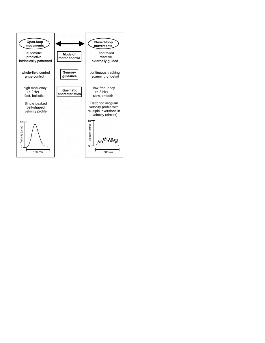
to the single-peaked, bell-shaped velocity pro®les of fast automatic
writing movements, controlled closed-loop writing movements show
¯attened velocity pro®les with an increased number of inversions in
velocity (NIV) per single stroke and a reduction in mean writing
velocity (Fig. 1; Eichhorn et al., 1996; Marquardt et al., 1999). The
®ner the adjustment required, the slower the movement and the higher
the NIV per stroke. This inherent reciprocal relation between
movement velocity and the mode of motor control (i.e. mode of
sensorimotor processing) implies that changes in the speed of
movements do not re¯ect a simple scaling of a kinematic variable,
but are invariably associated with a change in the mode of motor
control (Freund, 1986; Kunesch et al., 1989).
Switching from open-loop to closed-loop processing has several
implications for motor control. First, the motor output changes
dramatically, as indicated by different velocity pro®les (Fig. 1).
Second, closed-loop handwriting necessitates a continuous integra-
tion of sensory input into appropriate motor output, whereas open-
loop movements are run off automatically. Third, subjects need to
pay more attention to handwriting. Thus, similar to early stages of
motor learning, brain areas involved in the generation of the motor
output, in sensorimotor integration, and in attention are likely to
increase their functional activity when closed-loop processing is
required.
The mechanisms involved in open-loop and closed-loop processing
of skilled hand movements need to be considered when functional
brain mapping is employed to study patients with manual motor
de®cits. In patients, automatic open-loop performance of manual
skills is commonly impaired and hand movements are slowed down
(Phillips et al., 1994; Eichhorn et al., 1996; Siebner et al., 1999a). A
disease-related shift from fast open-loop processing to slow closed-
loop processing is likely to have a considerable impact on the
movement-related cerebral activation pattern in patients with manual
motor de®cits. Therefore, a closer understanding of the mechanisms
being involved in fast open-loop and slow controlled-loop processing
of skilled hand movements in healthy subjects will help the
Correspondence: Dr Hartwig Roman Siebner,
2
Sobell Department of
Neurophysiology, as above.
E-mail: h.siebner@ion.ucl.ac.uk
Received 19 January 2001, revised 20 June 2001, accepted 21 June 2001
European Journal of Neuroscience, Vol. 14, pp. 726±736, 2001
ã Federation of European Neuroscience Societies
understanding of disease-related cerebral reorganization patterns in
patients (Weiller et al., 1992; Jahanshahi et al., 1995; Bartenstein
et al., 1997; Samuel et al., 1997; Catalan et al., 1999). By combining
H
2
15
O positron emission tomography (PET) and kinematic analysis
of handwriting movements, the purpose of the present study was to
identify those brain areas that are particularly involved in either fast
open-loop processing or slow closed-loop processing of handwriting
movements in healthy subjects.
Methods
Subjects
Ten healthy right-handers (two females) between 25 and 56 years of
age (mean age, 41.3 6 10.9 years) participated in this study.
Handedness was assessed by the Edinburgh Handedness Inventory
(Old®eld, 1971). Written informed consent for the procedure was
given by all subjects prior to the PET study. Permission to administer
radioactive substances was obtained from the German radiation
protection authorities and the study had the approval of the Ethics
Committee of the Faculty of Medicine of the Technische UniversitaÈt
MuÈnchen.
Experimental design
Each participant underwent nine sequential H
2
15
O-PET measure-
ments within a single session. All subjects were scanned in supine
position in a dimly lit room. Three experimental conditions were
investigated: two writing conditions (i.e. condition `A' and condition
`B') and a baseline condition (i.e. condition `C'). For each
experimental condition, three PET scans were performed. The
experimental conditions were repeated in an alternating order (A-B-
C-A-B-C-A-B-C), which was counterbalanced across subjects. In the
baseline condition, subjects held the pencil on a writing tablet without
writing. In the writing conditions, participants were required to
repeatedly write the German verb `bellen' (i.e. `to bark') without
visual feedback (Fig. 2). This rather simple word was selected to
minimize the cognitive effort during the writing task and to facilitate
¯uent handwriting. In the ®rst writing task, the subjects were asked to
write at their own size and speed (i.e. fast open-loop handwriting,
condition A). In the second writing task, subjects were instructed to
write at approximately half of their normal speed (i.e. slow closed-
loop handwriting, condition B). In both conditions, participants were
instructed to ensure a constant writing velocity. Each subject was
asked to reposition their hand to the starting point after having written
the word. In order to keep the number of writing movements constant
across handwriting conditions, writing was paced by a tone every 6 s.
The background noise and the pacing tone were kept constant
throughout the experimental conditions.
Prior to PET scanning, subjects were trained to perform both
writing tasks in a lying position without visual feedback. Training
was carried out in the PET scanner and subjects wrote the same word
(i.e. the word `bellen') as they actually had to produce during PET
scanning. Practice continued until kinematic analysis of the writing
movements indicated that subjects correctly performed the tasks at a
constant level. Stable motor performance was achieved after about
10±20 min of practice.
Assessment of handwriting
During PET scanning, handwriting was recorded continuously using a
digitizing graphics tablet (UD-1212, Wacom Europe GmbH, Neuss,
Germany). The writing board was placed over the participant's legs
with the surface of the board angled at 45 degrees to the horizontal
plane. Care was taken that subjects could reach the digitizing tablet
without any effort. The ®rst pacing tone was given when starting the
injection of the radioisotope and participants continued to write until
the end of each 50-s PET scan. For each PET scan, only the last eight
words, which were written during the 50-s period of data acquisition,
were used for kinematic analysis. In order to minimize movements of
proximal joints and to match the posture of the hand as close as
possible to the normal position during handwriting, the right upper
limb was comfortably supported by foam plastic pads and partici-
pants were required to place the ulnar part of their right hand on the
writing board while they were writing.
Pen-tip position of an inking digitizing pen were sampled at a
frequency of 166 Hz with a spatial resolution of 0.05 mm and a
spatial accuracy of 0.025 mm. The CS software (MedCom, Munich,
Germany) was used for data collection and off-line analysis of the
writing movements. The curves of the vertical position, and vertical
velocity of the tip of the digitizing pen were calculated and smoothed
by nonparametric regression methods (Marquardt & Mai, 1994). The
CS software has been shown previously to be a suitable tool to assess
changes in kinematics during handwriting in patients with movement
disorders (Eichhorn et al., 1996; Siebner et al., 1999a, b). Kinematic
analyses focused on movement along the vertical axis (i.e. y-axis), as
this is the axis along which writing movements predominantly occur
(Fig. 2). Thus, for quantitative analysis, the writing movements were
segmented in subsequent vertical up- or down-strokes of the pencil,
which can be considered to be fundamental units of handwriting
movements (Hollerbach, 1981; Morasso & Mussa Ivaldi, 1982;
Plamodon, 1995). A single stroke is de®ned by the time segment
F
IG
. 1. Schematic illustration contrasting the characteristics of fast open-
loop movements with those of slow closed-loop movements. Note that a
change in velocity is not just a different scaling of a single kinematic
variable but involves a shift in the mode of motor control and a change in
the magnitude of sensorimotor guidance.
Central processing of writing velocity 727
ã 2001 Federation of European Neuroscience Societies, European Journal of Neuroscience, 14, 726±736
between two subsequent changes in vertical direction of handwriting
(Fig. 2). Only up- and down-strokes that exceeded a duration of
50 ms and an amplitude of 1 mm were included in the kinematic
analysis. The following dimensions of writing performance were
calculated: mean vertical stroke length, mean vertical stroke duration,
mean vertical writing velocity and mean vertical writing pressure.
Furthermore, the mean NIV per single stroke was estimated to
quantify the degree of automation of the handwriting movements
(Marquardt & Mai, 1994). A NIV of 1 per stroke is characteristic of
fast open-loop performance, whereas an increase in mean NIV per
stroke indicates continuous adjustments of writing velocity to the
incoming feedback information during slow closed-loop handwriting
(Eichhorn et al., 1996; Marquardt et al., 1999).
PET scanning
Regional changes in neuronal activity were indexed by monitoring
relative changes in normalized regional cerebral blood ¯ow (rCBF).
Normalized rCBF was measured by recording the regional distribu-
tion of radioactivity following the intravenous injection of
15
O-
labelled water (Fox & Mintun, 1989). PET measurements were
conducted using a Siemens ECAT 951 R/31 PET scanner (CTI,
Knoxville, TN, USA) in three-dimensional mode. After corrections
for randoms, dead time and scatter, all emission data were
reconstructed by ®ltered back projection (Hanning ®lter, 0.5 cycles
per pixel cut-off frequency) to 31 consecutive axial planes with an
interplane separation of 3.375 mm. Reconstructed slices were
displayed in a matrix consisting of 128 3 128 voxels. The PET
scanner had a total axial view of 10.5 cm and no interplane dead
space, ensuring coverage of the upper two-thirds of the brain from the
vertex to the upper cerebellum. Because the ®eld of view of the PET
scanner did not suf®ciently cover the cerebellum, the present study
provides no information on the speci®c involvement of the
cerebellum in the performance of fast open-loop or slow closed-
loop handwriting movements.
For each measurement of rCBF, 250 mBq of H
2
15
O were
administered in the left cubital vein as a semibolus injection using
an infusion pump. A 50-s PET scan was initiated with the appearance
of the tracer bolus in the brain, approximately 30 s after the start of
the infusion (Fox & Mintun, 1989). This procedure was repeated for
each PET scan with 10 min between scans to allow for adequate
decay of radioactivity. A 20-min headholder transmission scan with a
rotating
68
Ge/
68
Ga source was obtained prior to the ®rst relative rCBF
measurement in order to correct the relative rCBF measurements for
effects of radiation attenuation.
PET image reconstruction
All calculations and image transformations were performed on Sun
SPARC 2 workstations (Sun Computers Europe, Surrey, UK). PET
data were analysed using statistical parametric mapping software
(SPM 96b, Wellcome Department of Cognitive Neurology,
University College of London, UK) implemented in the PRO
Matlab environment (Mathworks, Natic, MA, USA). The scans
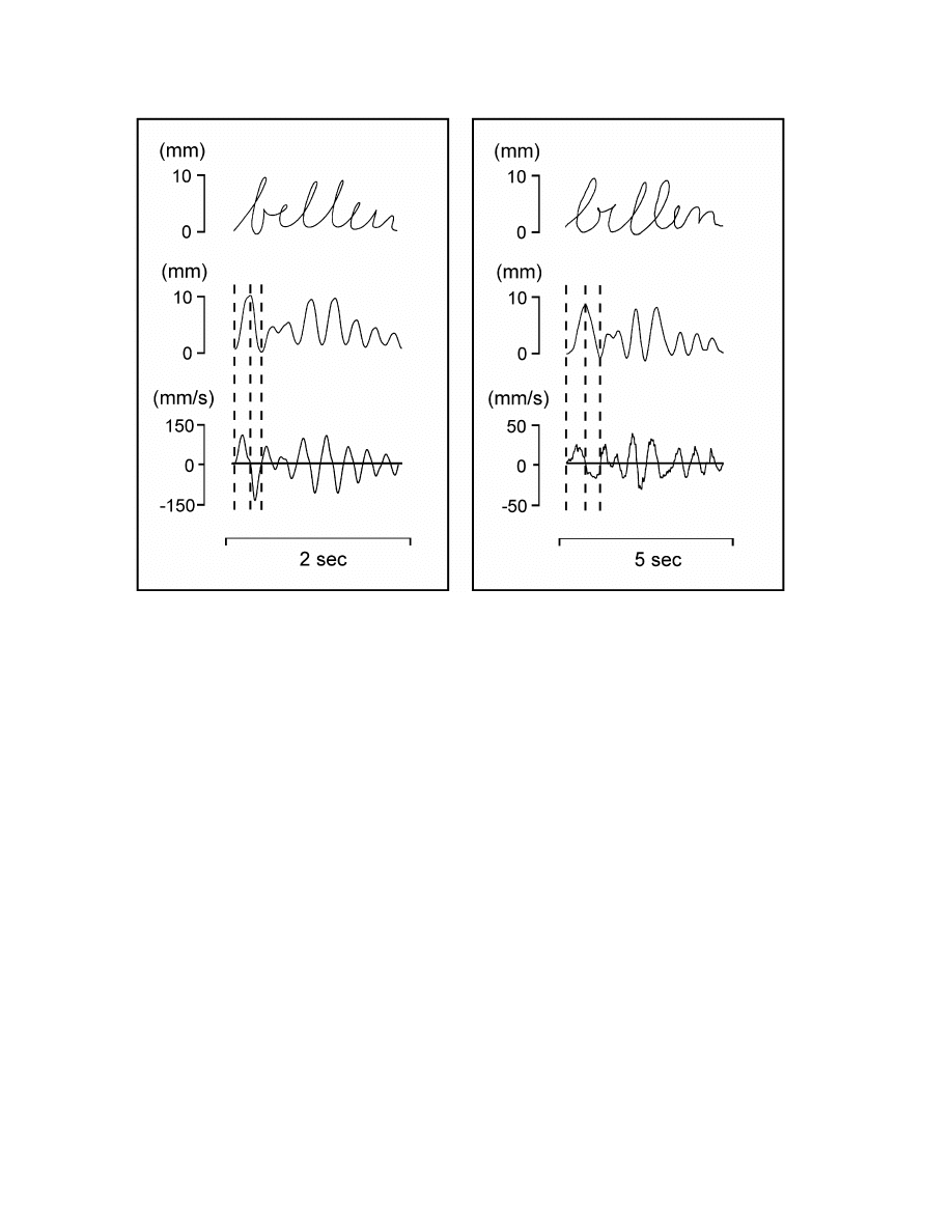
F
IG
. 2. Recording of writing movements in a single subject during fast open-loop handwriting (left panel) and slow closed-loop handwriting (right panel).
The top row of each panel gives the handwriting movements in space when writing the German word `bellen' (the verb `to bark'). The middle row illustrates
the vertical writing movements (i.e. strokes, with vertical position is shown as a function of time), while the bottom row demonstrates the vertical velocity
pro®les. Note the different time-scales for the left and right panels. Automatic open-loop handwriting shows smooth single-peaked velocity pro®les, whereas
velocity pro®les during slow closed-loop handwriting are characterized by many irregularities (i.e. inversions in velocity), indicating corrective adjustments of
vertical writing velocity.
728 H. R. Siebner et al.
ã 2001 Federation of European Neuroscience Societies, European Journal of Neuroscience, 14, 726±736

from each subject were realigned using the ®rst scan as a reference.
The six parameters of this rigid body transformation were estimated
using a least squares approach on a voxel-by-voxel basis (Friston
et al., 1995a). Following realignment, PET images were transformed
into standard stereotaxic space (Talairach & Tournoux, 1988). Spatial
normalization was performed using linear and nonlinear three-
dimensional transformations to match each scan to a reference image,
which already conformed to the standard stereotaxic space (Friston
et al., 1995a). As a ®nal preprocessing step, the normalized images
were smoothed using an isotropic Gaussian kernel of 12 mm full
width at half maximum (FWHM) for all directions to increase the
signal-to-noise ratio and reduce variance due to interindividual
differences in gyral anatomy (Friston et al., 1995a). Each voxel of the
resulting normalized and smoothed images was 2 3 2 3 4 mm in
size.
Data analysis
Statistical analysis of the kinematic data was performed with SPSS
Version 9 (SPSS Inc., Chicago, IL, USA). Analysis of variance
(
ANOVA
) for repeated measurements with `time' and `writing
condition' as within-subject factors was computed for each kinematic
variable to assess the effect of time and writing condition on motor
performance. Signi®cance was accepted at a P-value of 0.05.
Statistical analysis of the PET data was performed using statistical
parametric mapping software (SPM, Version 96b). To remove the
effect of variations in global cerebral blood ¯ow across subjects and
scans, an analysis of covariance (
ANCOVA
) was applied with global
cerebral blood ¯ow as the confounding variable (Friston et al., 1990).
Global blood ¯ow was normalized by scaling across the entire data
set to a global mean of 50 mL/100 mL/min. The adjusted voxel
values were then used for further statistical analysis.
In a ®rst set of analyses, a categorical comparison was performed
between the two writing conditions and the baseline condition in
order to delineate the brain areas involved in handwriting per se.
Using linear weighted contrasts, the main effects of both writing
conditions were estimated according to the general linear model and
the theory of Gaussian ®elds at each and every voxel (Friston et al.,
1991, 1995b; Worsley et al., 1992). The generated statistical
parametric [t] maps were subsequently transformed into normally
distributed statistical parametric [Z] maps (Friston et al., 1995b).
Signi®cance level was set at a P-value of 0.05 after correction for
multiple nonindependent comparisons; this corresponds to a Z-score
of 4.26. Brain areas showing increases in rCBF at an uncorrected
P-value of less than 0.001 (corresponding to a Z-score of 3.09), but
not surviving correction for multiple nonindependent comparisons,
were considered as trend activations. Given the aims of the study, the
subsequent analyses used these categorical comparisons as a mask
and restricted further interrogation of the data to brain areas showing
at least a trend activation during automatic or controlled handwriting
in comparison with the baseline condition.
In a second set of analyses, we directly compared the activation
patterns during fast open-loop and slow closed-loop handwriting. For
this purpose, baseline scans were not included. A categorical
comparison between both writing conditions was performed in
order to assess brain areas that were particularly activated during
either fast open-loop handwriting or slow closed-loop handwriting. A
categorical comparison is useful to demonstrate brain areas that
change their net activity in a stepwise fashion between two
experimental conditions. However, a categorical approach may fail
to detect those brain areas that gradually change their functional
activity according to the requirements of a given task. By contrast,
correlational analysis between regional functional activation and a
distinct variable of motor performance, such as velocity or com-
plexity, has been shown to provide a useful means to detect graded
changes in distinct brain areas related to certain aspects of motor
control (Boecker et al., 1998; Turner et al., 1998; van Mier et al.,
1998). Therefore, in addition to a categorical comparison between
fast and slow handwriting, an independent correlational analysis
between the NIV per stroke and the normalized rCBF was performed.
Because the mean NIV per stroke re¯ects the number of corrective
adjustments in velocity during a single up- or down-stroke, this
kinematic measure is thought to re¯ect directly the degree of closed-
loop processing during handwriting (Eichhorn et al., 1996; Marquardt
et al., 1999; Siebner et al., 1999a). Because data analysis was
restricted to brain areas that had already been identi®ed to participate
in handwriting, an uncorrected P-value of 0.001 was accepted as
threshold for signi®cance in the second set of analyses.
Results
Kinematic data
Mean total movement time for writing the target word was
2.24 6 0.64 s during fast handwriting, whereas mean total movement
time was 4.21 6 0.72 s during slow handwriting, indicating that
participants wrote about twice as long during the slow handwriting
condition. Figure 2 illustrates the recorded writing movements and
the corresponding velocity pro®les during fast open-loop handwriting
and slow closed-loop handwriting in a representative subject. Table 1
gives the averaged group values (mean 6 SD) of each kinematic
measure for both writing conditions.
ANOVA
for repeated measure-
ments revealed a highly signi®cant effect of `writing condition' on
mean stroke duration, mean vertical velocity and mean NIV per
stroke (Table 1). In accordance with the instruction, subjects wrote at
approximately half of their normal speed during slow, velocity-
controlled handwriting and velocity-controlled handwriting was
associated with a consistent increase in the mean NIV per stroke
T
ABLE
1. Group data of kinematic measurements during fast open-loop handwriting and slow closed-loop handwriting
Stroke-based kinematic measures of handwriting
Fast open-loop
handwriting
Slow closed-loop
handwriting
F
1,9
-value
P-value
Numbers of inversions in velocity (NIV) per stroke
1.38 6 0.36
3.62 6 1.06
51.5
P < 0.001
Vertical writing velocity (mm/s)
67 6 31
39 6 24
52.4
P < 0.001
Stroke duration (ms)
141 6 25
265 6 27
178.4
P < 0.001
Vertical writing pressure (N)
1.65 6 0.60
1.59 6 0.68
0.2
n.s.
Vetrical stroke length (mm)
7.1 6 3.2
8.4 6 4.7
2.9
n.s.
Data are given as mean 6 SD. The F- and P-values refer to multivariate analysis of variance for repeated measurements and describe the main effect of `writing
condition' on the dependent kinematic variable. n.s., not signi®cant (P > 0.05).
Central processing of writing velocity 729
ã 2001 Federation of European Neuroscience Societies, European Journal of Neuroscience, 14, 726±736
(Table 1). The increase in NIV per stroke con®rmed that writing
velocity was continuously adjusted and thus, a closed-loop mode of
motor control was employed during the slow handwriting task. There
was a highly signi®cant positive correlation between the mean NIV
per stroke and the mean stroke duration (Spearman, r = 0.89,
P < 0.0001), indicating that slow writing movements were associated
strongly with a more irregular velocity pro®le.
There was no signi®cant effect of `writing condition' on mean
vertical stroke length and mean vertical writing pressure, con®rming
a selective modi®cation of writing kinematics related to velocity
without concurrent modulation of scaling the stroke length or vertical
force production. Furthermore,
ANOVA
for repeated measurements
showed no signi®cant main effect of `time' on any of the kinematic
variables of interest, indicating that there were no systematic changes
of motor performance during the entire experiment. Moreover, there
was no signi®cant interaction term between `time' and `writing
condition'.
PET data
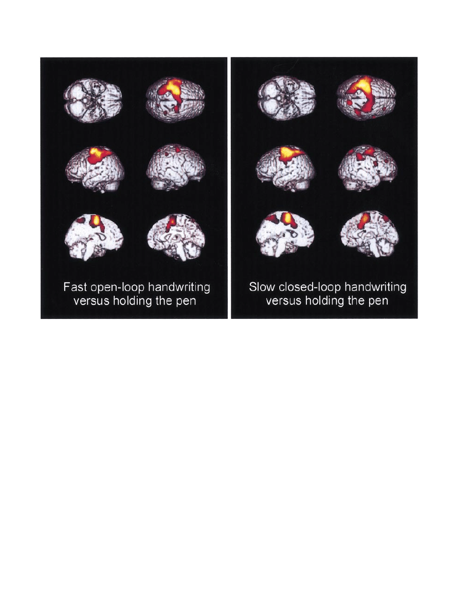
Identi®cation of brain regions involved in handwriting per se
Categorical comparison between handwriting and the baseline
condition demonstrated that the activation patterns were very similar
during fast open-loop handwriting and slow closed-loop handwriting
(Fig. 3, Table 2). Regardless of the mode of motor control,
handwriting was associated with a signi®cant relative increase in
normalized rCBF in a large bihemispheric cortical cluster with a
prevailing activation of left hemispheric regions (Fig. 3). Within this
cluster, peak activation occurred in the hand representation of the left
primary sensorimotor cortex (SM1) and the adjacent dorsal premotor
cortex (PMD), as indicated by the highest Z-scores on categorical
comparison (Table 2). In addition, the cluster covered large areas of
the left parietal lobe and the left ventral premotor cortex (PMV) on
the left hemispheric surface (Fig. 3). Within the interhemispheric
®ssure, the cluster extended into the supplementary motor area
(SMA), the anterior cingulate cortex (ACC) and the left precuneus.
On the right hemispheric surface, the cluster included mainly the
right PMD. A second cortical cluster, which showed an activation
during both writing conditions, was located at the border between the
precuneus and the superior parietal lobule in the right parietal cortex.
Subcortically, handwriting was associated with a bilateral activation
of the thalamus.
Despite the substantial overlap in the cerebral activations during
fast open-loop handwriting and slow closed-loop handwriting, there
were some notable differences in the cerebral activation patterns. The
spatial distribution of peak activations within the large frontoparietal
cluster (indexed as `cluster 1' in Table 2) differed between condi-
tions. For instance, only fast open-loop handwriting gave rise to
F
IG
. 3. Surface rendering of colour-coded statistical parametric maps superimposed on stereotactically normalized (Talairach & Tournoux, 1988) T1-weighted
magnetic resonance images. Surface projections showing categorical increases of regional cerebral blood ¯ow (rCBF) during fast open-loop handwriting (left)
compared with holding a pencil. Surface projections showing categorical increases of rCBF during slow velocity-controlled handwriting (right) compared with
holding a pencil. Maps are thresholded at an uncorrected P-value of 0.001. Pixels with lower Z-scores are represented in red, and pixels with higher Z-scores
in yellow.
730 H. R. Siebner et al.
ã 2001 Federation of European Neuroscience Societies, European Journal of Neuroscience, 14, 726±736

distinct peak activations in the left PMV and in the left anterior
inferior parietal cortex, whereas slow closed-loop handwriting caused
a selective peak of activation in the left lateral superior parietal
cortex. Moreover, slow closed-loop handwriting resulted in a larger
number of `activated voxels' and in a less lateralized activation
pattern with a relatively larger area of activation on the right
hemispheric surface. At a statistical threshold of P < 0.001
(uncorrected), » 10 000 voxels showed an increase in rCBF during
fast open-loop handwriting when compared with the baseline
condition, whereas » 15 000 voxels demonstrated a relative increase
in normalized rCBF during slow closed-loop handwriting (Table 2).
Only slow closed-loop handwriting caused a signi®cant relative CBF
increase in the right lateral prefrontal cortex and the right PMV, and a
trend activation in the left anterior putamen. By contrast, no
activation in the basal ganglia was observed during fast open-loop
handwriting, even at a low statistical threshold (P < 0.01, un-
corrected). Compared with slow closed-loop handwriting, no speci®c
cluster (i.e. brain region) was selectively activated in the `fast
handwriting' condition.
Fast open-loop handwriting vs. slow closed-loop handwriting
As outlined earlier, the search volume was limited to brain areas that
have shown at least a trend activation during either fast or slow
handwriting compared with holding the pencil. Please note that this
`mask' is not overly restrictive, as it incorporates the entire set of
brain areas that have previously been shown as being implicated in
the generation of skilled writing movements (Ceballos-Baumann
et al., 1997; Seitz et al., 1997; IbaÂnÄez et al., 1999). Within the regions
showing at least a trend activation during handwriting per se,
categorical comparison between fast open-loop handwriting and
slow closed-loop handwriting detected differences in rCBF in a
distinct set of brain areas (Table 3). Because fast and slow
handwriting resembles similar `activated states' of the cerebral
motor control system, the overall Z-scores were much lower than
those for the contrasts comparing each writing condition with the
baseline condition. The only area showing a signi®cant activation
during fast open-loop handwriting as compared with slow closed-loop
handwriting was a focus in the right SM1 dorsomedially to the
primary sensorimotor hand area (Table 3). Bilateral foci in the
inferior parietal lobule, the right PMD and the left anterior rostral
putamen were more active during slow closed-loop handwriting when
compared with fast open-loop handwriting (Table 3). The areas in the
left dorsolateral prefrontal cortex and the right PMV, which were
activated exclusively during slow handwriting but not during fast
handwriting (when compared with the baseline condition), did not
show signi®cant differences when performing a direct categorical
comparison between fast and slow handwriting. This was due to
subtle, nonsigni®cant rCBF increases in these areas during fast open-
loop handwriting compared with holding the pen.
Correlational analysis revealed a somewhat different regional
pattern of cerebral clusters, which demonstrated a linear relationship
between the rCBF and the degree of closed-loop performance, as
indexed by the mean NIV per stroke (Table 4). No cerebral area
within the ®eld of view of the PET scanner showed a negative linear
T
ABLE
2. Clusters of brain regions showing a signi®cant relative increase in normalized rCBF, indicating a functional activation during fast open-loop
handwriting or slow closed-loop handwriting compared with holding the pencil
Brain regions
(BA)
Voxels
per cluster
Z-score of
peak activation
Coordinates of peak activation
x
y
z
Areas showing relative increases in rCBF during automatic handwriting (condition A)
Cluster 1
9730
Left dorsal premotor cortex
(6)
7.24
±22
±10
66
Left primary sensorimotor cortex
(3/4)
7.22
±38
±24
58
Left ventral premotor cortex
(6)
5.97
±62
±12
38
Caudal supplementary motor area
(6)
5.87
±8
±8
56
Left anterior inferior parietal cortex
(2/40)
5.59
±54
±26
44
Right dorsal premotor cortex
(6)
(3.72)
30
±10
62
Cluster 2, left thalamus
±
28
(3.91)
±12
±22
4
Cluster 3, right inferior parietal cortex
(40)
93
(3.85)
38
±44
52
Cluster 4, right precuneus
±
77
(3.66)
16
±66
62
Cluster 5, right thalamus
±
30
(3.49)
14
±16
10
Areas showing relative increases in rCBF during controlled handwriting (condition B)
Cluster 1
14005
Left primary sensorimotor cortex
(3/4)
7.83
±38
±22
60
Left dorsal premotor cortex
(6)
7.55
±26
±14
62
Left lateral superior parietal cortex
(7)
6.87
±32
±52
60
Right dorsal premotor cortex
(6)
5.86
30
±12
60
Caudal supplementary motor area
(6)
5.55
0
±4
56
Cluster 2, right precuneus
(7)
440
5.40
14
±64
60
Cluster 3, right ventral premotor cortex
(6/44)
198
4.49
62
10
24
(3.67)
52
6
28
Cluster 4, right lateral prefrontal cortex
(9)
177
4.29
34
38
34
Cluster 5, left thalamus
±
29
(3.59)
±10
±20
4
Cluster 6, right thalamus
±
16
(3.57)
8
±2
22
Cluster 7, left anterior putamen
±
48
(3.55)
±24
6
16
Cluster 8, right anterior claustrum/putamen
±
43
(3.51)
32
16
2
A Z-score of 4.26 corresponds to a P-value of 0.05 after correction for multiple comparisons. Z-scores ranging from 3.09 to 4.26 were considered as trend
activations. Trend activations in this table are those in parentheses. Coordinates express the position of activation foci within the cluster relative to the anterior
commissure in the stereotactic space of Talairach & Tournoux (1988).
Central processing of writing velocity 731
ã 2001 Federation of European Neuroscience Societies, European Journal of Neuroscience, 14, 726±736

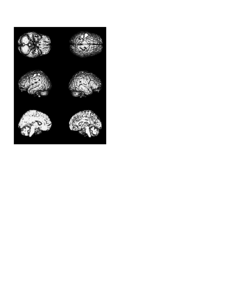
relationship between rCBF and the mean NIV per stroke. In contrast,
several regions within the distributed network, which participates in
handwriting, showed a positive relationship between rCBF and the
mean NIV per stroke, indicating a gradual increase in functional
activation with close-loop handwriting. These areas included distinct
foci in the left SM1, the left anterior inferior parietal cortex, the right
PMD, the left rostral SMA, the right posterior superior parietal
cortex, the right anterior inferior parietal cortex, and the left anterior
putamen (Fig. 4, Table 4).
Discussion
By combining PET scanning and continuous recording of writing
movements, the present study yielded two main results. First, a
distinct set of cortical and subcortical areas demonstrated a gradual
increase in functional activation with slow handwriting, involving
closed-loop control of writing velocity. Second, no cerebral region
within the ®eld of view of the PET scanner showed a graded
activation with fast automatic handwriting movements when match-
ing the numbers of movements per PET scan between `fast open-loop
handwriting' and `slow closed-loop handwriting'.
Methodological considerations
Automatic open-loop performance is characterized by fast move-
ments with a higher movement rate, whereas controlled closed-loop
performance is restricted to the range of slower movements with a
lower movement rate (Fig. 1; Freund et al., 1986; Kunesch et al.,
1989). The close relationship between movement velocity and the
mode of sensorimotor processing implies a methodological dilemma
in terms of matching total motor output between fast open-loop
handwriting and slow closed-loop handwriting. On one hand, when
matching the number of movements between writing conditions, total
movement time will differ, because subjects need less time to carry
out fast writing movements. On the other hand, by keeping total
movement time constant between writing conditions, the total motor
output (e.g. the length of the writing trace and the number of strokes)
will differ between writing conditions.
Previous studies on velocity control of skilled hand movements
(Turner et al., 1998; van Mier et al., 1998) investigated continuous
movements, keeping total movement time constant across PET scans.
Because an increase in movement velocity is invariably associated
with an increased number of submovements (i.e. switches in the
motor programme), as well as in an increase in the total length of the
T
ABLE
3. Clusters of brain regions showing signi®cant differences in normalized rCBF during both writing conditions when applying a categorical comparison
(i.e. fast open-loop vs. slow closed-loop handwriting)
Brain regions
(BA)
Voxels
per cluster
Z-score of
peak activation
Coordinates of peak activation
x
y
z
Areas showing relative rCBF increases during automatic handwriting compared with controlled handwriting
Right primary sensorimotor cortex
(3/4)
21
3.56
22
±20
76
Areas showing relative rCBF increases during controlled handwriting compared with automatic handwriting
Left inferior parietal cortex
(40)
153
3.95
±36
±40
54
Left rostral putamen
±
55
3.80
±24
10
14
Right inferior parietal cortex
(40)
93
3.73
38
±44
36
Right lateral premotor cortex
(6)
84
3.73
28
0
48
The data are presented as in Table 2. Please note that an uncorrected P-value of 0.001 was accepted as the statistical threshold for those brain areas, as data analysis
was restricted to those brain areas, which had shown at least a trend activation during either fast or slow handwriting as compared with holding the pencil.
T
ABLE
4. Clusters of brain regions showing a signi®cant correlation between normalized rCBF and the mean numbers of inversions in velocity (NIV) per
stroke (P < 0.001, uncorrected)
Brain regions
(BA)
Voxels
per cluster
Z-score of
peak activation
Coordinates of peak activation
x
y
z
Areas showing a negative correlation between rCBF and the mean NIV per stroke during handwriting
No cluster within the ®eld of view of the scanner
Areas showing a positive correlation between rCBF and the mean NIV per stroke during handwriting
Left primary sensorimotor cortex
(3/4)
258
4.57
±56
±14
52
Right lateral premotor cortex
(6)
208
4.34
3.59
16
20
±12
±8
58
48
Left inferior parietal cortex
(5/40)
208
3.99
±44
±34
58
Left rostral putamen
±
43
3.55
±22
8
12
Left supplementary motor area
(6)
33
3.37
±12
2
56
Right precuneus
(7)
15
3.37
20
±56
50
The mean NIV per stroke was taken as a kinematic measure to quantify the degree of closed-loop performance during handwriting. A mean NIV of 1 corresponds
to automatic open-loop handwriting, whereas an increase in mean NIV per stroke indicates a shift towards closed-loop performance. The higher the mean NIV per
stroke the greater the magnitude of closed-loop processing. The data are presented as in Table 3. Please note that an uncorrected P-value of 0.001 was accepted as
the statistical threshold for correlational analysis, as data analysis was restricted to those brain areas, which had shown at least a trend activation during either fast
or slow handwriting as compared with holding the pencil.
732 H. R. Siebner et al.
ã 2001 Federation of European Neuroscience Societies, European Journal of Neuroscience, 14, 726±736
movement trace, the reported differences in functional activation
were not exclusively related to movement velocity. In the present
study, we decided to keep the number of movements constant across
PET scans rather than matching total movement time. The rationale
for matching the number of writing movements was twofold. First, it
has been shown that differences in the number of movements per scan
have a profound effect on neuronal activation in executive motor
areas, especially in the primary sensorimotor cortex (Blinkenberg
et al., 1996; Sadato et al., 1997; Jancke et al., 1998; Kawashima et al.,
1999). Second, previous functional imaging studies on differences in
the motor activation pattern between `fast-moving' healthy controls
and `slow-moving' patients have generally matched the number of
movements per PET scan rather than total movement time (Weiller
et al., 1992; Jahanshahi et al., 1995; Bartenstein et al., 1997; Samuel
et al., 1997; Catalan et al., 1999). However, we like to emphasize that
total movement time was not matched between writing conditions, as
participants were engaged in slow closed-loop handwriting longer
than in fast open-loop handwriting.
Sensorimotor control of writing movements may involve different
sensory modalities and may focus on different aspects of handwriting,
such as velocity, stroke size or shape. The present study focused on
the kinaesthetic control of the velocity of handwriting. In order to
ensure that only kinaesthetic feedback was used to monitor writing
velocity, participants were deprived of visual feedback. In accordance
with Marquardt et al. (1999), kinematic analysis revealed that the
lack of visual feedback did not hamper open-loop processing of
handwriting movements. Moreover, the functional activation pattern
during handwriting was not in¯uenced by different levels of motor
learning, as stable task performance was con®rmed by kinematic
analysis during PET scanning.
Brain activation during handwriting per se
The cerebral activation pattern related to handwriting in comparison
with baseline condition was quite similar during fast open-loop and
slow-closed-loop performance, showing a widespread bilateral
increase in normalized rCBF in the SM1, the lateral premotor cortex,
the SMA, adjacent ACC, and anterior and posterior parts of the
parietal cortex with a left-hemispheric preponderance. Subcortically,
handwriting was associated with a consistent bilateral thalamic
activation. This cerebral activation pattern is in accordance with
previous functional imaging studies on healthy volunteers, which
have con®rmed the participation of these brain regions in the
generation of handwriting movements (Ceballos-Baumann et al.,
1997; Seitz et al., 1997; IbaÂnÄez et al., 1999). The close spatial
correspondence in writing-related cerebral activation patterns across
studies is somewhat surprising, given the fact that there are notable
differences in the writing tasks employed in these studies (Ceballos-
Baumann et al., 1997; Seitz et al., 1997; IbaÂnÄez et al., 1999). In the
study by Seitz et al. (1997), subjects were required to continuously
write letters or `nonsense' letters and visual feedback was provided,
whereas in the other studies subjects repeatedly wrote either a word
(`dog') paced by an auditory cue every 4 s (Ceballos-Baumann et al.,
1997) or a sentence (`the book is on the desk') in a self-paced manner
(IbaÂnÄez et al., 1999) without visual feedback. With the exception of
the study by Ceballos-Baumann et al. (1997), the number of writing
movements was not matched across PET scans. Moreover, the
instructions differed considerably across the studies. In the study by
Seitz et al. (1997), subjects were asked to write the letters either as
fast as possible or as exact as possible with respect to letter size,
requiring either an automatic open-loop mode of handwriting during
`fast writing' or a closed-loop mode during `exact writing'. Please
note that closed-loop performance in the study by Seitz et al. (1997)
referred to size control (i.e. spatial aspect of handwriting), whereas, in
the present study, closed-loop performance referred to velocity
control (i.e. temporal aspect of handwriting). In the study by IbaÂnÄez
et al. (1999), subjects were asked `not to write quickly' at their own
pace (presumably in an open-loop mode), whereas in the study by
Ceballos-Baumann et al. (1997), subjects were required to write
continuously and adapt their speed of handwriting to the interstimulus
interval of the cueing tone, demanding a continuous closed-loop
adjustment of writing velocity comparable with the `slow writing
condition' in the present study. With regard to the considerable
differences in the writing task within and between these studies, the
substantial overlap in the spatial activation pattern strongly suggests
that the generation of handwriting movements per se activates a
distinct set of cortical and subcortical regions.
Activation pattern associated with slow velocity-controlled
handwriting
Extending previous ®ndings by Seitz et al. (1997) on visually guided
handwriting, we found also notable differences in the cerebral
activation pattern during fast open-loop and slow closed-loop right
handwriting. During handwriting, the magnitude of closed-loop
control (as indexed by the mean NIV per stroke) was associated
with a graded increase in neuronal activity in a distinct subset of
cortical and subcortical motor control areas, including foci in the left
F
IG
. 4. Surface rendering of statistical parametric maps superimposed on
stereotactically normalized T1-weighted magnetic resonance images. The
white areas represent brain regions that showed a positive correlation
between normalized regional cerebral blood ¯ow (rCBF) and the mean
number of inversions in velocity (NIV) per stroke (see also Table 4),
indicating a weighted regional activation of these areas depending on the
amount of closed-loop processing. Maps are thresholded at an uncorrected
P-value of 0.001.
Central processing of writing velocity 733
ã 2001 Federation of European Neuroscience Societies, European Journal of Neuroscience, 14, 726±736

SM1, the right PMD, the left anterior superior parietal lobule, the left
anterior putamen, the left rostral SMA, and the right precuneus. As
already outlined in the introduction, these brain regions may be
involved in the generation of the motor output, in sensorimotor
integration or in attentional processes, when closed-loop processing is
required.
Left primary sensorimotor cortex (SM1)
Categorical and correlational comparison revealed an increased
neuronal activity in the left SM1 during slow velocity-controlled
handwriting. At a glance, this ®nding seems to be at odds with
previous imaging studies that have shown increasing movement
velocity results in a gradual increase in activation in SM1 (Turner
et al., 1998; van Mier et al., 1998). It needs to be pointed out that
these studies investigated continuous movements (Turner et al., 1998;
van Mier et al., 1998). As already outlined earlier, increasing the
velocity of continuous movements entails an increase in movement
rate and the total amount of ankle displacement per scan. This
increase in net motor output and net sensory feedback renders it
dif®cult to ascribe gradual changes in SM1 activation exclusively to
changes in movement velocity.
Three mechanisms may have contributed to the linear relationship
between functional activation in the left SM1 and closed-loop
performance in the present study. First and most importantly, subjects
were engaged for about twice as long in task performance during
slow closed-loop handwriting, resulting in a longer neuronal
processing time. Second, an increase in attention paid to kinaesthetic
input from the writing hand during closed-loop processing might have
contributed to increased functional activation of the left SM1 (Meyer
et al., 1991). Third, because the SM1 has been shown to be
particularly active during ®nger movements guided by somatosensory
information (Geyer et al., 1996), the gradual increase in SM1 activity
might also re¯ect increasingly higher demands for online processing
of kinaesthetic feedback during slow velocity-controlled handwriting.
Frontal premotor areas
Two circumscribed areas in the frontal premotor cortex showed a
graded increase in rCBF with an increase in sensorimotor guidance,
namely the right PMD and the left rostral SMA anterior to the vertical
anterior commissural plane. These premotor areas have shown a
progressive increase in rCBF with movement complexity (i.e. the
right PMD with increasing sequence length and left rostral-SMA with
complexity of sequence pattern) when subjects performed a sequence
of auditory paced ®nger movements with their dominant right hand,
suggesting a role of these areas in the control of sequential ®nger
movements (Sadato et al., 1996; Boecker et al., 1998; Catalan et al.,
1998). In the present study, subjects wrote the same word in both
writing tasks. Hence, differences in task complexity did not account
for the increased activity in these two premotor areas during slow
handwriting. Given that slow velocity-controlled handwriting de-
manded a high degree of temporal accuracy, the increased activation
in the left pre-SMA and right PMD suggests an involvement of these
areas in precise timing of over-learned movement sequences
(Halsband et al., 1993). The increased activation of the pre-SMA
during slow handwriting is probably related to additional timing
operations during handwriting at a frequency slower than the
subject's normal pace (Kawashima et al., 1999).
It has been suggested that the caudal part of the PMD is concerned
with online correction of movement execution (see Wise et al., 1997
for review, Grafton et al., 1998). In keeping with this notion, the
activation peak in the PMD, showing a graded increase in activation
with the amount of closed-loop handwriting, was located within the
more caudal part of the PMD (x, y and z of 16, ±12 and 58).
Moreover, in accordance with studies by Sadato et al. (1996) and
Catalan et al. (1998) on motor sequence control, activation of the
PMD was observed within the right hemisphere ipsilaterally to the
moving hand. This is most likely due to a right hemispheric
dominance for spatial attention (Gitelman et al., 1996; Winstein et al.,
1997).
Parietal cortex
An area in the left anterior inferior parietal cortex Brodmann area
(BA) 5, extending into BA 40, showed a graded functional activation
with slow closed-loop handwriting. It has been reported that the
activity in this anterior part of the parietal cortex gradually increases
with the degree of exerted force during ®nger movements (Dettmers
et al., 1995) and that this area is selectively active during execution
but not during preparation or imagination of freely selected joystick
movements (Stephan et al., 1995), indicating a role of this parietal
area in processing somatosensory feedback during hand movements.
This parietal area may correspond to the parietal area PE (area 5) in
monkeys, which is thought to be a higher-order somatosensory area
mostly devoted to the analysis of proprioceptive information (for
review see Rizzolatti et al., 1998). Area PE receives proprioceptive
and cutaneous input from the limb and responds to somaesthetic
stimuli, but it does not receive visual input (Sakata et al., 1973;
Mountcastle et al., 1975). Many neurons in area PE encode
movement kinematics of upper limb movements (Kalaska et al.,
1990) and the location of the arm in space in a body-centred
coordinate system (Lacquaniti et al., 1995). Furthermore, the PE area
is richly interconnected with the primary motor hand area of the
monkey (F1). These features of area PE and the PE±F1 circuits have
led Rizzolatti et al. (1998) to suggest that the `main role of the PE±F1
circuits appears to be that of providing F1 with information on the
location of body parts necessary for the control of movement'.
Because the velocity-controlled writing task involved continuous
sensorimotor processing of kinaesthetic feedback from the moving
hand, we would like to put forward the hypothesis that the increase in
the level of activation in the left SM1 and anterior parietal cortex
during controlled writing re¯ects the activation of parietofrontal
loops involved in kinaesthetic control of skilled hand movements,
which may be the human homologues to the PE±F1 circuits in the
monkey.
A gradual increase in the level of activation with the amount of
closed-loop handwriting was also observed in the right precuneus.
Apart from the importance of the precuneus in spatial awareness
(Corbetta et al., 1993), the precuneus is thought to be an integrative
transmodal area providing a sensory representation of extrapersonal
space (for review see Mesulam, 1998). Our data suggest that the
precuneus contributes to the integration of kinaesthetic information
into a complex movement sequence presumably by storing not only
spatial representations (Seitz et al., 1997), but also kinaesthetic
representations (Sirigu et al., 1996).
Basal ganglia
Subcortically, controlled handwriting was associated with a focus of
increased activation in the left rostral putamen anterior to the vertical
anterior commissural plane. The rostral putamen is thought to belong
to the `association striatal territory', which receives projections from
various frontal, temporal, and parietal areas, whereas the `sensori-
motor' striatal territory comprises the post-commissural portion of the
putamen, receiving projections from the somatosensory, motor and
premotor areas (Kunzle, 1975; Parent & Hazrati, 1995). The anterior
parts of the basal ganglia have been shown to be active during new
734 H. R. Siebner et al.
ã 2001 Federation of European Neuroscience Societies, European Journal of Neuroscience, 14, 726±736

learning or random generation of a ®nger sequence (Jenkins et al.,
1994; Jueptner et al., 1997), as well as during externally paced, but
not self-paced, sequences of arm movements (Menon et al., 1998).
The graded activation of the anterior putamen during slow closed-
loop handwriting demonstrates that the basal ganglia activity related
to motor performance is in¯uenced by the contextual setting and
plays a role in `optimizing the pattern of muscular activity in the light
of sensory feedback' (Brooks, 1997). Furthermore, the present ®nding
is in concordance with the view that the anterior part of the putamen
is activated during movement sequences with higher cognitive
demands (Jueptner et al., 1997), whereas the posterior parts may be
more related to motor performance itself (LeheÂricy et al., 1998).
Brain activation during fast open-loop handwriting
Fast open-loop handwriting was associated with a smaller extent of
brain activation compared with slow closed-loop handwriting.
Furthermore, the brain activation pattern was more lateralized,
showing a more preponderant activation of left-hemispheric parieto-
frontal motor areas. We attribute the smaller magnitude in brain
activation to several factors. Fast open-loop handwriting required a
lower level of sensorimotor integration and less attentional control
than slow closed-loop handwriting. More importantly, the manual
motor network needed only half of the neuronal processing time to
complete the writing task as opposed to slow velocity-controlled
handwriting.
Correlational analysis revealed no brain area within the motor
network related to handwriting, which showed a graded increase in
rCBF with the magnitude of movement automation of handwriting
movements. The basal ganglia, especially, did not show a graded
activation during automated handwriting. The lack of signi®cant
activation within the basal ganglia during automatic open-loop
handwriting is in accordance with recent PET studies by Seitz et al.
(1997) and IbaÂnÄez et al. (1999), who have observed no basal ganglia
activation during ¯uent automated writing in healthy subjects.
Interestingly, closed-loop velocity-controlled handwriting move-
ments resulted in a writing-related activation of the basal ganglia
(Ceballos-Baumann et al., 1997). Moreover, a recent fMRI study
found a consistent activation of the basal ganglia during self-paced
signing and zigzagging with the ®nger or toe (Rintjes et al., 1999).
However, Rintjes et al. (1999) performed a categorical comparison of
self-paced signing (and zigzagging) with rest, whereas the present
study compared externally paced handwriting with holding the pencil.
Thus, differences in the experimental conditions may explain the
discrepant activation pattern in the basal ganglia. The lack of an
automation-related increase in functional activity in the basal ganglia
during fast open-loop handwriting is in line with the notion that the
corticobasal ganglia±thalamocortical motor loops are not essential for
fast automated running of complex motor programmes in healthy
subjects once motor pro®ciency has been achieved (Brooks, 1997).
However, there are two points worth making about the lack of
negative covariance between NIV and rCBF. First, the cerebellum
was outside the ®eld of view of the PET scanner. Hence, we cannot
exclude that distinct parts of the cerebellum may show a signi®cant
negative covariance between NIV and rCBF. Second, a `ceiling'
effect may account for the absence of negative covariance with rCBF,
as handwriting per se resulted in a considerable activation of the
entire motor network regardless of the mode of motor control. Thus,
when comparing fast automatic with slow closed-loop handwriting, it
is conceivable that slight additional increases in functional activity in
a distinct motor control area related to fast open-loop performance
might have been missed in the present study. For instance, this may
apply to the caudal SMA, which is thought to be especially involved
in automatic open-loop motor performance (Jenkins et al., 1994).
Acknowledgements
The authors would like to express their gratitude to Ms C. Kruschke and Ms G.
Dzewas for their assistance during PET acquisition, to Ms N. Nguyen for
careful review of the manuscript, and to the staff of the Radiochemistry
Section. The study was supported by the German Research Council
(Sonderforschungsbereich `Sensomotorik' SFB 462, Teilprojekt C3).
Abbreviations
ACC, anterior cingulate cortex; NIV, number of inversions in velocity; PET,
positron emission tomography; PMD, dorsal premotor area; PMV, ventral
premotor area; rCBF, regional cerebral blood ¯ow; SMA, supplementary
motor area.
References
Bartenstein, P., Weindl, A., Spiegel, S., Boecker, H., Wenzel, R., Ceballos-
Baumann, A.O., Minoshima, S. & Conrad, B. (1997) Central motor
processing in Huntington's disease. Brain, 120, 1553±1567.
Blinkenberg, M., Bonde, C., Holm, S., Svarer, C., Andersen, J., Paulson, O.B.
& Law, I. (1996) Rate dependence of regional cerebral activation during
performance of a repetitive motor task: a PET study. J. Cereb. Blood Flow
Metab., 16, 794±803.
Boecker, H., Dagher, A., Ceballos-Baumann, A.O., Passingham, R.E., Samuel,
M., Friston, K.J., Poline, J., Dettmers, C., Conrad, B. & Brooks, D.J. (1998)
Role of human rostral supplementary motor area and the basal ganglia in
motor sequence control: investigations with H215O PET. J. Neurophysiol.,
79, 1070±1080.
Brooks, D.J. (1997) Neuroimaging of movement disorders. In Watts, R.L.,
Koller, W.C. (eds), Movement Disorders: Neurologic Principles and
Practice. McGraw-Hill, New York, NY, pp. 31±48.
Catalan, M.J., Honda, M., Weeks, R.A., Cohen, L.G. & Hallett, M. (1998) The
functional neuroanatomy of simple and complex sequential ®nger
movements: a PET study. Brain, 121, 253±264.
Catalan, M.J., Ishii, K., Honda, M., Samii, A. & Hallett, M. (1999) A PET
study of sequential ®nger movements of varying length in patients with
Parkinson's disease. Brain, 122, 483±495.
Ceballos-Baumann, A.O., Sheean, G., Passingham, R.E., Marsden, C.D. &
Brooks, D.J. (1997) Botulinum toxin does not reverse the cortical
dysfunction associated with writer's cramp. A PET study. Brain, 120,
571±582.
Corbetta, M., Miezin, F.M., Shulman, G.L. & Petersen, S.E. (1993) A PET
study of visuospatial attention. J. Neurosci., 13, 1202±1226.
Dettmers, C., Fink, G.R., Lemon, R.N., Stephan, K.M., Passingham, R.E.,
Silberzweig, D., Holmes, A., Ridding, M.C., Brooks, D.J. & Frackowiak,
R.S. (1995) Relation between cerebral activity and force in motor areas of
human brain. J. Neurophysiol., 74, 802±815.
Eichhorn, T.E., Gasser, T., Mai, N., Marquardt, C., Arnold, G., Schwarz, J. &
Oertel, W.H. (1996) Computational analysis of open loop handwriting
movements in Parkinson's disease: a rapid method to detect dopamimetic
effects. Mov. Disord., 11, 289±297.
Fox, P.T. & Mintun, M.A. (1989) Noninvasive functional brain mapping by
change-distribution analysis of averaged PET images of H215O tissue
activity. J. Nuclear. Med., 30, 141±149.
Freund, H.J. Time control of hand movements. Prog. Brain Res., 64, 287±294.
Friston, K.J., Ashburger, J., Poline, J.B., Frith, C.D., Heather, J.D. &
Frackowiak, R.S.J. (1995a) Spatial registration and normalization of
images. Hum. Brain Mapp., 2, 1±25.
Friston, K.J., Frith, C.D., Liddle, P.F., Dolan, R.J., Lammertsma, A.A. &
Frackowiak, R.S.J. (1990) The relationship between global and local
changes in PET scans. J. Cereb. Blood Flow Metab., 10, 458±466.
Friston, K.J., Frith, C.D., Liddle, P.F. & Frackowiak, R.S.J. (1991) Comparing
functional (PET) images: the assessment of signi®cant change. J. Cereb.
Blood Flow Metab., 11, 690±699.
Friston, K.J., Holmes, A., Worsley, K.J., Poline, J.B., Frith, C.D. &
Frackowiak, R.S.J. (1995b) Statistical parametric maps in functional
imaging: general linear approach. Human Brain Mapp., 2, 189±210.
Central processing of writing velocity 735
ã 2001 Federation of European Neuroscience Societies, European Journal of Neuroscience, 14, 726±736

Geyer, S., Ledberg, A., Schleichter, A., Kinomura, S., Schormann, T., Burgel,
U., Klingberg, T., Larsson, J., Zilles, K. & Roland, P.E. (1996) Two
different areas within the primary motor cortex of man. Nature, 382, 805±
807.
Gitelman, D.R., Alpert, N.M., Kosslyn, S., Daffner, K., Scinto, L., Thompson,
W. & Mesulam, M.M. (1996) Functional imaging of human right
hemispheric activation for exploratory movements. Ann. Neurol., 39,
174±179.
Grafton, S.T., Fagg, A.H. & Arbib, M.A. (1998) Dorsal premotor cortex and
conditional movement selection: a PET functional mapping study. J.
Neurophysiol., 79, 1092±1097.
Halsband, U., Ito, N., Tanji, J. & Freund, H.J. (1993) The role of the premotor
cortex and the supplementary motor area in the temporal control of
movement in man. Brain, 116, 243±266.
Hollerbach, J.M. (1981) An oscillation theory of handwriting. Biol. Cybern.,
39, 139±156.
IbaÂnÄez, V., Sadato, N., Karp, B., Deiber, M.P. & Hallett, M. (1999) De®cient
activation of the motor cortical network in patient's with writer's cramp.
Neurology, 53, 96±105.
Jahanshahi, M., Jenkins, I.H., Brown, R.G., Marsden, C.D., Passingham, R.E.
& Brooks, D.J. (1995) Self-initiated versus externally triggered movements.
I. An investigation using measurement of regional cerebral blood ¯ow with
PET and movement-related potentials in normals and Parkinson's disease
subjects. Brain, 118, 913±933.
Jancke, L., Specht, K., Mirazade, S., Loose, R., Himmelbach, M., Lutz, K. &
Shah, N.J. (1998) A parametric analysis of the `rate effect' in the
sensorimotor cortex: a functional magnetic resonance imaging analysis in
human subjects. Neurosci. Lett., 252, 37±40.
Jenkins, I.H., Brooks, D.J., Nixon, P.D., Frackowiak, R.S.J. & Passingham,
R.E. (1994) Motor sequence learning: a study with positron emission
tomography. J. Neurosci., 14, 3775±3790.
Jueptner, M., Frith, C.D., Brooks, D.J., Frackowiak, R.S.J. & Passingham,
R.E. (1997) Anatomy of motor learning. II. Subcortical structures and
learning by trial and error. J. Neurophysiol., 77, 1325±1337.
Kalaska, J.F., Cohen, D.A.D., Prud'Homme, M. & Hyde, M.L. (1990) Parietal
area 5 neuronal activity encodes movement kinematics, not movement
dynamics. Exp. Brain Res., 80, 351±364.
Kawashima, R., Inoue, K., Sugiura, M., Okada, K., Ogawa, A. & Fukuda, H.
(1999) A positron emission tomography study of self-paced ®nger
movements at different frequencies. Neuroscience, 92, 107±112.
Kunesch, E., Binkofski, F. & Freund, H.J. (1989) Invariant temporal
characteristics of manipulative hand movements. Exp. Brain Res., 78,
539±546.
Kunzle, H. (1975) Bilateral projections from the precentral motor cortex to the
putamen and other parts of the basal ganglia. An autoradiographic study in
Macaca fascicularis. Brain Res., 88, 195±209.
Lacquaniti, F., Guigon, E., Bianchi, L., Ferraina, S. & Caminiti, R. (1995)
Representing spatial information for limb movement: role of area 5 in the
monkey. Cereb. Cortex, 5, 391±409.
LeheÂricy, S., Van de Moortele, P.F., Lobel, L., Paradis, A.L., Vidalhet, M.,
Frouin, V., Neveu, P., Agid, Y., Marsault, C. & Le Bihan, D. (1998)
Somatotopical organization of striatal activation during ®nger and toe
movement: a 3-T functional magnetic resonance imaging study. Ann.
Neurol., 44, 398±404.
Marquardt, C., Gentz, W. & Mai, N. (1999) Visual control of automated
handwriting movements. Exp. Brain Res., 128, 224±228.
Marquardt, C. & Mai, N. (1994) Computational procedures for movement
analysis in handwriting. J. Neurosci. Meth., 52, 39±45.
Menon, V., Glover, G.H. & Pfefferbaum, A. (1998) Differential activation of
dorsal basal ganglia during externally paced and self paced sequences of
arm movements. Neuroreport, 9, 1567±1573.
Mesulam, M.M. (1998) From sensation to cognition. Brain, 121, 1013±1052.
Meyer, E., Ferguson, S.S., Zatorre, R.J., Alivisatos, B., Marrett, S., Evans,
A.C. & Hakim, A.M. (1991) Attention modulates somatosensory cerebral
blood ¯ow response to vibrotactile stimulation as measured by positron
emission tomography. Ann. Neurol., 29, 440±443.
Morasso, P. & Mussa Ivaldi, F.A. (1982) Trajectory formation and
handwriting. Biol. Cybern., 45, 131±142.
Mountcastle, V.B., Lynch, J.C., Georgopoulos, A., Sakata, H. & Acuna, C.
(1975) Posterior association cortex of the monkey: Command functions for
operations in extrapersonal space. J. Neurophysiol., 38, 871±908.
Old®eld, R.C. (1971) The assessment and analysis of handedness: the
Edinburgh inventory. Neuropsychologia, 9, 97±113.
Parent, A. & Hazrati, L.N. (1995) Functional anatomy of the basal ganglia. I.
The cortico-basal ganglia-thalamo-cortical loop. Brain Res. Rev., 20, 91±
127.
Phillips, J.G., Bradshaw, J.L., Chiu, E. & Bradshaw, J.A. (1994)
Characteristics of handwriting of patients with Huntington's disease. Mov.
Disord., 9, 521±530.
Plamodon, R. (1995) A kinematic theory of rapid human movements, part I.
Movement representation and generation. Biol. Cybern., 72, 295±307.
Rintjes, M., Dettmers, C., BuÈchel, C., Kiebel, S., Frackowiak, R.S.J. &
Weiller, C.A. (1999) Blueprint for movement: functional and anatomical
representations in the human motor system. J. Neurosci., 15, 8043±8048.
Rizzolatti, G., Luppino, G. & Matelli, M. (1998) The organization of the
cortical motor system: new concepts [Review]. Electroencephalogr. Clin.
Neurophysiol., 106, 283±296.
Sadato, N., Campbell, G., IbaÂnÄez, V., Deiber, M. & Hallett, M. (1996)
Complexity affects regional cerebral blood ¯ow change during sequential
®nger movements. J. Neurosci., 16, 2691±2700.
Sadato, N., IbaÂnÄez, V., Campbell, G., Deiber, M.P., Le Bihan, D. & Hallett, M.
(1997) Frequency-dependent changes of regional cerebral blood ¯ow during
®nger movements: functional MR compared to PET. J. Cereb. Blood Flow
Metab., 17, 670±679.
Sakata, H., Takaoka, Y., Kawarasaki, A. & Shibutani, H. (1973)
Somatosensory properties of neurons in the superior parietal cortex (area
5) of the rhesus monkey. Brain Res., 64, 85±102.
Samuel, M., Ceballos-Baumann, A.O., Blin, J., Uema, T., Boecker, H.,
Passingham, R.E. & Brooks, D.J. (1997) Evidence for lateral premotor and
parietal overactivity in Parkinson's disease during sequential and bimanual
movements. Brain, 120, 963±976.
Seitz, R., Canavan, A.G., Yaguez, L., Herzog, H., Tellmann, L., Knorr, U.,
Huang, Y. & Homberg, V. (1997) Representations of graphomotor
trajectories in the human parietal cortex: evidence for controlled
processing and automatic performance. Eur. J. Neurosci., 9, 278±289.
Siebner, H.R., Ceballos-Baumann, A., Standhardt, H., Auer, C., Conrad, B. &
Alesch, F. (1999a) Changes in handwriting due to bilateral high-frequency
stimulation of the subthalamic nucleus in Parkinson's disease. Mov. Disord.,
14, 964±971.
Siebner, H.R., Tormos, J.M., Ceballos-Baumann, A.O., Auer, C., Catala,
M.D., Conrad, B. & Pascual-Leone, A. (1999b) Low-frequency repetitive
transcranial magnetic stimulation of the motor cortex in writer's cramp.
Neurology, 52, 529±537.
Sirigu, A., Duhamel, J.R., Cohen, L., Pillon, B., Dubois, B. & Agid, Y. (1996)
The mental representation of hand movements after parietal cortex damage.
Science, 273, 1564±1568.
Stephan, K.M., Fink, G.R., Passingham, R.E., Silbersweig, D., Ceballos-
Baumann, A.O., Frith, C.D. & Frackowiak, R.S. (1995) Functional anatomy
of the mental representation of upper extremity movements in healthy
subjects. J. Neurophysiol., 73, 373±386.
Talairach, J. & Tournoux, P. (1988) Co-Planar Stereotaxic Atlas of the Human
Brain. Thieme, Stuttgart.
Turner, R.S., Grafton, S.T., Votaw, J.R., Delong, M.R. & Hoffman, J.M.
(1998) Motor subcircuits mediating the control of movement velocity: a
PET study. J. Neurophysiol., 80, 2162±2176.
van Mier, H., Tempel, L.W., Perlmutter, J.S., Raichle, M.E. & Petersen, S.E.
(1998) Changes in brain activity during motor learning measured with PET:
Effects of hand of performance and practice. J. Neurophysiol., 80, 2177±
2199.
Weiller, C., Chollet, F., Friston, K.J., Wise, R.J. & Frackowiak, R.S.J. (1992)
Functional reorganization of the brain in recovery from striatocapsular
infarction in man. Ann. Neurol., 31, 463±472.
Winstein, C.J., Grafton, S.T. & Pohl, P.S. (1997) Motor task dif®culty and
brain activity: investigation of goal-directed reciprocal aiming using
positron emission tomography. J. Neurophysiol., 77, 1581±1594.
Wise, S.P., Boussaoud, D., Johnson, P.B. & Caminiti, R. (1997) Premotor and
parietal
cortex:
corticocortical
connectivity
and
combinatorial
computations. Annu. Rev. Neurosci., 20, 25±42.
Worsley, K.L., Evans, A.C., Marrett, S. & Neelin, P. (1992) A three-
dimensional statistical analysis for CBF activation studies in human brain.
J. Cereb. Blood Flow Metab., 12, 900±918.
736 H. R. Siebner et al.
ã 2001 Federation of European Neuroscience Societies, European Journal of Neuroscience, 14, 726±736
Wyszukiwarka
Podobne podstrony:
Dannenberg et al 2015 European Journal of Organic Chemistry
Hua et al 2009 European Journal of Organic Chemistry
Grosser et al A social network analysis of positive and negative gossip
Vandeventer et al 2011 Mechanical disruption of lysis resistant bacterial cells by use of a miniatur
Beconyte, (2014) G , A Eismontaite & J Zemaitiene, Mythical creatures of Europe, Journal of Maps 10
Lester et al 2012 Comparative analysis of strawberry total phenolics via Fast Blue BB vs Folin–Cio
Huang et al 2009 Journal of Polymer Science Part A Polymer Chemistry
Li et al 2010 Chemistry A European Journal
Lebrini et al 2005 Journal of Heterocyclic Chemistry
new media and the permanent crisis of aura j d bolter et al
Rosie Sexton et al An Ordinal Indexed Hierarchy of Separation Properties
Mosna et al Z 2 gradings of CA & Multivector Structures (2003) [sharethefiles com]
Barret et al Templates For The Solution Of Linear Systems Building Blocks For Iterative Methods [s
new media and the permanent crisis of aura j d bolter et al
więcej podobnych podstron