Each of these bones can be explored further, using the related
images in the Atlas. Foramina and fissures are present for the
passage of vessels and nerves. The vessels are detailed in
Chapter 21; the nerves are shown in the Focus box on cranial
nerves in Chapter 14.
Cranial Bones
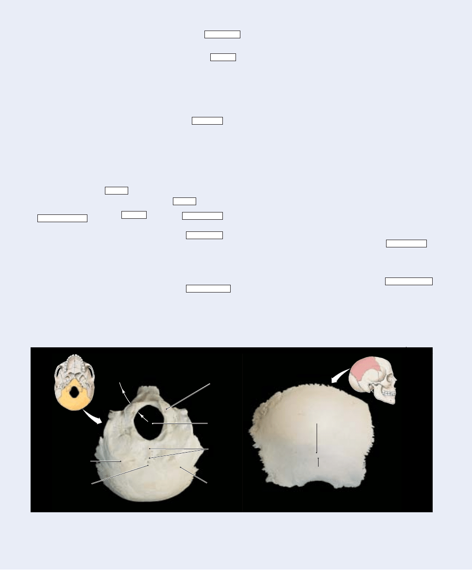
The Occipital Bone (Figure 7–5a)
General Functions:
The occipital bone forms much of the
posterior and inferior surfaces of the cranium.
Articulations:
The occipital bone articulates with the parietal
bones, the temporal bones, the sphenoid, and the first cervical
vertebra (the atlas) (Figures 7–3a–c,e and Figure 7–4).
Regions/Landmarks:
The external occipital protuberance
is a small bump at the midline on the inferior surface.
The external occipital crest, which begins at the external
occipital protuberance, marks the attachment of a ligament
that helps stabilize the vertebrae of the neck.
The occipital condyles are the site of articulation between
the skull and the first vertebra of the neck.
The inferior and superior nuchal (NOO-kul) lines are ridges
that intersect the occipital crest. They mark the attachment
sites of muscles and ligaments that stabilize the articulation at
the occipital condyles and balance the weight of the head over
the vertebrae of the neck.
The concave internal surface of the occipital bone
(
Figure 7–4a
) closely follows the contours of the brain. The
grooves follow the paths of major blood vessels, and the ridges
mark the attachment sites of membranes that stabilize the
position of the brain.
Foramina:
The foramen magnum (
Figure 7–4b
) connects
the cranial cavity with the spinal cavity, which is enclosed by
the vertebral column. This foramen surrounds the connection
between the brain and spinal cord.
The jugular foramen lies between the occipital bone and
the temporal bone (
Figure 7–3e
). The internal jugular vein
passes through this foramen, carrying venous blood from the
brain.
The hypoglossal canals (
Figure 7–4b
) begin at the lateral
base of each occipital condyle and end on the inner surface of
the occipital bone near the foramen magnum. The hypoglossal
nerves, cranial nerves that control the tongue muscles, pass
through these canals.
In this section, we classify cranial nerves as primarily sensory,
special sensory, motor, or mixed (sensory and motor). In this
classification, sensory nerves carry somatic sensory information,
including touch, pressure, vibration, temperature, or pain.
Inferior
nuchal line
External
occipital
protuberance
Foramen
magnum
Superior
nuchal
line
Occipital
condyle
External
occipital
crest
(a) Occipital bone, inferior view
(b) Right parietal bone, lateral view
Superior
temporal line
Inferior
temporal line
Hypoglossal
canal
Figure 7–5
The Occipital and Parietal Bones
166
FOC_FIRST
FOC_H1
FOC_H2
FOC_UL_FIRST
FOC_UL_MID
FOC_UL_MID_LP
FOC_UL_LAST
FOC_UL_LAST_LP
FOC_SUPTTL
FOC_UL_ITEM_TTL
FOC_KT
Focus
The Individual Bones of the Skull
FOC_TTL
Wyszukiwarka
Podobne podstrony:
Fundamentals of Anatomy and Physiology 8e DES MART5891 08 SE C0#2DDE9
Fundamentals of Anatomy and Physiology 8e M15 MART5891 08 SE C15
Fundamentals of Anatomy and Physiology 8e A01 MART5891 08 SE ESHT
Fundamentals of Anatomy and Physiology 8e M27 MART5891 08 SE C27
Fundamentals of Anatomy and Physiology 8e A01 MART 5891 08 SE FM
Fundamentals of Anatomy and Physiology 8e Z03 MART 5891 08 SE ANS
Fundamentals of Anatomy and Physiology 8e Z05 MART 5891 08 SE PCRED
Fundamentals of Anatomy and Physiology 8e Z02 MART 5891 08 SE App
Fundamentals of Anatomy and Physiology 8e Z07 MART 5891 08 SE INDX
Fundamentals of Anatomy and Physiology 8e Z08 MART 5891 08 SE END
Fundamentals of Anatomy and Physiology 8e Z04 MART 5891 08 SE GLOS
Fundamentals of Anatomy and Physiology 8e ZO6 MART 5891 08 SE S#2DE0F
Fundamentals of Anatomy and Physiology Glossary 2
Fundamentals of Anatomy and Physiology 22 Chapter
Fundamentals of Anatomy and Physiology FM
Fundamentals of Anatomy and Physiology Appendix III
Fundamentals of Anatomy and Physiology Appendix I
Fundamentals of Anatomy and Physiology Appendix II
Fundamentals of Anatomy and Physiology ENDPAP
więcej podobnych podstron