
Sterile Supply Specialist Training Course
Level II
SPECIAL MICROBIOLOGY
T. Miorini
D. Percin
2010

Level 2 Script of the wfhss education group
Special Microbiology
Page 2/27
TABLE OF CONTENTS
1
BACTERIAL INFECTIONS
3
1.1
Tuberculosis
3
1.2
Salmonellosis (enteritis salmonellae)
4
1.3
EHEC infection
5
1.4
Infections by Staphylococcus aureus, with special attention to MRSA
7
1.5
Legionellosis (legionnaires disease)
9
1.6
Antibiotic-associated diarrheae and pseudomembranous colitis
10
1.7
Multidrug-Resistant Organisms (MDROs)
11
2
VIRAL INFECTIONS
12
2.1
Blood-borne viruses (BBV)
12
2.2
Viruses spreading via faecal-oral route
19
2.3
Other important viruses
24
3
PRION DISEASES
26
3.1
Creutzfeldt-Jakob disease (CJD and vCJD)
26
4
AUTHORS
27
5
REFERENCES
27
6
LEARNING OBJECTIVES
27

Level 2 Script of the wfhss education group
Special Microbiology
Page 3/27
Special Microbiology
Here we present a number of special microorganisms of particular importance in medical
device reprocessing or occupational safety and which cause major concerns.
1 Bacterial Infections
1.1 Tuberculosis
1.1.1 Causative organism
The main causative organism of tuberculosis in humans is Mycobacterium tuberculosis.
1.1.2 Incidence
Worldwide. The areas most affected are Sub-
Saharan African countries, South and East Asia, a
number
of
Latin
American
countries
and
increasingly also the former republics of the Soviet
Union. Humans are the only relevant reservoir for
M. tuberculosis.
1.1.3 Route of infection
Infection is caused almost always by very fine
expired droplets (aerosols) that are released, in
particular,
when
coughing
and
sneezing.
Transmission through unpasteurized milk of
infected cattle is possible in principle, however, this
is no longer of importance, e.g. in Central Europe,
since cattle herds are to a large extent free of
tuberculosis.
1.1.4 Clinical manifestations
The incubation period can range between weeks and several months. Pulmonary
tuberculosis is at its most contagious for as long as acid-fast bacilli (rods) can be detected on
microscopy (in sputum, aspirated bronchial secretions or gastric juice). Conversely, patients
for whom bacteria can be detected only in culture or using molecular biology techniques are
essentially less infectious.
The general symptoms manifested can include a feeling of malaise, weight loss,
concentration difficulties, fever, increased perspiration (especially at night), loss of appetite,
tiredness, general weakness, signs of flu infection. Respiratory complaints can occur in the
form of cough, chest pain and breathing difficulties.

Level 2 Script of the wfhss education group
Special Microbiology
Page 4/27
1.1.5 Treatment
Tuberculosis can be treated only with a combination of medications because tuberculosis
infection always involves bacteria with proven resistance to a certain drug. Treatment is
being hampered by the spread of increasingly more common multi-resistant tuberculosis
strains (MDRTB = multi-drug-resistant tuberculosis).
1.1.6 Hygiene (infection control) rules
Isolation poses a considerable challenge to both the patient and staff. Therefore, on the one
hand, this should not be resorted to without justification but, on the other hand, in justified
cases it should be rigorously imposed. The problem often encountered in practice is that
when tuberculosis is clinically suspected, no microbiological results are available to diagnose
infection or the existing results are not sufficiently conclusive.
1.1.7 Instrument reprocessing
There is no increased resistance to thermal processes.
Mycobacteria are highly resistant to chemical disinfectant processes, and products with
demonstrated tuberculocidal properties must be used (instruments and surfaces:
1.2 Salmonellosis (enteritis salmonellae)
1.2.1 Causative organisms
Salmonella spp, primarily S. Enteritidis und S. Typhimurium.
1.2.2 Incidence
Worldwide
1.2.3 Transmission route
Mainly
through
consumption
of
contaminated
foodstuffs, e.g. raw or inadequately cooked eggs, raw
milk, meat and poultry products. Group infections or
even epidemic outbreaks are common. Faecal-oral
person-to-person transmission is also possible but
this tends to be very rare because of the "requisite"
infectious dose of 10
3
-10
5
bacteria). Infected young
children and incontinent persons pose a particular
risk in this respect. The main reservoir is various domestic and working animals (in particular
poultry).
1.2.4 Diagnosis
The causative organism is detected by growing cultures from stools or rectal swabs.

Level 2 Script of the wfhss education group
Special Microbiology
Page 5/27
1.2.5 Clinical manifestations
Onset of infection is acute with abdominal pain, headache, nausea, vomiting and watery,
mainly non-bloody, diarrhoea. Almost all patients develop fever of around 39-40 ºC. Severe
dehydration can occur especially in young children and elderly people. Symptoms generally
last for a few days. Overall mortality tends to be low. But because of dehydration young
children and elderly people are particularly at risk.
1.2.6 Treatment
Symptomatic. Only in special cases are patients treated with antibiotics.
1.2.7 Precautionary measures
Good kitchen hygiene and well-trained kitchen staff are indispensable for prevention. For
example, no raw eggs may be used for communal catering in many countries.
1.2.8 Hygiene rules
General hygiene (infection control) rules, in particular hand hygiene.
1.2.9 Instrument reprocessing
No special requirements.
1.3 EHEC infection
1.3.1 Causative organisms
Enterohaemorrhagic Escherichia coli strains (EHEC)
1.3.2 Incidence
Worldwide. Ruminants, in particular cattle, sheep and goats, but also game ruminants
(especially deer and stags) are thought to be the principle reservoirs for EHEC.
1.3.3 Transmission route
The number of ingested bacteria needed to cause
infection appears to be very small (approx. 100
bacteria!), and this can occur when consuming certain
foodstuffs, such as inadequately cooked beef
mincemeat and unpasteurized milk. But other
foodstuffs, too, such as yoghurt, salami, cheese, raw
vegetables or unpasteurized apple juice have been
found to be the source of outbreaks. These bacteria
have been detected as part of the intestinal flora of
around .8% of cattle, and inappropriate slaughter
processes can result in spread of the bacteria. Less
common sources of infections are direct contact with

Level 2 Script of the wfhss education group
Special Microbiology
Page 6/27
animals (petting zoo) or transmission within the family.
1.3.4 Diagnosis
In the presence of bloody diarrhoea and fever, a stool test should definitely be carried out.
1.3.5 Clinical manifestations
The incubation period is generally 1–3 days, but can be as long as 8 days.
Infection can be spread for as long as EHEC bacteria are detected in stools. In general,
bacteria are shed for 5–10 days but this can continue for one month (especially in the case of
young children).
Many EHEC infections manifest no clinical symptoms and hence often go undetected.
Around one-third of infections manifest as mild diarrhoea. Onset of infection generally
involves watery diarrhoea, which as infection progresses increasingly is of a watery-bloody
nature, with dysentery-like manifestations. Concomitant symptoms include nausea, vomiting
and increasing abdominal pain, rarely fever. Young children, elderly people and
immunosuppressed persons are known to have more severe courses of infection, and
infection can result in death.
1.3.6 Treatment
Antibacterial treatment is not indicated. This can prolong bacterial shedding and lead to
production of toxin. Infection is treated symptomatically.
1.3.7 Precautionary measures
Ensure foodstuffs, such as beef mincemeat and unpasteurized milk, are adequately heated.
1.3.8 Hygiene rules
General hygiene rules. Hand hygiene!
1.3.9 Instrument reprocessing
No special requirements; the bacterium is killed by disinfection measures.

Level 2 Script of the wfhss education group
Special Microbiology
Page 7/27
1.4 Infections by Staphylococcus aureus, with special attention to
MRSA
1.4.1 Causative organism
Staphylococcus aureus
Resistance is developed relatively fast in staphylococci. This is
seen mainly in hospitals and nursing homes. The best known multi-
resistant bacterium is MRSA (Methicillin-resistant Staphylococcus
aureus). Multiple resistance, as manifested by the classic MRSA
strains, is aimed at a number of different substance groups, making
treatment extremely difficult to impossible.
1.4.2 Incidence
Worldwide. These bacteria play a pivotal role in causing healthcare-
associated (hospital-acquired/nosocomial) infections. The human being is the main reservoir
for S. aureus as a human pathogen. Carriage rate in adults ranges between 15 % and 40 %.
Like S. aureus in general, MRSA can also colonise e.g. the nasal-throat region.
1.4.3 Transmission route
1. Onset of infections
Like S. aureus in general, MRSA infections in the persons concerned can also originate from
the patient’s own flora or infection is spread from one person to another, most commonly via
the hands of nursing or medical personnel.
2. Intoxications in the form of food poisoning
Around 30 % of all S. aureus strains produce toxins. Once these have multiplied in
foodstuffs, in particular in meat products and milk, the amount of toxin present can be
enough to cause food poisoning. While subsequent heating will kill the bacteria, it will not
destroy the already formed heat-resistant toxins.
1.4.4 Diagnosis
Bacteriological investigation. Detection of the bacterium in culture is needed for diagnosis.
1.4.5 Clinical manifestations
The incubation period is only a few hours (around 2-6 hours) in the case of food poisoning,
and 4-10 days for infections. The infection can be spread for as long as clinical symptoms
are manifested. But the bacteria can also be spread by clinically healthy persons who are
colonised by staphylococci.
Diseases caused by S. aureus: furuncles, carbuncles, abscesses, wound infections, middle
ear infection, sinusitis, (secondary) meningitis, pneumonia, osteomyelitis, endocarditis,
sepsis..

Level 2 Script of the wfhss education group
Special Microbiology
Page 8/27
1.4.6 Treatment
Treatment of MRSA is difficult and calls for close interaction with the bacteriology laboratory.
Appropriate treatment must be administered on the basis of the bacteriology results and in
collaboration with microbiologists/infectiologists.
1.4.7 Precautionary measures
Appropriate kitchen hygiene must be observed to prevent food poisoning. MRSA patients
should be isolated if airborne transmission is possible (e.g. colonisation of the respiratory
tract).
1.4.8 Hygiene rules
Stringent hygiene rules must be observed when dealing with MRSA patients.
Hand disinfection: before and after contact with MRSA patients or their immediate
surroundings and after removing gloves.
Shaking hands should definitely be avoided.
An individual-patient gown and disposable shoes must be worn for all episodes of nursing
and medical care given to the patient as well as if there is risk of contamination.
Contaminated waste (e.g. gloves, dressings, handkerchiefs, etc.) and textiles (laundry, hand
towels, nigh dresses, etc) must be packed into bags in the patient’s room, sealed and
disposed of in the usual manner; while ensuring that no dust is raised.
Healthy persons, medical personnel and their relatives are not endangered!
1.4.9 Instrument reprocessing
No special reprocessing requirements apply; the bacterium is killed by disinfection
measures.

Level 2 Script of the wfhss education group
Special Microbiology
Page 9/27
1.5 Legionellosis (legionnaires disease)
1.5.1 Causative organism
The most important species is Legionella pneumophila.
1.5.2 Incidence
Legionellae are bacteria that are widespread in freshwater, but
even here they are mainly found only in very low
concentrations. Legionellae reproduce at temperatures
between 25 °C and 50 °C and encounter such conditions
especially in hot-water systems. The bacteria can survive
temperatures of up to 55 °C without any damage, and are
killed only as from 60 °C.
1.5.3 Transmission
Infection is contracted through inhalation of aerosols (droplets) harbouring legionellae, e.g.
while taking a shower, via the open cooling towers of air conditioning systems, room air
humidifiers, whirlpools, etc.
Person-to-person transmission has not been reported so far.
1.5.4 Diagnosis
Diagnosis is made by culture of bronchial secretions or using other laboratory diagnostic
techniques.
1.5.5 Clinical manifestations
The incubation period is mainly between 5 and 6 days. In legionnaires disease, flu-like
symptoms are followed by high fever, often with shaking chills, dry cough and muscle pain
and headache. Involvement of other organs apart from the lungs can result in diarrhoea,
confusion as well as liver and kidney disorders. Infection leads to death in around 15- 20 %
of cases.
1.5.6 Treatment
Antibiotics that are effective against legionellae are used.
1.5.7 Precautionary measures
Prevention of legionnaires disease is based on measures that counter the growth of
legionellae in water.
1.5.8 Hygiene rules
No special rules apply
1.5.9 Instrument reprocessing
No special requirements.

Level 2 Script of the wfhss education group
Special Microbiology
Page 10/27
1.6 Antibiotic-associated diarrheae and pseudomembranous colitis
1.6.1 Causative organisms
Clostridium difficile that
is a spore-forming gram positive anaerobic bacillus
.
1.6.2 Incidence
Worldwide
1.6.3 Transmission route
This pathogen is a major cause of healthcare-associated diarrhea and has been responsible
for many large outbreaks in healthcare settings that were extremely difficult to control.
Important factors that contribute to healthcare-associated outbreaks include environmental
contamination, persistence of spores for prolonged periods of time, resistance of spores to
routinely used disinfectants and antiseptics, hand carriage by healthcare personnel to other
patients, and exposure of patients to frequent courses of antimicrobial agents.
1.6.4 Diagnosis
The causative organism is detected by growing cultures from stools or detection of toxins in
stool or molecular methods.
1.6.5 Clinical manifestations
Onset of infection is diarrhoea associated with antibiotic usage. In some cases
pseudomembranous colitis may occur which is more severe. Symptoms generally last for a
few days after antibiotic treatment is stopped.
1.6.6 Treatment
Symptomatic and supportive therapy is important. All antibiotics must be stopped. Only in
severe cases, patients can be treated with metronidazole or vancomycin.
1.6.7 Precautionary measures
Prevention of transmission focuses on application of Contact Precautions for patients with
diarrhea, accurate identification of patients, environmental measures (e.g., rigorous cleaning
of patient rooms) and consistent hand hygiene.
1.6.8 Hygiene rules
Use of soap and water, for mechanical removal of spores from hands as well as alcohol
based handrubs (to kill the vegetative forms), and a bleach-containing disinfectant (5000
ppm) for environmental disinfection, may be valuable when there is transmission in a
healthcare facility.
1.6.9 Instrument reprocessing
No special requirements.

Level 2 Script of the wfhss education group
Special Microbiology
Page 11/27
1.7 Multidrug-Resistant Organisms (MDROs)
1.7.1 Causative organisms
•
Methicillin-resistant Staphylococcus aureus [MRSA],
•
Vancomycin resistant enterococcus [VRE],
•
Multidrug-resistant gram-negative bacteria,
o
Acinetobacter baumannii
o
Pseudomonas aeruginosa
o
Carbapenem-resistant Klebsiella pneumoniae
•
S. aureus that are intermediate or resistant to vancomycin (i.e., VISA and VRSA)
.
1.7.2 Incidence
Worldwide
1.7.3 Transmission route
Patient-to-patient transmission in healthcare settings, usually via hands of Healthcare
Workers (HCWs), has been a major factor accounting for the increase in MDRO
incidence and prevalence
.
1.7.4 Diagnosis
The causative organisms are detected by growing cultures from clinical specimens
1.7.5 Clinical manifestations
Clinical manifestations are not different than the manifestations with susceptible ones.
1.7.6 Treatment
Antibiotics are very limited.
1.7.7 Precautionary measures
Preventing the emergence and transmission of these pathogens requires a
comprehensive approach that includes administrative involvement and measures (e.g.,
nurse staffing, communication systems, performance improvement processes to ensure
adherence to recommended infection control measures), education and training of
medical and other healthcare personnel, judicious antibiotic use, comprehensive
surveillance for targeted MDROs, application of infection control precautions during
patient care, environmental measures , and decolonization therapy when appropriate
.
1.7.8 Hygiene rules
Hand hygiene, cleaning and disinfection of the patient care environment and equipment,
dedicated single-patient-use of non-critical equipment
.are the most important hygiene
rules

Level 2 Script of the wfhss education group
Special Microbiology
Page 12/27
1.7.9 Instrument reprocessing
Dedicated single-patient-use of non-critical equipment
.must be preferred.
2 Viral Infections
2.1 Blood-borne viruses (BBV)
The BBVs present most cross-infection hazard to HCWs. Occupational risks of transmission
of BBVs to HCWs arise from the possibility of exposure to blood and exceptionally to certain
other body fluids or body tissues from an infected patient.
Body fluids and tissues which carry risk for BBV
• Blood
• Cerebrospinal fluid
• Peritoneal fluid
• Pleural fluid
• Pericardial fluid
• Synovial fluid
• Amniotic fluid
• Semen
• Vaginal secretions
• Breast milk
• Saliva including visible blood,
• Unfixed tissues and organs
2.1.1 Hepatitis B
2.1.1.1 Causative organism
Hepatitis B virus (HBV)
HBV is highly resistant and continues to be infectious for a very
long time, for example in serum at a temperature of 30 to 32° C
for at least 6 months or at a temperature of -20° C for 15 years.
Nor does exposure to temperatures of 60° C for 4 hours result in
any loss of infectiousness. HBV is definitely inactivated only
when exposed to temperatures of 90° C or over for around 5 min.
2.1.1.2 Incidence
Worldwide.
2.1.1.3 Transmission
HBV occurs in humans and some other primates. As such, the human being is virtually the
only relevant source of infection.
HBV is transmitted primarily from person to person through sexual contact, direct contact
with blood and other body fluids as well during childbirth from mother to child. Indirect routes
of infection are transmission via blood transfusions and blood products as well as through
contaminated syringes and instruments. Infections have also been reported from tattooing,
piercings, including ear piercing, with inadequately reprocessed instruments.

Level 2 Script of the wfhss education group
Special Microbiology
Page 13/27
2.1.1.4 Diagnosis
Hepatitis B is diagnosed by detection of antibodies in blood in a virology/serology laboratory.
2.1.1.5 Clinical manifestations
The incubation period in most cases is 60-90 days.
In cases of an acute course of HBV infection, this stage lasts 3-4 weeks and, with chronic
courses of infection, for several years or decades and can lead to cirrhosis and other
complications.
2.1.1.6 Treatment
As in other acute forms of viral hepatitis, the most important measures include avoidance of
physical exertion, of alcohol and of fatty foods.
2.1.1.7 Precautionary measures
Active immunisation is the most important protection against hepatitis B infection.
2.1.1.8 Hygiene rules
See Level 1 Script
2.1.1.9 Instrument reprocessing
The most reliable way of inactivating HBV is heating, therefore as far as possible
thermal processes must be used for instrument disinfection:
Thermal disinfection at 80 °C / 50 min or 85 °C / 16 min or 90 °C / 5 min
If chemical instrument disinfection is required, substances with proven efficacy
against HBV must be used For surface disinfection use disinfectants based on active
chlorine, percompounds or aldehydes, while for hand disinfection use skin-
compatible disinfectants based on alcohol or active chlorine.
Level 2 Script of the wfhss education group
Special Microbiology
Page 14/27
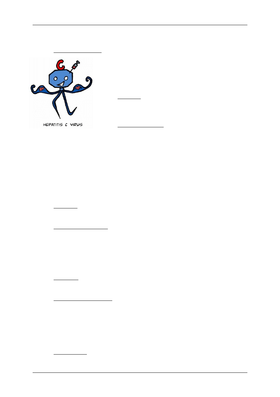
2.1.2 Hepatitis C
2.1.2.1 Causative organism
Hepatitis C virus (HVC)
The human being is the only relevant source of infection. HCV is
found in blood as well as in other body fluids such as saliva,
perspiration, tears, sperm and mother’s milk.
2.1.2.2 Incidence
Worldwide. The incidence is higher in Mediterranean countries
than in other EU states.
2.1.2.3 Transmission route
Transmission is mainly through blood. The risk of transmission rises in line with the viral load.
Typically hepatitis C is a form of posttransfusion hepatitis. Transfusion of HCV-positive blood
conserves or administration of contaminated blood products was the most common route of
transmission until the introduction of serology test systems. Now intravenous drug use
involving the sharing of needles or the use of unsterile implements are the most important
sources. Other potential sources of infection are poor hygiene conditions in tattooing and
piercing studios, in manicure and pedicure establishments, hairdressing salons, acupuncture
or dental treatment leading to bleeding. The transmission route is unknown in up to around
one-third of HCV infections, hence the risk factor in unclear.
2.1.2.4 Diagnosis
Hepatitis C is diagnosed by detection of antibodies in blood in a virology laboratory.
2.1.2.5 Clinical manifestations
The incubation period is on average 40-50 days. The majority of infections are
asymptomatic. At most 20 % of patients develop clinical symptoms. The most common
manifestations are mild, in particular, fatigue, nausea and/or signs of flu. Since a proportion
of HCV infections do embark on a chronic course, infected persons can act as a source of
infection for decades.
2.1.2.6 Treatment
Combination therapy increases the ongoing response to treatment by up to 50%.
2.1.2.7 Precautionary measures
Since at present there is no immunisation, the following precautionary measures represent
the only protection. In general the same precautionary measures apply as for HBV (see
Specialist Course 1 Script).
The following point should be borne in mind: the risk of transmission is very low within the
family or among members of a household.
2.1.2.8 Hygiene rules
See Level 1 Script

Level 2 Script of the wfhss education group
Special Microbiology
Page 15/27
2.1.2.9 Instrument reprocessing
See hepatitis B
2.1.3 Other hepatitis viruses
2.1.3.1 Causative organism
Hepatitis D virus (HDV)
HDV causes infection only in those who have active HBV infection. HDV infection can occur
either as co-infection with HBV or as superinfection of an HBV carrier.
GB virus-type C (Hepatitis G virus)
Recently a further BBV has been described, provisionally designated either as GBV-C agent
or hepatitis G virus.
Incidence, transmission route, diagnosis, clinical manifestations and treatment are the same
as Hepatitis B and C.
Since HDV depends on an HBV-infected host for replication, prevention of HBV infection by
immunisation will also prevent HDV infection.
2.1.3.2 Hygiene rules
See Level 1 Script
2.1.3.3 Instrument reprocessing
See hepatitis B

Level 2 Script of the wfhss education group
Special Microbiology
Page 16/27
2.1.4 Human immunodeficiency virus (HIV)
2.1.4.1 Causative organism
Human immunodeficiency virus (HIV) which causes defects in the immune system, whose
most severe form is acquired immunodeficiency syndrome (AIDS).
2.1.4.2 Incidence
Worldwide.
More than 95 % of all HIV-infected persons live in developing countries.
2.1.4.3 Transmission route
Every infected person will continue to be potentially infectious throughout their lifetime. The
risk of spreading infection is particularly high within the first weeks of contracting infection.
After this, infectiousness declines in general but increases once again as immunodeficiency
progresses with onset of clinical symptoms.
The highest concentrations of HIV are found in the blood, seminal fluid and vaginal
secretions. Transmission in mother’s milk is also possible. With the exception of the few
cases described in the literature, HIV infections can be imputed to one of the main three
transmission routes:
♦ Unprotected sexual intercourse: anal sex, vaginal sex, oral sex (orogenital contact) 85% of
all infections are contracted in this way; the risk is increased when where is a frequent change
of partner.
♦ Blood or blood products (sharing syringes among several persons - "needle exchange"
among drug addicts, transfusion of contaminated blood conserves or coagulation products).
Blood donors are tested for HIV antibodies. Blood donations containing HIV antibodies are
discarded. Furthermore, persons who cannot definitely rule out that they do not pose a risk of
infection are called upon not to donate blood. By taking these measures, it was possible to
reduce the statistical risk of HIV transmission, posed by undetected HIV infection of the donor
at the time of donation (diagnostic window), to around one case per 1,000.000 donations.
♦ Pre -, peri – or postnatal spread from infected mother to her child. European studies have
shown that the risk of HIV transmission from an infected mother to her child was between 15
% and 25 % before the introduction of preventive measures. Today the probability of
transmission can be reduced to less than 2 % through treatment during pregnancy and opting
for caesarean section. HIV can also lead to infection of children in mother’s milk. In countries
in which formula milk is readily available, HIV infected mothers should not breastfeed their
babies.
♦ Everyday bodily contact, sharing of crockery, cutlery, etc, or use of communal sanitary
facilities do not pose a risk of infection. HIV is not transmitted in droplets or insect bites.
2.1.4.4 Diagnosis
HIV diagnosis can be initiated only after providing the patient with information and advice.
Diagnosis of HIV infection is based essentially on detection of specific antibodies. These

Level 2 Script of the wfhss education group
Special Microbiology
Page 17/27
specific antibodies generally appear within four weeks to three months of contracting
infection (diagnostic window).
To date, there have only been isolated reports in the literature of cases where antibodies
were detectable only after three months. If antibodies cannot be detected even six months
after possible infection, infection can be ruled out with a great margin of certainty.
2.1.4.5 Clinical manifestations
The problem is that the infected body is unable to eliminate the HIV virus, and its spread
within the body cannot be prevented in the long term. Immunodeficiency, with its attendant
clinical manifestations, continues unabated – even if the rate at which this happens varies
from one patient to another. The most common causes of death are infection complications
that can no longer be controlled.
2.1.4.6 Treatment
In the meantime a number of substances are available for treatment of HIV infection. In view
of the rapid pace at which insights are gained into this topic, please consult the regularly
updated consensus recommendations on treatment of HIV infection. In Germany, the current
recommendations can be consulted, for example, on the Robert Koch Institute website
(http://www.rki.de).
Diagnosis of HIV infection can give rise to major psychosocial problems. In many places
there are special services available to help overcome these problems, e.g. self-help groups,
psychosocial advisory services, etc. The treating physician should try to promote close
cooperation with such services.
2.1.4.7 Prevention
Both non-infected and infected persons must avoid the risk of contracting and spreading
infection, respectively, and must protect against these. Both parties must know how to
behave such that infection is avoided and the available knowledge is put to use. Attention
has been drawn repeatedly to the fact that HIV is spread only through sexual intercourse,
inoculation (introduction) of virus-containing material, or from mother to child. On the other
hand, the risks posed by sexual contact with new or changing partners must be clearly
addressed. Drug addicts are made aware of the risks of sharing syringes and of the need to
dispose safely of used syringes.
Prevention and limitation of discrimination of HIV infected persons or those at risk for HIV are
important.
2.1.4.8 Hygiene rules
Observance of well-established hygiene rules is indispensable for treatment of HIV infected
persons and of AIDS patients. The same precautionary measures apply as those which have
proved their merit in prevention of hepatitis B virus infection. See Specialist Course 1 Script
The virus can be inactivated (destroyed) through disinfection measures since, strictly
speaking, viruses cannot be killed because they are not living entities.

Level 2 Script of the wfhss education group
Special Microbiology
Page 18/27
Disinfectants and disinfection processes with proven efficacy against HIV must be used. For
hygienic hand disinfection disinfectants that have been approved as drugs and contain 70 to
85 vol. % alcohol are suitable.
2.1.5 Instrument reprocessing
See hepatitis B

Level 2 Script of the wfhss education group
Special Microbiology
Page 19/27
2.2 Viruses spreading via faecal-oral route
2.2.1 Norovirus (=Norwalk virus) Infection
2.2.1.1 Causative organism
Noroviruses (formerly Norwalk and Norwalk-like viruses)
2.2.1.2 Incidence
Worldwide. In infants and young children, after rotaviruses, they are the second most
common cause of acute gastroenteritis. Noroviruses are often the cause of acute
gastroenteritis outbreaks in communal catering institutions such as homes for the elderly,
nursing homes and childcare establishments, but they can also cause sporadic
gastroenteritis. Infections caused by viruses belonging to the norovirus group can occur
throughout the year, but clusters of such infections have been observed in the winter months.
The human being is the only known reservoir for this virus.
2.2.1.3 Route of infection
The viruses are excreted in the stools of infected persons in very large quantities.
Transmission takes place primarily via the faecal-oral route, with direct person-to-person
transmission playing a pivotal role. But infections or outbreaks can also originate from
contaminated foodstuffs (salads, crabs, mussels, etc.) or drinks (contaminated water).
Contaminated objects can also give rise to transmission.
Infectiousness is very high, with the minimum infectious dose being between 10-100 virus
particles.
The very rapid spread of infection within communities suggests that, in addition to the faecal-
oral route, other routes of transmission are also possible, e.g. airborne spread through
formation of virus-containing aerosols as released during vomiting.
2.2.1.4 Diagnosis
Detection of noroviruses in stools is possible only in special laboratories.
Clinical manifestations: The incubation period is between 12 and 48 hours. Noroviruses
cause gastroenteritis of acute onset, accompanied by projectile vomiting and profuse
diarrhoea, which can lead to considerable fluid loss. In general, there are well-pronounced
clinical manifestations of malaise, abdominal pain, nausea and fatigue.

Level 2 Script of the wfhss education group
Special Microbiology
Page 20/27
2.2.1.5 Treatment:
In general, outpatient treatment is adequate. The symptoms are treated by restoring the, in
some cases, widespread loss of fluids and electrolytes. No antiviral treatment is available.
2.2.1.6 Hygiene rules
In outbreaks it is important to identify the source as quickly as possible. If contaminated
foodstuffs or drinks are a possible source of the outbreak, measures must be initiated
immediately to stop infection from this source.
To prevent faecal-oral transmission, extensive hygiene measures must be initiated (wearing
of gloves and gowns, isolation of infected persons, extra scrupulous cleaning of toilets, more
intensive hand hygiene, frequent disinfection of bed linen). However, in view of the highly
contagious nature of noroviruses these measures are effective only to a certain extent. In
practice it has been observed time and again that even meticulous hygiene measures are not
able to prevent further spread.
In communal establishments such as hospitals and homes for the elderly, movement of
patients, residents and personnel within wards should be limited as far as possible to prevent
spread between different wards and areas of the establishment. Infected personnel should
be released from their duties even if they suffer from only slight gastrointestinal complaints
and should resume work by the earliest only 2 days after clinical symptoms have resolved.
2.2.1.7 Instrument reprocessing
No special reprocessing requirements

Level 2 Script of the wfhss education group
Special Microbiology
Page 21/27
2.2.2 Rotavirus Infections
2.2.2.1 Causative organism
Rotaviruses
2.2.2.2 Incidence
Worldwide, rotaviruses trigger more than 70 % of cases
of severe diarrhoea in children and, as such, are the
most common cause of intestinal infections in this age
group. In Western industrialised countries infants and
children between the age of 6 months and 2 years are
the most commonly affected. In neonates and young
children rotaviruses are the main cause of healthcare-
associated intestinal infections. The incidence of
infection is highest in the winter months because the virus is more easily transmitted in
enclosed spaces, in particular in dry room air. In adults, infections – which generally have a
mild course – occur mainly as travellers’ diarrhoea, in parents of infected children or during
outbreaks in homes for the elderly. The human being is the main reservoir for rotaviruses.
Rotaviruses have also been detected in domestic and working animals, however, the viruses
found apparently are not implicated to any great extent in human infections.
2.2.2.3 Route of infection
Rotaviruses are spread, in particular, as smear infections via the faecal-oral route but also
through infected water and foodstuffs. Although the viruses cannot reproduce in the
respiratory tract, during the acute phase of infection they can also be shed in respiratory tract
secretions, hence airborne transmission is also possible. The virus is easily spread, with as
few as 10 virus particles sufficing to infect a child. In the case of persons suffering from acute
infection, between 10
9
–10
11
viruses per g stools are shed.
2.2.2.4 Diagnosis:
The laboratory diagnostic method of choice entails detection of an antigen from stools.
2.2.2.5 Clinical manifestations
The incubation period is between 1 and 3 days.
Symptoms of rotavirus infections range from subclinical infections through mild diarrhoea to
severe infections. Infection begins with acute watery diarrhoea and vomiting. Mucus is often
found in stools. Fever and abdominal pain can occur. Infection resulting in dehydration gives
rise to complications which, if not properly treated in a timely manner, can lead to death.
2.2.2.6 Treatment
In general, administration of fluids and electrolytes suffices. Only in rare cases are
intravenous fluids needed.
2.2.2.7 Hygiene rules

Level 2 Script of the wfhss education group
Special Microbiology
Page 22/27
The spread of rotavirus infections in children’s hospitals, kindergartens and similar
establishments can be countered only through strict observance of hygiene rules. The aim
here is to break the faecal-oral transmission chain. Special emphasis must be put on hand
hygiene! Practical experience shows that it is very difficult to prevent secondary infections.
The virus survives in an infectious state for a long time on contaminated surfaces and hands.
In the hospital setting, infected children should be isolated and cared for by specific nursing
staff.
In the home, scrupulous hand hygiene suffices, and gloves are needed only when changing
nappies / diapers.
2.2.2.8 Instrument reprocessing
No special reprocessing requirements
2.2.3 Hepatitis A
2.2.3.1 Causative organism
Hepatitis A virus (HAV)
2.2.3.2 Incidence
This virus is found mainly in tropical and subtropical regions, i.e. in
Central and Southern Asia, Central Africa, the Far East and Middle
East but also in parts of South American and Central America and
in various Mediterranean countries. There is also a high risk in the
former republics of the Soviet Union. Humans are the principle host
and, possibly, the only reservoir for hepatitis A viruses.
2.2.3.3 Route of infection
Transmission is normally via the faecal-oral route, mainly through
contaminated foodstuffs, water or everyday use utensils. Outbreaks are mainly caused by
contaminated drinking water or foodstuffs, especially mussels or oysters as well as
vegetables and salads for which faecal fertiliser was used.
2.2.3.4 Diagnosis
Hepatitis A is diagnosed by detection of antibodies in blood in a virology/serology laboratory.
2.2.3.5 Duration of infectiousness
Viral shedding - and hence infectiousness – begins around 1-2 weeks before onset of
symptoms and continues for around a further week. Viral shedding is greatest during the first
phase, i.e. during the incubation period and then continues to decrease. To date, there has
been no evidence of ongoing viral shedding.
2.2.3.6 Clinical manifestations
The incubation period lasts on average 30 days. In children infection is often asymptomatic
compared to adults. The symptoms are: fever, malaise, weakness, loss of appetite, nausea,

Level 2 Script of the wfhss education group
Special Microbiology
Page 23/27
vomiting. In general, patients will have made a complete recovery within 3-6 months. In some
cases symptoms maybe very severe like hepatic coma.
2.2.3.7 Treatment
Only the symptoms can be treated.
2.2.3.8 Precautionary measures
Hepatitis A virus immunisation is available and is recommended for travellers to areas with
increased risk as well as for healthcare workers or, for example, laboratory staff engaged in
testing of stool specimens.
2.2.3.9 Hygiene rules
The usual hygiene measures apply, with special emphasis naturally on hand disinfection.
There is also a risk of cross-infection when using communal toilets.
2.2.3.10 Instrument reprocessing
No special measures are required

Level 2 Script of the wfhss education group
Special Microbiology
Page 24/27
2.3 Other important viruses
2.3.1 Severe Acute Respiratory Syndrome (SARS)
2.3.1.1 Causative organism
SARS is caused by SARS CoV, a previously unrecognized member of the coronavirus
family
2.3.1.2 Incidence
SARS is a newly discovered respiratory disease that emerged in China late in 2002 and
spread to several countries. Mainland China, Hong Kong, Hanoi, Singapore, and
Toronto were affected significantly.
2.3.1.3 Route of infection
Droplet and contact transmission are important. Aerosolization of small infectious
particles generated during these and other similar procedures could be a risk factor for
transmission to others within a multi-bed room or shared airspace.
2.3.1.4 Diagnosis
Detection of antibodies to SARS-CoV or detection of SARS-CoV using RT_PCR in
virology laboratories.
2.3.1.5 Clinical manifestations
Signs and symptoms usually include fever >38.°C and chills and rigors, sometimes
accompanied by headache, myalgia, and mild to severe respiratory symptoms. Fatality
rate is 6%.
2.3.1.6 Treatment:
Treatmant should be done in hospital.
2.3.1.7 Hygiene rules:
CDC recommends Standard Precautions, with emphasis on the use of hand hygiene,
Contact Precautions with emphasis on environmental cleaning due to the detection of
SARS CoV RNA by PCR on surfaces in rooms occupied by SARS patients, Airborne
Precautions, including use of fit-tested NIOSH-approved N95 or higher level respirators,
and eye protection.
2.3.1.8 Instrument reprocessing
No special reprocessing requirements but if possible single used items should be used.
Personell protective equipment with eye protection is very important during handling of
the instruments.

Level 2 Script of the wfhss education group
Special Microbiology
Page 25/27
2.3.2 Hemorrhagic fever viruses (HFV)
2.3.2.1 Causative organism
The more commonly known HFVs are Ebola and Marburg viruses (Filoviridae), Lassa
virus (Arenaviridae), Crimean-Congo hemorrhagic fever and Rift Valley Fever virus
(Bunyaviridae), and Dengue and Yellow fever viruses (Flaviviridae)
2.3.2.2 Incidence
These viruses are endemic in areas of Africa, Asia, the Middle East, and South America.
2.3.2.3 Route of infection
These viruses are transmitted to humans via contact with infected animals or via
arthropod vectors. Person-to-person transmission is associated primarily with direct
blood and body fluid contact. Percutaneous exposure to contaminated blood carries a
particularly high risk for transmission and increased mortality. Airborne transmission of
naturally occurring HFVs in humans has not been seen.
2.3.2.4 Diagnosis
Detection of antibodies to causative organism or detection of viruses using RT_PCR in
virology laboratories.
2.3.2.5 Clinical manifestations
They cause serious disease with high fever, skin rash, bleeding diathesis, and in some
cases, high mortality.
2.3.2.6 Treatment:
Treatmant should be done in hospital.
2.3.2.7 Hygiene rules:
In less developed countries, outbreaks of HFVs have been controlled with basic hygiene,
barrier precautions, safe injection practices, and safe burial practices. Contact and
Droplet Precautions with eye protection are effective in protecting healthcare personnel
2.3.2.8 Instrument reprocessing
No special reprocessing requirements but if possible single used items should be used.
Personell protective equipment with eye protection is very important during handling of
the instruments.
Level 2 Script of the wfhss education group
Special Microbiology
Page 26/27
3 Prion Diseases
3.1 Creutzfeldt-Jakob disease (CJD and vCJD)
3.1.1 Causative organism
This disease is caused by prions. Prions are not
living creatures but rather “infectious” protein
particles.
Creutzfeldt-Jakob disease (CJD), which was first
described in 1920, belongs to the prion diseases
and is a rare disease. This disease occurs
sporadically or in families (around between 10 and
15 % of all CJD cases are genetically mediated) and
inevitably leading to death.
The sporadic form is the most common, with a worldwide, similar incidence of around 1-2
cases per million inhabitants per year. In recent years this disease has been increasingly the
focus of public interest because of the occurrence of bovine spongiform encephalopathy
(BSE) in cattle in the United Kingdom and the probability of transmission through foodstuffs
to people.
3.1.2 Incidence
In general CJD occurs sporadically, i.e. without any demonstrable cause; the average age for
onset of disease is 64 years, and the average duration of disease is 4 months. Among the
acquired forms of this disease is kuru, which is a neurodegenerative disease found in a
group of people with a specific language in Papua-New Guinea following consumption of
human brain during cannibalistic rituals. There is also the possibility of unintentional
transmission when performing medical procedures. The new variant of Creutzfeldt-Jakob
disease (vCJD), which is associated with BSE in cattle, has occurred mainly in the United
Kingdom and France.
3.1.3 Transmission route
The disease can be transmitted through administration of human hypophyseal hormones,
corneal eye transplants or through neurosurgical instruments. The interval between exposure
and onset of the first clinical symptoms is between 1 and 30 years.
3.1.4 Diagnosis
With onset of disease, patients have concentration and attention disorders, progressing later
to impaired movements, personality changes, impaired vision and gait. After, in general rapid
progression of symptoms, the disease inevitably leads to death. Apart from clinical
symptoms, the diagnostic methods used include electroencephalogram (EEG), cerebrospinal

Level 2 Script of the wfhss education group
Special Microbiology
Page 27/27
fluid (CSF) tap test and magnetic resonance imaging (MRI). CJD can be definitively
diagnosed only through post mortem examination.
At present there is no treatment.
3.1.5 Precautionary measures
Today in industrialised countries hypophyseal hormones can be safely produced using
recombinant technologies, and there are also strict safety regulations and restrictions in
place for transplantations.
There is no evidence of a risk of transmission in routine nursing and when dealing with
infected persons, and conventional hygiene measures suffice.
3.1.6 Instrument reprocessing
See “CJD” chapter in Instrument Reprocessing
4 Authors
Mag. Dr. Tillo Miorini, Institute for Applied Hygiene, Graz
Prof. Duygu Percin, Department of Clinical Microbiology, Erciyes University Faculty of
Medicine, Kayseri, Turkey.
The script has been proof read and authorized by the wfhss education group
5 References
1) Steirischer Seuchenplan (O. Feenstra Hrsg.), 2002
(
http://www.verwaltung.steiermark.at/cms/ziel/2651878/DE/)
2) RKI Mitteilung: Die Variante der Creutzfeldt-Jakob-Krankheit (vCJK): Epidemiologie,
Erkennung, Diagnostik und Prävention unter besonderer Berücksichtigung der
Risikominimierung einer iatrogenen Übertragung durch Medizinprodukte, insbesondere
chirurgische Instrumente – Abschlussbericht der Task Force vCJK zu diesem Thema.
Bundesgesundheitsbl -Gesundheitsforsch – Gesundheitsschutz 2002, 45:376–394,
Springer-Verlag 2002. (www.rki.de)
6 Learning Objectives
Understand and be able to cite the mentioned special microorganisms of particular
importance in medical device reprocessing or occupational safety their most important
characteristics
Be able to tell, which of the pathogens require special treatment of MDs and why
Wyszukiwarka
Podobne podstrony:
wfhss training 2 05 en
wfhss training 2 02 en
wfhss training 2 08 en
wfhss workshop20071206 lecture05 01 en
wfhss conf20091007 lecture sp s401 training programme en
wfhss workshop20071206 lecture06 01 en
Ćwiczenie 01 EN DI
Ćwiczenie 01 EN DI
161TEC ELEC 01 EN
wfhss conf20070503 lecture10 en
wfhss guideline 03 en
wfhss conf20070503 lecture03 en
wfhss conf20070503 lecture09 en
Ćwiczenie 01 EN DI
wfhss training 1 04 pl
wfhss training 1 06 pl
Papcie(01) EN
wfhss conf20070503 lecture05 en
więcej podobnych podstron