
Crystal structure and properties of the copper(II) complex of sodium monensin A
Ivayla N. Pantcheva
a,*
, Petar Dorkov
a
, Vasil N. Atanasov
a
, Mariana Mitewa
a
, Boris L. Shivachev
b
,
Rosica P. Nikolova
b
, Heike Mayer-Figge
c
, William S. Sheldrick
c
a
Department of Analytical Chemistry, Faculty of Chemistry, Sofia University, 1, J. Bourchier Blvd., 1164 Sofia, Bulgaria
b
Central Laboratory of Mineralogy and Crystallography, Bulgarian Academy of Sciences, Acad. G. Bonchev Str., Build. 107, 1113 Sofia, Bulgaria
c
Lehrstuhl für Analytische Chemie, Ruhr-Universität Bochum, D-44780 Bochum, Germany
a r t i c l e
i n f o
Article history:
Received 30 December 2008
Received in revised form 6 May 2009
Accepted 7 May 2009
Available online 25 August 2009
Keywords:
Monensin
Monovalent polyether ionophorous
antibiotic
Copper(II) complex
Crystal structure
Antibacterial activity
SOD-like activity
a b s t r a c t
The preparation and structural characterization of a new copper(II) complex of the polyether ionophor-
ous antibiotic sodium monensin A (MonNa) are described. Sodium monensin A binds Cu(II) to produce a
heterometallic complex of composition [Cu(MonNa)
2
Cl
2
]H
2
O, 1. The crystallographic data of 1 show that
the complex crystallizes in monoclinic space group C2 with Cu(II) ion adopting a distorted square–planar
geometry. Copper(II) coordinates two anionic sodium monensin ligands and two chloride anions produc-
ing a neutral compound. The sodium ion remains in the inner cavity of the ligand retaining its sixfold
coordination with oxygen atoms. Replacement of crystallization water by acetonitrile is observed in
the crystal structure of the complex 1. Copper(I) salt of the methyl ester of MonNa, 2, was identified
by X-ray crystallography as a side product of the reaction of MonNa with Cu(II). Compound 2, [Me–Mon-
Na][H–MonNa][CuCl
2
]Cl, crystallizes in monoclinic space group C2 with the same coordination pattern of
the sodium cation but contains a chlorocuprate(I) counter [CuCl
2
]
, which is linear and not coordinated
by sodium monensin A. The antibacterial and antioxidant properties as two independent activities of 1
were studied. Compound 1 is effective against aerobic Gram(+)-microorganisms Bacillus subtilis, Bacillus
mycoides and Sarcina lutea. Complex 1 shows SOD-like activity comparable with that of the copper(II) ion.
Ó 2009 Elsevier Inc. All rights reserved.
1. Introduction
The term ‘‘ionophore” was introduced in 1967 for a large group
of naturally occurring compounds able to transport cations as neu-
tral complexes through the cell membranes
. The discovery
that monensin – the first known ionophore – is effective as an
anticoccidial and antimicrobial agent prompted a search for other
compounds possessing similar properties. Nowadays members of
the ionophorous family such as monensin, lasalocid, salinomycin,
maduramicin, etc. are widely used as anticoccidial drugs and anti-
biotics in the poultry industry
. From a chemical point of view
these ionophores are polyether derivatives of monocarboxylic
acids and consist of heterocyclic ether-containing rings. When
present as deprotonated anions, they form stable neutral com-
plexes with alkali metal cations and for that reason are known as
‘‘monovalent polyether ionophores”. When applied to the cell,
the carboxylic acid ionophores promote perturbations in the intra-
cellular cation balance, which consecutively disturb a variety of
homeostatic processes leading to cell death
.
The complexes of ionophorous antibiotics, and especially of
monensin, known up to date, are those obtained with alkali metal
ions
and Ag
+
. The metal compounds were
characterized by single crystal X-ray diffraction and their struc-
tures were determined both in solid state and in solution using
various spectroscopic methods. Data on the possible formation of
complexes of the monovalent ionophores with alkali–earth and
other divalent metal ions are also available in the literature
although single crystals of the reported compounds were not
obtained for subsequent structural elucidation and questions
concerning the coordination mode of the ligands are still arising
Recently a lot of attention has been paid to chemical modifica-
tions of monensin in order to improve its ability and selectivity of
binding metal ions
. Although there is some evidence that
monensin reacts with divalent metal ions, only a limited number of
research teams are studying the antibiotic reactions with metal
ions other than monovalent representatives
.
In our previous studies we had shown that the polyether iono-
phore monensin A binds Mn(II) or Co(II) producing unique metal
complexes of different content and structure depending on the
form of the antibiotic applied (monensin acid, MonH or sodium
monensin, MonNa)
. The results on antibacterial screening
showed that [Co(Mon)
2
(H
2
O)
2
] possesses high cytotoxicity in
comparison to the free ligands probably due to unusual coordina-
tion mode of monensin
. In order to confirm the ability of
0162-0134/$ - see front matter Ó 2009 Elsevier Inc. All rights reserved.
doi:
10.1016/j.jinorgbio.2009.08.007
*
Corresponding author. Tel.: +359 28161446; fax: +359 29625438.
E-mail address:
(I.N. Pantcheva).
Journal of Inorganic Biochemistry 103 (2009) 1419–1424
Contents lists available at
Journal of Inorganic Biochemistry
j o u r n a l h o m e p a g e : w w w . e l s e v i e r . c o m / l o c a t e / j i n o r g b i o

monovalent polyether ionophore monensin A for binding divalent
metal ions we have extended our investigations towards its com-
plexation with copper(II). In the present paper we report the re-
sults on structure elucidation and some biological properties of
the new copper(II) complex of sodium monensin A.
2. Experimental
2.1. Materials
All chemicals and solvents were of reagent grade and were used
as purchased. Sodium monensin A was supplied by BIOVET Ltd.
and CuCl
2
2H
2
O – by Riedel de Häen AG, respectively. The solvents
(MeCN, MeOH, DMSO) were received from Merck and were used
without further purification. Xanthine, buttermilk xanthine oxi-
dase, bovine erythrocyte superoxide dismutase (SOD), cytochrome
c from equine heart and nitroblue tetrazolium chloride (NBT) were
purchased from Fluka. In all experiments deionized water was
used.
2.2. Preparation of [Cu(MonNa)
2
Cl
2
]H
2
O, 1
To a solution containing MonNa (1 mmol, 693 mg in MeCN/
MeOH (10/1, 10 mL)) CuCl
2
2H
2
O was added (1 mmol, 170 mg in
10 mL MeCN/MeOH = 10/1). The slow evaporation of the resulting
yellow–brown mixture afforded the precipitation of [Cu(Mon-
Na)
2
Cl
2
]H
2
O, 1 as a green solid, insoluble in MeCN (483 mg, 63%
yield). Anal. Calcd. for C
72
H
124
Na
2
Cl
2
O
23
Cu (MW = 1538.19): H,
8.13; C, 56.22; O, 23.92; Cl, 4.61; Na, 2.99; Cu, 4.13. Found: H,
7.95; C, 56.67; O, 23.50; Cl, 4.70; Na, 3.35; Cu, 4.03%. The complex
is soluble in MeOH and DMSO. Green single crystals of composition
[Cu(MonNa)
2
Cl
2
]MeCN were obtained by slow concentration of a
diluted reaction mixture.
As a side product of the above reaction, a chlorocuprate(I) salt of
the methyl ester of sodium monensin, [Me–MonNa][H–Mon-
Na][CuCl
2
]Cl, 2, was also isolated and analyzed by single crystal
X-ray diffraction.
2.3. Physical measurements
Infrared spectra (4000–400 cm
1
) were recorded on a Specord
75-IR in a Nujol mull. The electronic spectra were registered on a
UV–visible (UV–Vis) Spectrometer T80+(PG Instruments Ltd.).
The X-band EPR spectra were obtained on a Bruker-ER 420 spec-
trometer, using Mn/ZnS as a standard. The experimental data were
processed with Spectracalc PC program. Elemental analysis data (C,
H, O) were obtained with a VarioEL V5.18.0 Elemental Analyzer.
Chlorine was determined by titration with Hg(NO
3
)
2
after wet
digestion of the sample. Metal content was determined by AAS
on a Perkin Elmer 1100 B using a stock standard solution (Merck,
1000
l
g/mL) and working reference solutions were prepared after
suitable dilution.
2.4. Crystallographic studies
Details concerning data collection, structure solution and
refinement are given in
. X-ray diffraction measurements
were performed on a CAD diffractometer at 290 K (1) and on an
Oxford Diffraction Xcalibur 2 diffractometer at 293 K (2), both
operating with Mo–K
a
(k = 0.71073 Å) radiation and equipped
with graphite monochromators. The structures were solved by di-
rect methods and were refined by full-matrix least-square proce-
dures on F
2
. All non-H atoms were refined isotropically with
a riding model.
2.5. Antimicrobial (antibacterial) activity assay
Three Gram-positive microorganisms were used as test strains
to evaluate the antimicrobial properties of copper(II) complex 1
and copper(II) chloride. The microorganisms Bacillus subtilis (ATCC
6633), Bacillus mycoides spp. and Sarcina lutea FDA strain PCI 1000
(ATCC 10054) were obtained from the National Bank for Industrial
Microorganisms and Cell Cultures (NBIMCC, Bulgaria). The double
layer agar diffusion method was applied for the screening per-
formed in accordance with literature procedures
.
2.6. Superoxide dismutase (SOD) assay
Superoxide dismutase activity was assayed using both the
indirect xanthine–xanthine oxidase–cytochrome c
and xan-
thine–xanthine oxidase–nitroblue tetrazolium chloride (NBT)
methods
. The system xanthine–xanthine oxidase was the
source of superoxide anion, which causes reduction of cytochrome
c or NBT reduction to formazan, respectively. The kinetic of reduc-
tion of cytochrome c and formazan formation was followed by
continuous spectrophotometric method at 550 nm and 530 nm,
respectively. The SOD activity of compounds (IC
50
value and
the corresponding k
McCF
) is the concentration that causes
50% inhibition of the reduction of cytochrome c or NBT, respec-
tively. The SOD activity of the native SOD enzyme was also
measured.
All compounds tested (CuCl
2
2H
2
O, sodium monensin and com-
plex 1) were studied as DMSO solutions. The total amount of DMSO
solution of compounds at different concentrations added to the
enzymic reaction was 50
l
L in a final volume of 1000
l
L. First
we completed several control tests to evaluate the influence of
DMSO (5%) on the reactions tested. The initial rate of: (i) the
xanthine conversion to urate; (ii) the cytochrome c reduction,
(iii) the NBT reduction to formazan, (iv) the SOD assay of the
bovine erythrocyte SOD enzyme (IC
50
5.5 nM, kMcF 1.2 10
9
)
is not affected by the presence of 5% DMSO. Next, we performed
control tests with sodium monensin A, complex 1 and copper(II)
chloride (in 50
l
L DMSO) to verify that the studied compounds
by themselves do not affect the xanthine conversion to urate and
the cytochrome c or NBT reduction, respectively
.
Table 1
Crystal data and structure refinement for copper(II) complex 1 and chlorocuprate(I)
salt, 2.
Compound
Complex 1
Copper(I) salt, 2
Formula
C
74
H
125
O
22
Na
2
NCuCl
2
C
73
H
126
O
22
Na
2
CuCl
3
M
1561.23
1571.67
Crystal system
Monoclinic
Monoclinic
Space group
C2
C2
a (Å)
19.062(7)
19.0639(11)
b (Å)
15.841(6)
15.5761(11)
c (Å)
13.384(5)
13.4409(8)
b
(°)
90.787(11)
90.695(7)
V (A
3
)
4041.1(3)
3990.9(4)
Z
2
2
D
c
(Mg/m
3
)
1.293
1.303
F (000)
1681
1672
l
(mm
1
)
0.418
0.455
Crystal size (mm)
0.24 0.32 0.35
0.29 0.30 0.47
h
min
–h
max
(°)
1.52–25.97
3.03–27.76
Dataset (h, k, l)
23 23, 19 19, 16 16
22 24, 20 19, 17 17
Total Refl./Unique Refl.
8200/7954
20,292/8427
Obs. Refl. [I > 2
r
(I)]
5219
4253
Data/restraints/
parameters
7954/1/462
8427/3/475
R1, wR2 [I > 2s(I)]
0.0684, 0.1548
0.0915, 0.2259
Residuals/eÅ
3
0.402/0.511
1.100/0.735
GOF
1.026
0.903
1420
I.N. Pantcheva et al. / Journal of Inorganic Biochemistry 103 (2009) 1419–1424
2.6.1. Xanthine to urate conversion
The influence of compounds on the xanthine–xanthine oxidase
reaction was examined by kinetic measurement of urate formation
at 295 nm. The reaction was initiated by addition of 50
l
L xanthine
oxidase (0.13 U/mL) to 900
l
L phosphate buffer (54 mM, pH 7.8)
which contains 0.5 mM xanthine and the tested compound (in
50
l
L DMSO) at different concentrations. The absorbance change
in the absence and in the presence of the tested compounds was
measured.
2.6.2. Cytochrome c/NBT assay
The amount of superoxide ions generated by xanthine/xanthine
oxidase reaction was measured using cytochrome c or NBT as sub-
strates and following their reduction at 550 nm or at 530 nm,
respectively. The cytochrome c assay
was performed in a final
volume of 1000
l
L (930
l
L 54 mM phosphate buffer, pH 7.8), con-
taining 20
l
M cytochrome c, 0.5 mM xanthine and 50
l
L DMSO
(with/without the tested compounds). The NBT test
was also
carried out in a phosphate buffer (50 mM, pH 7.8) containing xan-
thine (2.5 mM), NBT (112
l
M) and the tested compounds (in
DMSO) in a final volume of 1000
l
L. The amount of xanthine oxi-
dase was adjusted to produce a rate of cytochrome c/NBT reduction
(
D
absorbance) at 550 nm/530 nm, respectively, of 0.010–0.025
per minute in the presence of DMSO (5%) (blank sample).
3. Results and discussion
3.1. X-ray structure and spectral properties of 1
The green complex [Cu(MonNa)
2
Cl
2
]H
2
O, 1 was isolated as a
main product of the reaction of sodium monensin A with
CuCl
2
2H
2
O at metal-to-ligand molar ratio = 1:1 from MeCN/MeOH
solutions. Slow concentration of the diluted reaction mixture at
room temperature leads to the formation of green single crystals
of composition [Cu(MonNa)
2
Cl
2
]MeCN suitable for X-ray diffrac-
tion analysis.
The crystal structure of 1 including one MeCN molecule as crys-
tallization solvent has been determined by X-ray crystallography
(
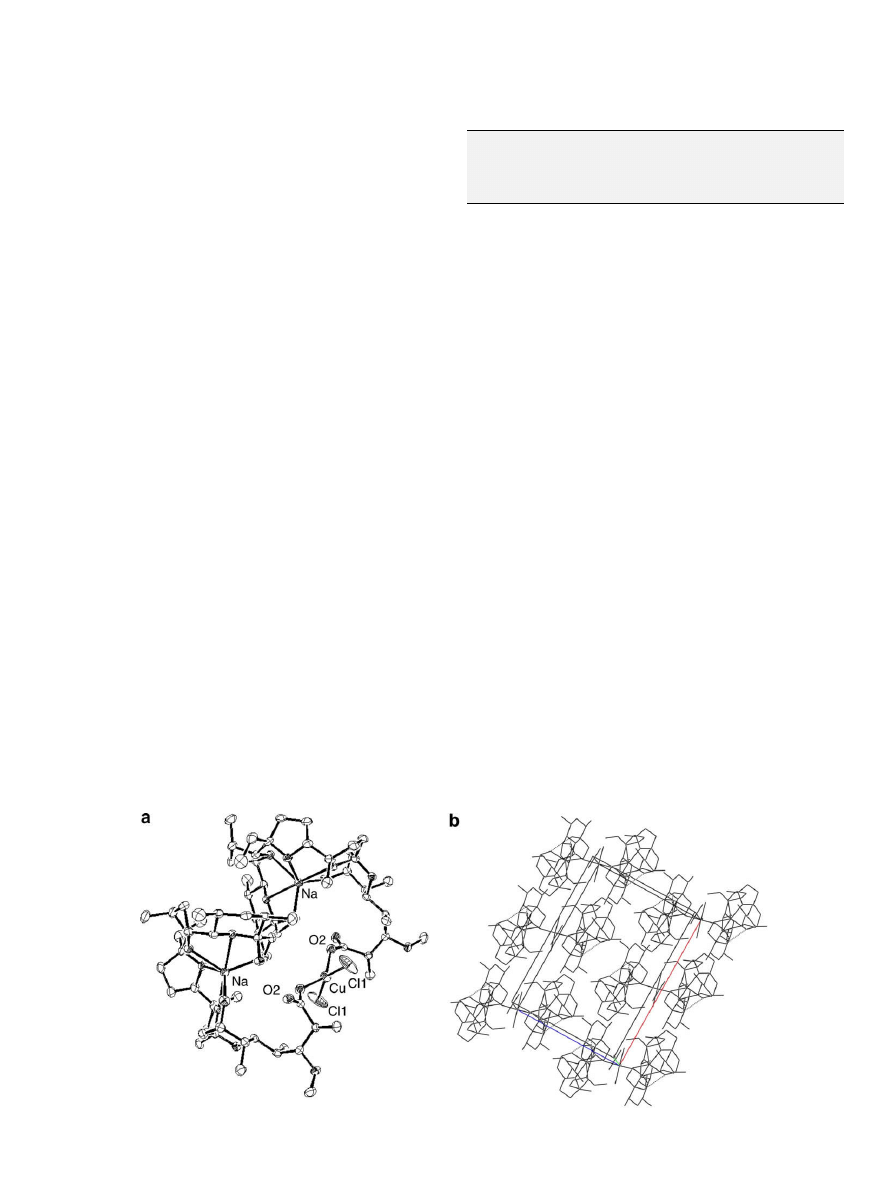
). The ORTEP diagram and crystal packing of the complex
are presented in
. The data reveal that 1 is a heterometallic
compound containing both sodium and copper(II) ions. The
complex consists of two sodium monensin ligands bound
monodentately to a single copper(II) ion via their carboxylate func-
tions. Additionally, Cu(II) reacts with two chloride anions yielding
a neutral mononuclear compound with respect to the transition
metal center. The copper(II) ion is four-coordinated forming two
metal–oxygen and two metal–chloride bonds. The ligands bind
the transition metal center in a distorted square–planar environ-
ment. The metal–ligand bond lengths and angles lie in typical
ranges for square–planar Cu(II) complexes containing both mono-
dentate carboxylate functions and chloride anions (
). The X-
ray data confirm that sodium ion of sodium monensin remains in
the cavity of the ligand and its sixfold coordination with oxygen
atoms is retained during the complexation. The sodium–oxygen
bond lengths and angles (
) are similar to those found in
non-coordinated sodium monensin and in the corresponding
Mn(II)/Co(II) complexes previously reported
. The X-ray
crystal structure of 1 exhibits intramolecular hydrogen bonds of
various origin (
) and no intermolecular H-bonds were
observed.
The comparison of crystallographic data for Mn(II), Co(II)
and Cu(II) complexes of sodium monensin shows that the transi-
tion metal center reacts in a similar manner both with monensin
ligands and with chloride ions. The M–O and M–Cl bond lengths
decrease in the order of Mn(II) > Co(II) > Cu(II) following the
decrease of the corresponding metal ionic radii. The main differ-
ence between the three structures determined was found in the
ligand environment around the transition metal center. Thus,
while Mn(II) and Co(II) ions possess a slightly distorted tetrahedral
geometry with bond angles varying from 105.82° to 109.74°,
the copper(II) ion is surrounded in a distorted square–planar
environment with bond angles in the range of 94.12–95.66°. The
difference in the geometry of the transition metal center may influ-
ence the reactivity of copper(II) complex of sodium monensin in
solution, where additional axial coordination of solvent molecules
could take a place changing the square–planar environment of
Cu(II) ion to an elongated octahedral one.
The properties of the paramagnetic copper(II) complex of sodium
monensin were studied by UV–Vis, EPR and IR spectroscopies. The
Fig. 1. (a) ORTEP drawing of 1, [Cu(MonNa)
2
Cl
2
]MeCN, at the 30% probability level (protons and MeCN molecule are omitted for clarity); (b) crystal packing of complex 1.
Table 2
Selected bond lengths (Å) and bond angles (°) for complex 1.
Cu–O2
1.960(4)
Na–O8
2.403(5)
Cu–Cl1
2.181(2)
Na–O9
2.474(4)
Na–O4
2.331(5)
Na-O11
2.338(5)
Na–O6
2.339(4)
Cl1
i
–Cu–Cl1
152.6(3)
Na–O7
2.448(5)
O2
i
–Cu–O2
137.9(3)
Symmetry position: [i] 2 x, y, 2 z.
I.N. Pantcheva et al. / Journal of Inorganic Biochemistry 103 (2009) 1419–1424
1421

spectral data obtained for 1 are presented below and are in agree-
ment with the solid state structure of the compound solved by single
crystal X-ray diffraction.
The X-band EPR spectra of 1 were recorded in solution and in
solid state both at room (293 K) and liquid nitrogen (77 K) temper-
atures. In the EPR spectrum of copper(II) complex in MeOH (293 K)
the isotropic signal typical for mononuclear Cu(II) compounds was
not observed due to low solubility of 1. The EPR spectra of 1 in fro-
zen MeOH solution (77 K) and in solid phase (293 K, 77 K) consist
of hyperfine structure resulting from the interaction of the un-
paired electron of Cu(II) ðd
x
2
y
2
Þ with the nuclear spin of
63,65
Cu.
The g- and A-values of 1 are characteristic of mononuclear oxygen-
and chloro-containing Cu(II) species possessing distorted tetrago-
nal symmetry and are in agreement with the crystallographic data
(solid state, 77 K: g
||
= 2.38, A
||
= 96 10
4
cm
1
, g
\
= 2.09; MeOH
solution, 77 K: g
||
= 2.42, A
||
= 127 10
4
cm
1
, g
\
= 2.08). In the
electronic spectrum of 1 in MeOH a broad band at 840–870 nm
(
e
= 33 M
1
cm
1
) is observed, while DMSO solution of 1 shows
absorbance maximum at 900 nm (
e
= 200 M
1
cm
1
). The EPR
and UV–Vis spectral data correspond to d–d transitions in the cop-
per(II) chromophore CuO
2
Cl
2
.
In the IR spectrum of the free ligand the stretching vibrations of
hydroxyl groups appear as a broad band in the 3500–3300 cm
1
range due to the complexation of internal OH-groups to Na
+
and
to the participation of corresponding ‘‘tail” OH-groups in H-bond
formation with the ‘‘head” carboxylate moiety. IR spectrum of 1
displays three stretching vibrations,
m
(OH), in the range from
3600 cm
1
to 3100 cm
1
which are in agreement with the pres-
ence of crystallization water (3550 cm
1
), of non-coordinated hy-
droxyl groups (3490 cm
1
) and of OH-groups bound to sodium
ions (3190 cm
1
). The monodentate coordination mode of carbox-
ylate group of sodium monensin A to Cu(II) is confirmed by the
appearance of asymmetric
m
(CO
2
)
asym
at 1590 cm
1
and symmetric
m
(CO
2
)
sym
bands at 1410 cm
1
in the spectrum of 1 (
D
m
= 180 cm
1
,
D
m
=
m
(CO
2
)
asym
–
m
(CO
2
)
sym
), while the COO
-function of the free
ligand, engaged in intramolecular H-bonds, absorbs at 1540 cm
1
and 1390 cm
1
.
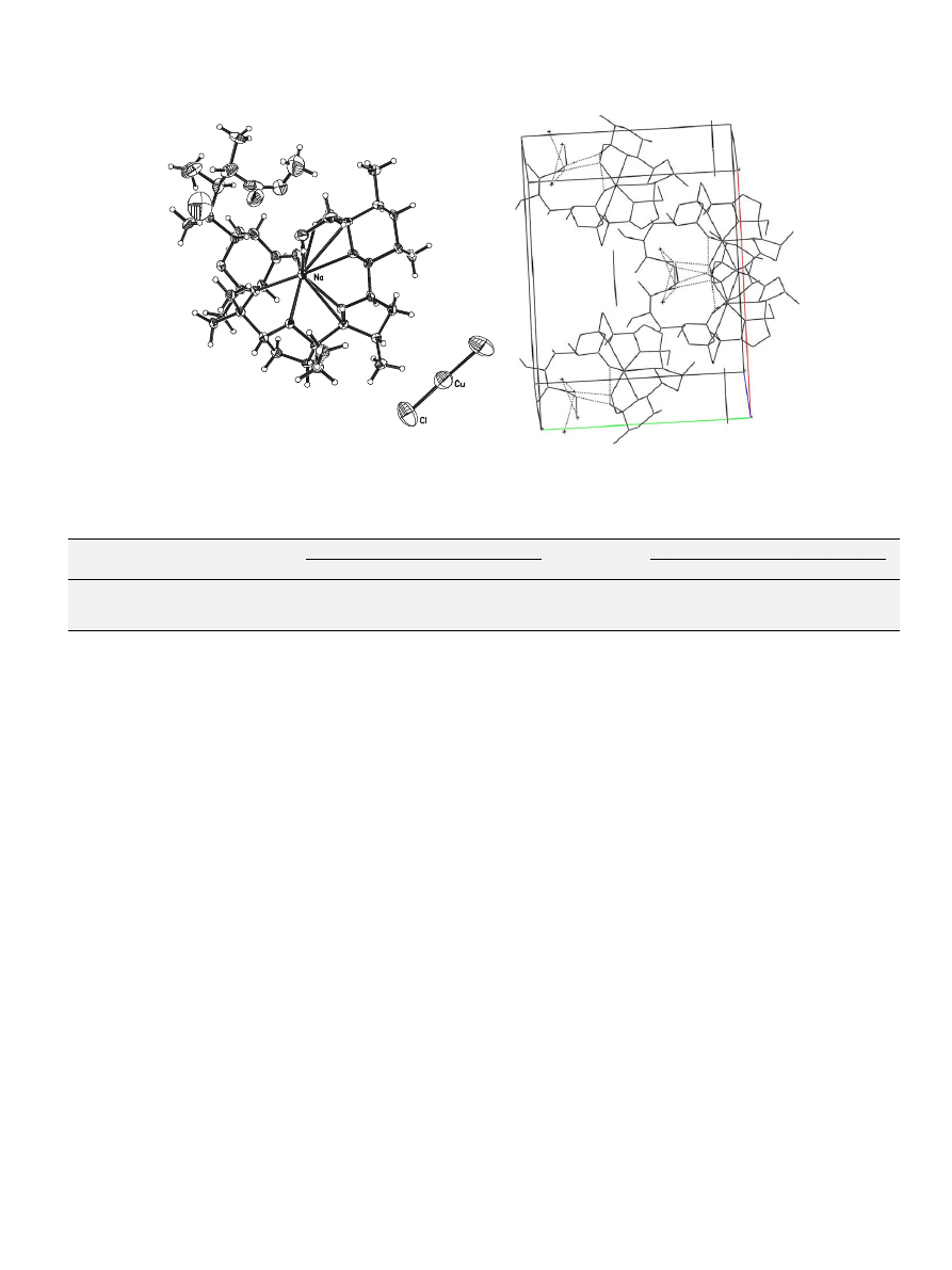
3.2. Crystal structure of compound 2
Compound 2 was isolated in a minor amount as a side product
of the reaction of MonNa with Cu(II). The X-ray crystallography
established its formulation as a copper(I) salt of the methyl ester
of sodium monensin A with the composition [Me-MonNa][H-Mon-
Na][CuCl
2
]Cl (
). 2 Crystallizes in the monoclinic space group
C2 and is isomorphous (isotype) to the copper(II) complex 1. The
monensin ligands and sodium cations exhibit effectively the same
positions in both complexes but the copper(I) compound contains
a discrete chlorocuprate(I) anion [CuCl
2
]
, which is linear and not
coordinated by monensin (the Cu–Cl bond length is 2.089(3) Å,
the Cl–Cu–Cl angle is 178.4(3)°). Charge neutrality is achieved by
the presence of a disordered chloride ion with a site occupation
factor of 0.5. Coordination of a copper(II) atom through two car-
boxylate oxygen atoms as in complex 1 is no longer possible as half
the monensin A ligands are present as methyl ester in this minor
side product. Methanol is probably the source of the methyl func-
tion and together with acetonitrile is presumably responsible for
the partial Cu(II) ? Cu(I) reduction to the dichlorocuprate(I) anion.
Selected bond lengths and angles of 2 and intramolecular H-bonds
observed are presented in
, respectively. The crystal
structure and crystal packing of 2 are displayed in
3.3. Biological properties of sodium monensin A and complex 1
The experimental results on isolation and structure character-
ization of copper(II) complex of sodium monensin and data ob-
tained for the previously reported transition metal complexes of
the ligand
confirm the suggestion that polyether ionophorous
antibiotic reacts not only with monovalent metal ions but also
with divalent metal cations. The mixed-metal complexes of so-
dium monensin can be discussed as possibly formed biological
compounds resulting of the coordination of MonNa to the corre-
sponding transition metal ions and chloride anions existing both
in the extra- and intra-cellular environment. The new copper(II)
complex of sodium monensin is the first copper(II) complex of nat-
ural occurring polyether ionophores and comprises both the prop-
erties of the biologically effective ligand and of the transition metal
ion, that why we evaluated its biological activity in two indepen-
dent directions. First, due to the antimicrobial mode of action of
the ligand, we studied the antibacterial activity of 1 in order to
estimate the influence of Cu(II) on the properties of MonNa. On
the other hand, taking into account the fact that various cop-
per(II)-containing compounds of low molecular weight possess
in vitro SOD-like activity
, we screened the SOD-mimetic
properties of the copper(II) complex of sodium monensin.
3.3.1. Antimicrobial (antibacterial) activity
It is well known that polyether ionophores possess an antimi-
crobial activity against Gram(+)- and have no effect on growth of
Gram()-bacteria, as also confirmed by our experiments using
monensins (MonNa, MonH) and their Mn(II)/Co(II) complexes
. In the present work we tested the same Gram-positive
bacteria strains studied previously to evaluate the antimicrobial
activity of copper(II) complex 1 and to compare the results ob-
tained with those already reported. It was found that copper(II)
chloride has no effect on the visible growth of bacteria strains be-
low concentrations of 3 10
3
l
M while sodium monensin A is
effective against B. subtilis, S. lutea (MIC 23.8
l
M) and B. mycoides
(MIC 11.9
l
M). The experimental results showed that 1 exhibits
cytotoxic activity against tested bacteria strains with MIC values
of 10.7
l
M (B. subtilis, S. lutea) and of 5.4
l
M (B. mycoides), respec-
tively. The data are of the same order as those for manganese and
cobalt complexes of sodium monensin A. The antimicrobial assay
of 1 is in agreement with our hypothesis that the effect of hetero-
metallic complexes of sodium monensin A on the growth of
Gram(+)-microorganisms can be probably explained by the intro-
duction of two active ligand moles per one mole of the complex
Table 3
Intramolecular H-bonds (Å) for complex 1.
O1–O10
2.643
O2–O11
2.679
O4–O10
2.900
Table 4
Selected bond lengths (Å) and bond angles (°) for the chlorocuprate(I) salt of the
methyl ester of sodium monensin A, 2.
Cu–Cl1
2.089(3)
Cl1
i
-Cu–Cl1
178.4(3)
Na-O4
2.340(5)
Na–O8
2.408(4)
Na–O6
2.359(4)
Na–O9
2.473(4)
Na–O7
2.458(4)
Na–O11
2.374(5)
Symmetry position: [i] 2 x, y, 2 z.
Table 5
Intramolecular H-bonds (Å) for compound 2.
O1–O10
2.574
O2–O11
2.684
O4–O10
2.829
O2–Cl1
2.984
1422
I.N. Pantcheva et al. / Journal of Inorganic Biochemistry 103 (2009) 1419–1424
. The compounds discussed in
and in the present paper are
the only representatives of polyether ionophore complexes con-
taining both sodium and transition metal ions that is why in our
opinion it is still early to make a final conclusion regarding the
antibacterial mode of action of mixed-metal complexes of sodium
monensin.
3.3.2. SOD-mimetic activity
To the best of our knowledge, a single study on SOD assay of
metal complexes of polyether ionophores was performed in
2005. Fisher et al.
reported an enzymatic study on SOD-mi-
metic activity of lipophilic complexes of monensin A with Cu(II),
Mn(II) and Fe(II). The authors inferred the composition of com-
pounds from solution studies only, suggesting the formation of
two copper(II) complex species with composition of 2:1 and 1:1
metal-to-ligand molar ratio.
In the present study the structure of the first copper(II) complex
of polyether ionophores, 1 was reliably established in the solid
state. The availability of well defined Cu(II) compound with known
structure and stoichiometry allows us to examine adequately its
possible antioxidant properties, performing a SOD assay using
two indirect xanthine–xanthine oxidase based cytochrome c and
NBT methods
The IC
50
values and the corresponding kinetic constant values
k
McCF
of the compounds tested are summarized in
. As it
can be seen, the non-coordinated ligand sodium monensin A does
not possess SOD-like activity by itself and its influence is negligible
compared to that of copper(II) compounds.
The SOD-like activity of complex 1 is of the same order as the
k
McCF
value determined for CuCl
2
at the selected reaction
conditions (
). The results obtained in this study show that
complex 1 retains the SOD-like activity of the copper(II) ion. At
the same time it is proven that in contrast to Cu(II) ions the poly-
ether ionophore is able to penetrate cell membranes due to its lipo-
philicity and to transfer metal ions into the intracellular space
In this respect, it could be suggested that at real conditions com-
pound 1 would reveal its SOD-mimic properties while the cop-
per(II) chloride (or its aqua complex, respectively) will be
inactive in such a system. The close data obtained both for 1 and
copper(II) ion arouse questions concerning the stability of the com-
plex in solution and its possible dissociation as well as its redox
properties which will be a subject of further detailed elucidation.
4. Conclusion
The first copper(II) complex of polyether ionophores with so-
dium monensin, [Cu(MonNa)
2
Cl
2
]H
2
O was prepared and its struc-
ture in solid state was solved by X-ray crystallography. The
complex is heterometallic, containing two sodium and one cop-
per(II) ions. Two sodium monensin anions are bound monoden-
tately to Cu(II) center through their carboxylate functions, and
two chloride ions participate in the inner coordination sphere of
the transition metal ion determining the neutral character of the
complex. [Cu(MonNa)
2
Cl
2
]H
2
O possesses an antibacterial activity
against Gram(+)-bacteria due to insertion of two moles of the ac-
tive ligand per mole of the complex. Complex 1 shows SOD-like
activity comparable with that of the copper(II) ion.
5. Appendix A. Supplementary data
CCDC 714412 (1) and CCDC 713238 (2) contain the supplemen-
tary crystallographic data for this paper. These data can be ob-
tained free of charge via
(or an
a
b
Fig. 2. (a) ORTEP drawing of [Me-MonNa][H-MonNa][CuCl
2
]Cl, 2, at the 30% probability level (protons are omitted for clarity); (b) crystal packing of compound 2.
Table 6
SOD-like activity of sodium monensin A, complex 1 and copper(II) chloride.
Compound
Cytochrome c assay
NBT assay
IC
50
(M)
k
McCF
(M
1
s
1
)
IC
50
(M)
k
McCF
(M
1
s
1
)
MonNa
>4.0 10
4
<1.2 10
4
>4.0 10
4
<1.7 10
4
[Cu(MonNa)
2
Cl
2
]H
2
O, 1
2.7 10
7
1.9 10
7
3.4 10
7
2.0 10
7
CuCl
2
2H
2
O
2.9 10
7
1.8 10
7
3.9 10
7
1.7 10
7
a
Sodium monensin A does not affect the rate of cytochrome c/NBT reduction at the studied concentration.
I.N. Pantcheva et al. / Journal of Inorganic Biochemistry 103 (2009) 1419–1424
1423

application from Cambridge Crystallographic Data Center, 12 Un-
ion Road, Cambridge CB2 1EZ, UK; fax: +44 01223 336033; or e-
mail: deposit@ccdc.cam.ac.uk.
Acknowledgements
This research work is partially supported by the Research Fund
of Sofia University (Project No. 26/2008). The authors are grateful
to Rumyana Zhorova, MSci and Mr. Hristo Kolev, Sofia University,
for their assistance performing antimicrobial and enzymic tests
on compounds studied. M.M. is thankful to Alexander von Humboldt
Foundation for the opportunity for carrying out research in the lab-
oratory of Prof. W.S. Sheldrick at the University of Bochum.
References
[1] A. Agtarap, J.W. Chamberlin, M. Pinkerton, L.K. Steinrauf, J. Am. Chem. Soc. 89
(1967) 5737–5739.
[2] B.C. Pressman, E.J. Harris, W.S. Jagger, J.H. Johnson, Proc. Natl. Acad. Sci. USA 58
(1967) 1949.
[3] P.H. Stern, Clin. Orthop. Rel. Res. 122 (1977) 273–298.
[4] S.C. Gad, C. Reilly, K. Siino, F.A. Gavigan, G. Witz, Drug Chem. Toxicol. 8 (1985)
451–468.
[5] H.H. Mollenhauer, D.J. Morre, L.D. Rowe, Biochim. Biophys. Acta 1031 (1990)
225–246.
[6] B.C. Pressman, M. Fahim, Ann. Rev. Pharmacol. Toxicol. 22 (1982) 465–490.
[7] J.W. Westley, in: J.W. Westley (Ed.), Polyether antibiotics: naturally occurring
acid ionophores, Chemistry, vol. 2, Marcel Dekker Inc., New York, 1983, pp. 51–
87.
[8] W.L. Duax, G.D. Smith, P.D. Strong, J. Am. Chem. Soc. 102 (1980) 6725–6729.
[9] P.Y. Barrans, M. Alleaume, G. Jeminet, Acta Cryst. B 38 (1982) 1144–1149.
[10] B.G. Cox, N. Vantruong, J. Pzeszotarska, H. Schneider, J. Am. Chem. Soc. 106
(1984) 5965–5969.
[11] D.M. Walba, M. Hermsmeier, R.C. Haltiwanger, J.H. Noordik, J. Org. Chem. 51
(1986) 245–247.
[12] M. Mimouni, S. Perrier, Y. Pointud, J. Juillard, J. Sol. Chem. 22 (1993) 769–785.
[13] M. Mimouni, M. Hebrant, G. Dauphin, J. Juillard, J. Chem. Res. 6 (1996) 278–
279.
[14] Y. Pointud, C. Bernard, S. Touzain, L. Astier, B. Sabatier, J. Juillard, J. Sol. Chem.
26 (1997) 479–495.
[15] T. Martinek, F.G. Riddell, C. Wilson, C.T. Weller, J. Chem. Soc. Perkin Trans. 2
(2000) 35–41.
[16] L. Wittenkeller, W.R. Lin, C. Diven, A. Ciaccia, F. Wang, D.M. de Freitas, Inorg.
Chem. 40 (2001) 1654–1662.
[17] F.G. Riddell, Chirality 14 (2002) 121–125.
[18] F.A.A. Paz, P.J. Gates, S. Fowler, A. Gallimore, B. Harvey, N.P. Lopes, C.B.W. Stark,
J. Staunton, J. Klinowski, J.B. Spencer, Acta Cryst. E 59 (2003) m1050–m1052.
[19] S.O. Yildirim, V. McKee, F.-Z. Khardli, M. Mimouni, T.B. Hadda, Acta Cryst. E 64
(2007) m154–m155.
[20] M. Pinkerton, L.K. Steinrauf, J. Mol. Biol. 49 (1970) 533–546.
[21] J. Garcla-Rosas, H. Schneider, B.G. Cox, J. Phys. Chem. 87 (1983) 5467–5472.
[22] R.D. Williams, D.R. Bright, V.V. Young, J.L. Martin, US Patent No. 4478935,
1984.
[23] J.L. Martin, US Patent No. 5043333, 1991.
[24] N. Aouad, G. Miquel-Mercier, E. Bienvenue, E. Tronel-Peyroz, G. Jeminet, J.
Juillard, P. Seta, J. Membr. Sci. 139 (1998) 167–174.
[25] A. Nakamura, S. Nagai, T. Ueda, N. Murakami, J. Sakakibara, H. Shibuya, I.
Kitagawa, Chem. Pharm. Bull. (Tokyo) 39 (1991) 1726–1730.
[26] A. Nakamura, S. Nagai, T. Takahashi, R. Malhan, N. Murakami, T. Ueda, J.
Sakakibara, M. Asano, Chem. Pharm. Bull. (Tokyo) 40 (1992) 2331–2337.
[27] A. Nagatsu, T. Takahashi, M. Isomura, S. Nagai, T. Ueda, N. Murakami, J.
Sakakibara, K. Hatano, Chem. Pharm. Bull. (Tokyo) 42 (1994) 2269–2275.
[28] A. Nagatsu, Y. Tabunoki, S. Nagai, T. Ueda, J. Sakakibara, H. Hidaka, Chem.
Pharm. Bull. (Tokyo) 45 (1997) 966–970.
[29] A. Nagatsu, J. Sakakibara, Yakugaku Zasshi 117 (1997) 583–596.
[30] A. Nagatsu, R. Tanaka, M. Hashimoto, H. Mizukami, Y. Ogihara, J. Sakakibara,
Tetrahedr. Lett. 41 (2000) 2629–2632.
[31] R. Tanaka, A. Nagatsu, H. Mizukami, Y. Ogihara, J. Sakakibara, Chem. Pharm.
Bull. (Tokyo) 49 (2001) 711–715.
[32] R. Tanaka, A. Nagatsu, K. Hatano, H. Mizukami, Y. Ogihara, J. Sakakibara, Tenn.
Yuki Kag. Tor. Koen Yosh. 43 (2001) 623–628.
[33] T.H. Elsasser, J. Anim. Sci. 59 (1984) 845–853.
[34] M. Mimouni, Y. Pointud, J. Juillard, Bull. Soc. Chim. Fr. 131 (1994) 58–65.
[35] M. Hebrant, M. Mimouni, M. Tissier, Y. Pointud, J. Juillard, G. Dauphin, New J.
Chem. 16 (1992) 999–1008.
[36] S.A. Hamidinia, O.I. Shimelis, B. Tan, W.L. Erdahl, C.J. Chapman, G.D. Renkes,
R.W. Taylor, D.R. Pfeiffer, J. Biol. Chem. 277 (2002) 38111–38120.
[37] A. Huczynski, P. Przybylski, B. Brzezinski, J. Mol. Struct. 788 (2006) 176–
183.
[38] A. Huczynski, P. Przybylski, G. Schroeder, B. Brzezinski, J. Mol. Struct. 829
(2007) 111–119.
[39] A. Huczynski, B. Brzezinski, F. Bartl, J. Mol. Struct. 886 (2008) 9–16.
[40] P. Dorkov, I.N. Pantcheva, W.S. Sheldrick, H. Mayer-Figge, R. Petrova, M.
Mitewa, J. Inorg. Biochem. 102 (2008) 26–32.
[41] I.N. Pantcheva, M. Mitewa, W.S. Sheldrick, I.M. Oppel, R. Zhorova, P. Dorkov,
Curr. Drug Discov. Technol. 5 (2008) 154–161.
[42] G. Sheldrick, SHELXL-97 Program for Crystal Structure Refinement, Institut fur
Anorganische Chemie der Universitat, Gottingen, Germany, 1997.
[43] J.M. McCord, I. Fridovich, J. Biol. Chem. 244 (1969) 6049–6055.
[44] L.W. Oberley, D.R. Spitz, Meth. Enzym. 105 (1984) 457–464.
[45] R.F. Pasternack, B. Halliwell, J. Am. Chem. Soc. 101 (1979) 1026–1031.
[46] S. Durot, C. Policar, F. Cisnetti, F. Lambert, J.-P. Renault, G. Pelosi, G. Blain, H.
Korri-Youssoufi, J.-P. Mahy, Eur. J. Inorg. Chem. (2005) 3513–3523.
[47] R.H. Weiss, A.G. Flickinger, W.J. Rivers, M.M. Hardy, K.W. Aston, U.S. Ryan, D.P.
Riley, J. Biol. Chem. 268 (1993) 23049–23054.
[48] K. Nakamoto (Ed.), Infrared and Raman Spectroscopy of Inorganic and
Coordination Compounds, fifth ed., Wiley, Toronto, 1997.
[49] J.R.J. Sorenson (Ed.), Inflammatory Diseases and Copper, The Humana Press,
Clifton, 1982.
[50] J.R.J. Sorenson, in: H. Sigel (Ed.), Metal Ions in Biological Systems, vol. 14,
Marcel Dekker, New York, 1982, pp. 77–124.
[51] R.G. Bhirud, T.S. Srivastava, J. Inorg. Biochem. 40 (1990) 331–338.
[52] R.G. Bhirud, T.S. Srivastava, Inorg. Chim. Acta 179 (1991) 125–131.
[53] A.E.O. Fisher, G. Lau, D.P. Naughton, Biochem. Biophys. Res. Commun. 329
(2005) 930–933.
[54] J.B. Russell, J. Anim. Sci. 64 (1987) 1519–1525.
1424
I.N. Pantcheva et al. / Journal of Inorganic Biochemistry 103 (2009) 1419–1424
Document Outline
- Crystal structure and properties of the copper(II) complex of sodium monensin A
Wyszukiwarka
Podobne podstrony:
Structure and properties of Ti
Walker The Production, Microstructure, and Properties of Wrought Iron (2002)
Structure and reactivity of alkenes and alkynes
71 1021 1029 Effect of Electron Beam Treatment on the Structure and the Properties of Hard
Guide to the properties and uses of detergents in biology and biochemistry
Fibrillar Structure and Mechanical Properties of Collagen
Derrida, Jacques Structure, Sign And Play In The Discourse Of The Human Sciences
Guide to the properties and uses of detergents in biology and biochemistry
Aspects of the development of casting and froging techniques from the copper age of Eastern Central
The Structure and the Unity of Beowulf Arthur G Brodeur
The Structure and Heat Treatment of Low Carbon Steel
SCHAFER, Christian The Philosophy of Dionysius the Areopagite an introduction to the structure and
Energy and Nutritional Properties of the White Mulberry
Spinks, Lee Genesis And Structure And The Object Of Postmodernism
African Filmmaking North and South of the Sahara
Modeling of Polymer Processing and Properties
więcej podobnych podstron