
Research paper
Acta Neurobiol Exp 2010, 70: 196–208
© 2010 by Polish Neuroscience Society - PTBUN, Nencki Institute of Experimental Biology
INTRODUCTION
Autism spectrum disorders (ASDs) represent a
group of neurodevelopmental disorders typified by
impairments in verbal and non-verbal communication,
social withdrawal and stereotypical behaviors, which
may or may not be associated with cognitive deficits,
self-injurious behaviors and other neurological comor-
bidities. The current world-wide epidemic of ASDs
and other neurodevelopmental disorders, including
attention deficit hyperactivity (ADHD), learning dis-
abilities, and mental retardation constitute the most
disturbing public health problems (Robison et al. 1999,
Merrick et al. 2004, Altarac and Saroha 2007, Shayer
et al. 2007, Hertz–Picciotto and Delwiche 2009). Its
magnitude is best illustrated by a dramatic rise in inci-
dences of ASDs in the past 25 years. In many countries
current ASDs prevalence is about 1 in 100, whereas in
the 1970s and early 1980s it was about 1 in 2500-3000
(Merrick et al. 2004, Baird et al. 2006, Gillberg 2009).
It is estimated that only about 5% of the autistic popu-
lation carries identifiable genetic /chromosomal defects
(Newbury et al. 2009). The increase of ASDs preva-
lence cannot be fully explained by advances in diag-
nostics or sudden genetic shifts. There is a growing
consensus among scientists and clinicians that ASDs
ensue from an interaction between biological vulnera-
bility factors and environmental or iatrogenic insults
(James et al. 2006, Gillberg 2009).
The contemporaneous emergence of the ASDs epi-
demic and the introduction of several new infant vac-
cines in the late 1980s and the 1990s, generated a
suspicion that these events might be linked. One of the
agents suspected in autism etiology is an organomer-
cury compound, thimerosal (THIM; sodium ethyl-
mercurithiosalicylate containing approximately 49%
Age-dependent lower or higher levels of hair mercury
in autistic children than in healthy controls
Maria Dorota Majewska
1
*, Ewa Urbanowicz
3
, Paulina Rok-Bujko
3
, Irena Namysłowska
3
,
Paweł Mierzejewski
2
1
Marie Curie Chair, Department of Pharmacology and Physiology of the Nervous System, Institute of Psychiatry
and Neurology, Warsaw; *Email : majewska@ipin.edu.pl;
2
Department of Pharmacology and Physiology of the Nervous
System, Institute of Psychiatry and Neurology, Warsaw, Poland;
3
Department of Child and Adolescent Psychiatry,
Institute of Psychiatry and Neurology, Warsaw, Poland;
An association between autism and early life exposure to mercury is a hotly debated issue. In this study, 91 autistic Polish
children, male and female, 3-4 and 7-9 years old, were compared to 75 age- and sex-matched healthy children with respect
to: demographic, perinatal, clinical and developmental measures, parental age, birth order, morphometric measures,
vaccination history, and hair mercury content. In demographic and perinatal measures there were no consistent differences
between the autistic and control groups. Autistic children had a significantly greater prevalence of adverse reactions after
vaccinations and abnormal development than controls. Between 45 and 80% of autistic children experienced developmental
regress. Autistic children significantly differed from healthy peers in the concentrations of mercury in hair: younger autistics
had lower levels, while older – higher levels than their respective controls. The results suggest that autistic children differ
from healthy children in metabolism of mercury, which seems to change with age.
Key words: autism, mercury, hair, thimerosal, vaccines, development
Abbreviations: THIM - thimerosal
Correspondence should be addressed to M. D. Majewska,
Email: majewska@ipin.edu.pl
Received 19 January 2010, accepted 1 June 2010

Hair mercury in autistic children
197
of Hg by weight), which has been used as a vaccine
preservative for decades without being comprehen-
sively tested for its safety in developing organisms. A
large body of research and at least two centuries of
human experience show that all forms of mercury are
highly toxic to vertebrates. In the body THIM is
metabolized to ethylmercury and then into inorganic
mercury compounds (Qvarnstrom et al. 2003).
Significant amounts of mercury have been measured
in the blood of infants after inoculations with THIM-
containing vaccines, with premature infants accumu-
lating over 3 times more mercury than the mature
ones (Stajich et al. 2000, Pichichero et al. 2008).
Studies conducted with infant monkeys injected with
THIM-containing vaccines showed that a few days
after vaccinations, concentrations of mercury in the
brain were several times higher than those in blood.
Mercury levels in the brain may remain markedly
increased for many months or years, considering con-
tinuous re-exposure to vaccines (Burbacher et al.
2005). Mid-nanomolar concentrations of mercury,
which are likely to be reached in the infant brain after
inoculation with THIM-containing vaccines, are neu-
rotoxic (Parran et al. 2005, Yel et al. 2005).
The preclinical study of Hornig and coauthors
(2004) documented multiple neurodevelopmental dis-
turbances in mice prone to autoimmune diseases after
exposure to THIM doses analogous to those used in
pediatric vaccines. Our recent study showed that
administration of similar doses of THIM to suckling
rats causes persistent disruption of endogenous opioid
system (Olczak et al. 2009), which resembles opioid
dysfunction in autism (Sandyk and Gillman 1986,
Sandman 1988). Based on numerous analogies of bio-
logical and clinical abnormalities associated with mer-
cury poisoning and autism, the hypothesis emerged
linking this disorder with early life exposure to mercu-
rials (Bernard et al. 2001, Mutter et al. 2005). Some
epidemiological and ecological studies associated
autism and other neurodevelopmental disorders with
THIM present in infant vaccines (Geier and Geier
2003, 2006, Young et al. 2008, Gallagher and Goodman
2008, 2009). Other studies denied such a link (Hviid et
al. 2003, Madsen et al. 2003), but they were criticized
for flawed design and clear conflict of interests (Mutter
et al. 2005,
Isaacs 2010).
Measurement of heavy metal content in hair is often
used as a marker of exposure, because it correlates
with past blood levels (Clarkson 1993, Magos and
Clarkson 2008). As a non-invasive procedure, it is
especially useful for testing children. A few studies
compared mercury levels in hair of autistic and healthy
children, reporting divergent results. Holmes and
coauthors (2003) and Adams and colleagues (2008)
demonstrated significantly lower levels of mercury in
first baby haircuts of American children diagnosed
with autism, than in healthy controls, which was inter-
preted as possibly impaired mercury and other toxin
elimination by autistic children. Reduced levels of
heavy metals such as arsenic, cadmium, lead and mer-
cury in hair of autistic children 1-6 years old were also
measured by Kern and others (2007). In contrast,
strikingly higher levels of hair mercury in autistic
Kuwaiti boys (4 to 7 years old) than in healthy controls
were detected by Fido and Al-Saad (2005). Higher
concentrations of this metal were also found in baby
teeth and blood of autistic children than in controls
(Adams et al. 2007, Desoto and Hitlan 2007).
Although several studies point to a link between
autism and mercury exposure (Mutter et al. 2005,
Geier and Geier 2006, Windham et al. 2006, Young et
al. 2008, Palmer et al. 2009), the critical sources of this
metal and its role in autism pathogenesis are a subject
of hot debates, particularly in reference to possible
iatrogenic effects of THIM from vaccines and mercury
from amalgam fillings. Continuing controversy and
public apprehension regarding this issue impelled us to
conduct our own study in Polish children, who con-
tinue to be inoculated with THIM-containing vac-
cines. We were particularly interested in identifying
possible demographic, perinatal and/or vaccination-
related factors, which distinguish autistic from healthy
children. In order to assess them we analyzed several
birth-related measures such as Apgar scores, body
weight, head and chest circumference, Rh conflict, as
well as abnormal development in autistic and healthy
children of both sexes from two age groups (3-4 and
7-9 years old). We also compared their history of vac-
cinations, adverse reactions to them, and levels of
mercury in hair.
METHODS
Study Participants
The study was carried out in accordance with the
Declaration of Helsinki of the World Medical
Association. The protocol was approved by the Ethics

198
M. D. Majewska et al.
Committee for Human Studies at the Institute of
Psychiatry and Neurology. Participation in the study
was voluntary. The parents of participants read and
signed informed consent forms after the study proce-
dures had been fully explained to them.
Autistic and control children of both sexes from two
age groups, 3-4 and 7-9 years old were enrolled into
the study. The subjects were not compensated for their
participation. The autistic participants were recruited
from the children earlier diagnosed with autism in out-
patient clinics in Poland. Healthy control children were
recruited from 6 preschools and 3 primary Warsaw
schools. Recruitment took place from November 2007
to April 2009. All participants were Caucasians, about
40% of autistic children were from Warsaw metropoli-
tan area, others were from different, mostly urban, but
some from rural regions located at distances less than
180 km from Warsaw. The autistic children had to
fulfill DSM IV criteria for autistic disorder, and score
at least 30 points in the CARS scale (see later).
Participants were excluded if they had: a neurological
and psychiatric disorder other than autism and comor-
bid disorders; history of liver, renal or endocrine disor-
der; current infection of respiratory tract or fever state
of any origin; and mental retardation. Mental retarda-
tion or behavioral disorders, including hyperkinetic
disorder in children over 6 years old or significant
symptoms of hyperactivity, impulsiveness or restless-
ness in younger children were exclusion criteria only
for the group of healthy control children, but were
allowed as comorbid condition in the autistic cohort.
Children diagnosed with Asperger’s syndrome were
excluded from the study. Study participants were
divided into two groups. Group I was male and female,
autistic and control children 3-4 years old, and group
II – similar children 7-9 years old.
Clinical evaluation
Each autistic child was once more diagnosed by a
group of three specialists (2 psychologists and one
child psychiatrist), who had over five years experi-
ence in autism diagnosis. The examination consisted
of semi-structured interview with parents and 1 hour
observation of the child’s behavior. Extensive medical
histories of the autistic and control children were
taken. The parents were asked about: detailed history
of pregnancy and labor, mental and motor develop-
ment of the child, any illnesses and traumatic events,
history of vaccinations, the occurrence of vaccine-
associated adverse effects (if present, parents were
asked about their subjective appraisal of observed
symptoms and about results of medical professional
consultations). Additional information about birth
morphometric measures, Apgar scores, vaccinations
and development was taken from Child Health
Notebooks, which every child born in Poland receives
at the hospital and which carries his/her health infor-
mation until the age of 18 years. The parents of autis-
tic children were also questioned about the first
symptoms of autism, which occurred in their child,
and the results of previous diagnostic tests. The clini-
cal evaluation of autistic children was based on a one-
hour observation of their behavior by two experienced
psychologists. The diagnosis of autism was made
according to the Diagnostic and Statistical Manual of
Mental Disorders (DSM–IV) criteria for autism or
pervasive developmental delay (PDD) by a trained
professional. The activity and functioning of an autis-
tic child was also assessed according to Childhood
Autism Rating Scale (CARS) (Schopler et al. 1980)
and the Clinical Global Impression Scale. Children
who scored 30 or more points in CARS were diag-
nosed as autistic.
All control study participants were assessed with
use of the Abbreviated Parent-Teacher Questionnaire
(IOWA-Conners; version for scientific research
(Conners 1969, Rowe and Rowe 1997) in order to
exclude children with symptoms of ADHD (diagnosis
was made according to DSM IV criteria).
Measurement of mercury concentration in hair
Haircut samples of autistic and control study par-
ticipants were obtained from occipital area of head,
from the proximal (up to 3 centimeters from scalp) part
of hair. The blinded analysis of mercury content in hair
by atomic absorption spectrometry was performed at
the Chemical Laboratory of Multi-Elemental Analyses
at Wroclaw University of Technology. A single-pur-
pose atomic absorption spectrometer based on in situ
dry washing followed by gold amalgamation cold
vapor AAS method was used for analysis using an
Advanced Mercury Analyzer (AMA-254, ALTEC,
Czech Republic). This cold vapor AAS method is one
of the most widely used techniques for determination
of trace amounts of total mercury in environmental
materials.
Hair mercury in autistic children
199
Table I
Demographic data on study participants
Demographics
Males
Females
p (t-Student, U-M-W
or χ
2
)
GROUP I (age 3-4 years)
Autistic
Control
Autistic
Control
N
30
19
25
19
Mean age
3.6 ± 0.1
3.5 ± 0.1
3.8 ± 0.1
3.6 ± 0.1
Weight at birth (g)
3441 ± 103 3358 ± 159
3295 ± 73
3281 ± 108
Head circumference at birth (cm)
34.3 ± 0.3
33.7 ± 0.5
34.0 ± 0.3
33.3 ± 0.4
Gestational age at birth (weeks)
38.7 ± 0.3
38.8 ± 0.7
39.2 ± 0.3
39.2 ± 0.4
Apgar score
9.5 ± 0.3
9.3 ± 0.3
9.6 ± 0.4
9.9 ± 0.1
Birth order
1.4 ± 0.1
1.5 ± 0.2
1.6 ± 0.2
1.3 ± 0.1
Mother’s age at birth
28.9 ± 0.6
28.4 ± 0.8
27.3 ± 0.9**
31.4 ± 0.8
p<0.01
Father’s age at birth
30.9 ± 0.7
31.3 ± 0.9
28.9 ± 1.0*
33.2 ± 1.3
p=0.01
GROUP II (age 7-9 years)
Autistic
Control
Autistic
Control
N
23
18
13
19
Mean age
8.2 ± 0.1
8.4 ± 0.2
7.9 ± 0,2
8.4 ± 0.1
Weight at birth (g)
3329 ± 183 3486 ± 151
3066 ± 183*
3402 ± 108
p=0.02
Head circumference at birth (cm)
33.9 ± 0.7
34.3 ± 0.5
33.2 ± 0.6
33.7 ± 0.4
Gestational age at birth (weeks)
38.2 ± 0.7
38.8 ± 0.4
38.7 ± 0.6
39.4 ± 0.4
Apgar score
8.7 ± 0.5
9.4 ± 0.2
9.0 ± 0.4
9.6 ± 0.2
Birth order
1.4 ± 0.1
1.7 ± 0.2
2.3 ± 0.8*
1.2 ± 0.1
p=0.03
Mother’s age at birth
27.3 ± 0.7
28.3 ± 1.0
28.5 ± 1.4
27.3 ± 0.9
Father’s age at birth
29.5 ± 0.8
30.0 ± 0.9
30.3 ± 1.3
28.7 ± 0.8
Autistic and control study participants were divided into two age-groups, as indicated. Statistically significant
differences between autistic and control groups are denoted by (*) and (**).

200
M. D. Majewska et al.
Statistics
The STATISTICA software package for Windows
(StatSoft, Tulsa, OK, USA) was used to analyze all data.
Student’s t-test was used when means of data from two
groups were compared. U–Mann-Whitney test was
used for comparisons of nonparametric data, McNemar’s
test with χ
2
statistics was applied for categorical vari-
ables (‘yes’ or ‘no’). For comparisons of mercury levels
in hair 2-way ANOVA (disease x age) was utilized.
Newman-Keuls test was used for individual post-hoc
comparisons. Results with p-level less than 0.05 were
considered significant. The results are presented as
mean ± standard error of mean (SEM).
RESULTS
Demographics and birth-related measures
Altogether 91 autistic children and 75 age- and sex-
matched healthy control children were enrolled into
the study. The demographic data on participants is
shown in Table I. There were no statistically signifi-
cant age differences at the time of psychiatric exami-
nation and collection of specimens between the autistic
and control groups. Most autistic and control children
did not differ significantly with respect to their birth
weights, except for a slightly lower weights of autistic
girls from group II (p=0.02). Also head circumference
at birth was not significantly different between autistic
and control children, although in group I, both in
males and females, there was a trend for slightly larger
head size in autistics than in controls. This difference
did not reach statistical significance (p=0.07). Likewise,
Apgar scores were not statistically significantly differ-
ent between autistic and control children, except that
there appeared to be a tendency for slightly lower val-
ues for autistic children from group II. The only statis-
tically significant difference in Apgar scores was
between control males and females, with females hav-
ing higher scores (p=0.04). The autistic and control
groups did not vary significantly with respect to their
Table II
Comparison or clinical features between autistic and control children and between autistic males and
females within each age-group
Clinical Features
Males
Females
p (t-Student , U-M-W
or χ
2
)
GROUP I (age 3-4 years)
Autistic
Control
Autistic
Control
Allergies (%)
50
22.7
38.5
36.8
Number of vaccines till Year 2
24.5 ± 0.9
23.6 ± 0.7
24.6 ± 0.6
24.2 ± 0.6
Maximal number vac. at once
5.0 ± 0.2
4.8 ± 0.1
4.8 ± 0.1
5.1 ± 0.2
Vaccine complications (%)
38.5*
4.5
15.4
5.3
p=0.03
Abnormal development (%)
26.2**
0
32**
0
p<0.01
Regress (%)
80.8
81
Hyperactivity (%)
26.9
34.6
CARS total scores
43.6 ± 1.3
45.5 ± 1.2
DSM IV A
9.2 ± 0.3
9.6 ± 0.2
Autism-vaccine connection (%)
19.2
15.4

Hair mercury in autistic children
201
birth order, except again for the females from group II,
who were of higher order (2.3) than controls (1.2;
p=0.03). Autistic and healthy groups also did not
diverge significantly with respect to parental age at
child’s birth, except for autistic girls from group I, who
had slightly younger parents than the controls (p≤
0.01).
Clinical parameters
Comparisons of major clinical features of autistic
and control children are shown in Table II. There was
a good correlation between CARS and DSM IV autism
diagnostics (r=0.74). In the younger group of autistic
children CARS and DSM IV scores were not notably
dissimilar between males and females. However, in the
older group of autistic children, the females appeared
to be more impaired, as evidenced by their statistically
significantly higher CARS scores than those in males
(p<0.02). Generally, autistic and control children did
not diverge significantly in the number of vaccinations
received up to the 24
th
month of life, except for the
autistic girls from the group II, who received fewer
vaccinations (p<0.001) due to more frequent vaccine
adverse events.
Autistic children from combined groups I and II
experienced significantly more vaccine complications
(20.4%) than controls (6.5%). This difference was sta-
tistically significant (χ2=6.75; p=0.009 (Table III). It
was particularly pronounced in the males from group
I, where 38.5% of autistics had adverse reactions to
vaccines, while in the control group only 4.5% mani-
fested such reactions (p=0.03, Table II). Vaccine com-
plications reported by parents of autistic children
included: high fever, prolonged crying, extended hypo-
activity and hypotonicity, loss of contact, loss of facial
mimicry, sleepiness, circling around, loss or ability to
walk, point or talk, developmental regress, emergence
of autistic behaviors. The vaccines most frequently
associated with these adverse reactions, were: DTP,
DTP-polio, DTP-Hib, DTP-polio-Hib, MMR, pneu-
mococcal vaccine. A few adverse vaccination events
reported by parents of control children included: skin
reaction, crying and fever. Autistic patients also
Clinical Features
Males
Females
p (t-Student , U-M-W
or χ
2
)
GROUP II (age 7-9 years)
Autistic
Control
Autistic
Control
Allergies (%)
50
40
33.3
33.3
Number of vaccines till Year 2
22.3 ± 0.7
24.2 ± 0.7
20.2 ± 0.9** 24.2 ± 0.5
p<0.001
Maximal number vac. at once
4.6 ± 0.1
4.8 ± 0.1
4.3 ± 0.1*
4.9 ± 0.1
p=0.03
Vaccine complications (%)
16.7
10
26.7
9.2
Abnormal development (%)
52
11
66.6**
0
p<0.01
Regress (%)
45.8
0
80
0
Hyperactivity (%)
50
40
CARS total scores
38.7 ± 1.2*
44.3 ± 1.8
p=0.02
DSM IV A
10.0 ± 0.3
10.0 ± 0.3
Autism-vaccine connection (%)
12.5
26.7
Autistic and control study participants were divided into two age-groups, as indicated. Statistically significant
differences between autistic and control groups, or between autistic males and females are denoted by (*) and (**).

202
M. D. Majewska et al.
diverged from controls in developmental characteris-
tics, where 40.9% of autistics versus 3.9% of controls
were reported to have abnormal development (χ2=30.6;
p<0.001). Among autistic cohort developmental regress
was noted in about 80%, except for the males from
group II, where it was 46% (Table II). Comorbid
hyperactivity was diagnosed in approximately 30% of
the autistic children from group I, and in 45% of such
children from group II. Among autistic boys the fre-
quency of allergies (reported by parents) also appeared
to be slightly higher than in controls, but the distinc-
tion was not statistically significant.
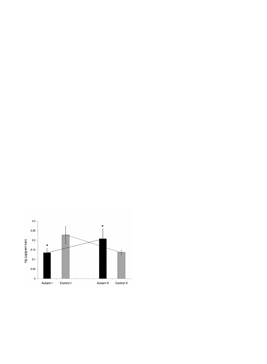
Hair mercury content
Autistic and control children from both age-groups dif-
fered noticeably in the concentration of mercury in hair
(Fig. 1). In group I, hair mercury levels were lower in
autistic than in control children, but the situation was
opposite in group II, where autistics had higher levels than
controls (p=0.01). Consequently, there appeared to be
opposing developmental trends between autistic and con-
trol children with respect to change of hair mercury levels.
In autistics these levels increased with advancing age
(from 3-4 to 7-9 years), whereas in controls – decreased.
DISCUSSION
This study compared autistic and healthy control
children of both sexes aged 3-4 and 7-9 years with
respect to perinatal morphometric and clinical mea-
sures, abnormal development, vaccination history and
mercury content in hair. The results point to statisti-
cally significant differences between autistic and con-
trol cohorts in three major categories. Autistic children
had: 1) greater prevalence of abnormal development;
2) more frequent vaccine complications; 3) different
concentrations of mercury in hair (younger autistics
had lower levels, while older – higher levels than their
age-matched controls).
For three out of four experimental (age-sex) com-
parison groups, the demographic and birth morpho-
metric measures of autistic children were not statisti-
cally significantly different from the controls. Only the
Table III
Comparison of combined groups of autistic and control children
Autistic (M+F)
Groups I + II
Controls (M+F)
Groups I + II
p
Vaccine complications (%)
20.4*
6.5
p=0.009
Abnormal development (%)
40.9*
3.9
p<0.001
Caesarian or pathological birth (%)
32
29
NS
Epilepsy (%)
5.5
1.2
NS
Potential Rh conflict (%)
8
12
NS
Genetic load (%)
12
5
NS
Nonparametric measures: Comparison of nonparametric measures between combined groups of autistic and control
children. Information about epilepsy, potential genetic load and potential Rh conflict is based on parental interviews. M
= males, F = females. Statistically significant differences are denoted by (*).
Hair mercury in autistic children
203
autistic girls from group II appeared at birth to be
somewhat more disadvantaged, as they weighed less
and were of a greater birth order than the controls.
Greater impairment of this group was also evidenced
by their higher CARS scores, when compared to autis-
tic boys from the same age-group. Moreover, in group
II, both autistic boys and girls had slightly lower Apgar
scores than controls, but these differences did not
reach statistical significance. Thus in the present
study, obvious perinatal disadvantage did not appear to
be a universal feature distinguishing the autistic from
the control children, although a slight tendency for
greater weakness at birth of some autistic children was
noted. Even though this difference was not statistically
significant for all experimental groups, its clinical sig-
nificance for autism development cannot be entirely
ruled out.
The most intriguing observation of this study is a
significant difference in concentrations of hair mer-
cury between autistic and control children, which was
present in both age-groups, albeit with opposite devel-
opmental trends. In the autistic children, hair mercury
levels were lower at a younger age and increased with
development, whereas in the control children these
levels were higher at a younger age and declined with
development. In humans mercury content in hair is a
biomarker of past exposure (Clarkson 1993, Gosselin
et al. 2006), although hair appears to be a minor route
of elimination of heavy metals from the body (Magos
and Clarkson 2008). Typically hair mercury levels cor-
relate with blood levels, but not necessarily with the
burden to various tissues and the whole body (Nielsen
et al. 1994). Studies with infant monkeys and rats
showed that particularly organomercurials, which eas-
ily penetrate the blood brain-barrier and cell mem-
branes, accumulate in the brain and other vital organs
in much larger amounts than are present in blood, and
they can stay in these organs for months or years
(Burbacher et al. 2005, Olczak et al. 2009). The cor-
relation or ratios of blood and hair mercury levels may
be lessened in persons with inefficient cellular mecha-
nisms of metals’ elimination. Such a pattern was
reported in autistic children (DeSoto and Hitlan
2007).
Our finding of lower concentrations of mercury in
the hair of younger autistic children than in that of the
healthy controls are qualitatively similar to the data of
Holmes and colleagues (2003) and Adams and coau-
thors (2008) concerning first baby hair of American
children. Lower hair levels of heavy metals, including
mercury, in autistic children (1-6 years old) were also
measured by Kern and others (2007). On the other
hand, our results pertaining to older children – demon-
strating higher mercury levels in the hair of autistic
than control children – are comparable to the findings
of Fido and Al-Saad (2005) in Kuwaiti boys (4-7 years
old). It is important to stress, however, that the simi-
larities with the latter study are only qualitative. While
the concentrations of mercury in hair of control
Kuwaiti boys (0,3 μg/g) were of the same order as in
our study participants, the levels measured in autistic
Kuwaiti children were 15 times higher (4,5 μg/g).
These children must have been exposed to an extreme-
ly toxic environment, as they also had greatly increased
levels of lead and uranium in their hair. Opposite
developmental trends of hair mercury levels in autistic
and healthy children may explain why in some studies,
which used children of mixed ages, the difference in
hair mercury levels between these cohorts was statisti-
cally insignificant (Kern et al. 2007, Ip et al. 2004).
DeSoto and Hitlan (2007), who reanalyzed the dataset
of Ip and coworkers (2004) that was originally ana-
lyzed with error (Wong 2007) found a significant cor-
relation of autism diagnosis with higher levels of mer-
cury in blood, but not in hair in Chinese children
approximately 7 years old.
Fig. 1 Different levels of mercury in hair of autistic and
healthy children from age groups I and II. The histogram
shows mean values ± SEM. Statistically significant differ-
ences between autistic and control groups are denoted by
(*), (p=0.01). Crossing lines point to divergent develop-
mental trends of change in hair mercury levels between the
autistic and control groups.

204
M. D. Majewska et al.
We did not scrutinize in detail the sources of mer-
cury exposure in our study participants. It could be
both prenatal and postnatal. Only one child – an autis-
tic girl from the group II – had 5 amalgam fillings,
which was highly unusual, as such procedures have
been rarely used in children in Poland during the past
10 years. One of the greatest sources of prenatal mer-
cury exposure, which increases vulnerability to autism,
is the number and the age of maternal amalgam fill-
ings (Drasch et al. 1994), because these fillings for
years release significant amounts of mercury vapor,
which is easily absorbed by the lung tissue into the
body. Also, mercury level in breast milk is influenced
by the number, size and age of maternal dental amal-
gams. Over time of amalgam exposure, mercury accu-
mulates in body tissues (Mutter et al. 2007).
Furthermore, dental treatments during pregnancy
(cleaning, polishing, insertion or removal of amalgam
fillings) markedly increase maternal and fetal expo-
sure to mercury.
From our data, there appeared to be no significant
difference in the numbers of amalgam fillings between
mothers of autistic and healthy children. Fifty-five
percent of mothers of healthy children and 58% of
mothers of autistic children did not have any amal-
gams. Others had from 1 to 8, but there was no sig-
nificant difference between these two groups. We have
no data regarding the age and size of maternal amal-
gam fillings, nor dental treatments during pregnancy.
Therefore a possible influence of dental amalgam
exposure during pregnancy and body burden of the
infants at the time of birth cannot be excluded. In the
studies of Holmes and coauthors (2003) and Geier and
coworkers (2009b), dental amalgams during pregnancy
increased the risk for autism or the risk for a high
severity of autism. Nonetheless, it seems rather unlike-
ly that maternal amalgams would significantly influ-
ence mercury levels in hair of children 3-9 years old.
The most significant source of mercury exposure in
the studied population is from THIM-containing vac-
cines, the environment, including foodstuffs, and
breast milk for the younger group. In this study, we
have not examined the influence and duration of breast
feeding of amalgam bearing mothers. Even though we
did not scrutinize the diets of autistic and healthy chil-
dren, they were not reported to be markedly different
with respect to potential methylmercury sources,
although some autistic children were on gluten, casein
and sugar free diets as part of their therapy. None of
the children have undergone chelation treatment.
Evaluation of the types of vaccines received by autistic
and control children also did not show significant dif-
ferences, suggesting that both groups were probably
exposed to comparable doses of mercury from vacci-
nations.
Distinct levels of hair mercury in autistic and con-
trol children from the same age groups may result
either from dissimilar environmental exposure or dif-
ferences in efficiency of elimination of this metal.
While the first possibility cannot be entirely ruled out,
the second appears more probable. Since older chil-
dren receive fewer vaccinations than younger, hair
mercury content in 7-9 years old would be expected to
be lower than in the younger group. Such a pattern was
indeed observed in the control, but not in the autistic
children, where it was opposite. Our data seem consis-
tent with the notion that young autistic children might
be poor eliminators of heavy metals – hence showing
lower mercury levels in the hair – but may retain
greater amounts of mercury in their body tissues,
including the brain (Holmes et al. 2003, Adams et al.
2007, 2008). At adrenarche their toxin elimination
capacity may improve, as reflected by higher levels of
mercury in hair of older autistic children.
Vertebrates have several mechanisms of elimina-
tion and detoxification of heavy metals. They include
a system of sulfur containing molecules, such as
sulfhydryl- aminoacids and peptides – cysteine and
reduced glutathione – as well as sulfates (Clarkson
1993, Bernard et al. 2001). Glutathione, synthesized
by all mammalian cells is believed to serve as a pri-
mary heavy metal detoxifying molecule, which is
excreted in bile as glutathione-metal complexes
(Refsvik and Norseth 1975, Ballatori and Clarkson
1985). These sulfur-compounds are synthesized in
various tissues, predominantly in the liver via the
methionine transmethylation and transsulfuration
metabolic pathways (Clarkson 1993, James et al.
2005). The mechanisms of mercury binding by
cysteine and glutathione and its detoxification are
complex, regulated by sex, age, genetic factors, and
diets (milk diet decreases mercury excretion)
(Rowland et al. 1984, Thomas et al. 1986, Clarkson
1993). Other heavy metal detoxifying molecules are
cysteine rich proteins, metallothioneins (Piotrowski
et al. 1974, West et al. 2008), the expression of
which changes during postnatal development reach-
ing adult levels in prepubertal age (Waalkes and

Hair mercury in autistic children
205
Klassen 1984). These factors may explain lower
rates of heavy metals’ excretion by suckling animals
than by adults (Doherty et al. 1977, Ballatori and
Clarkson 1982, Lok 1983), as well as our finding of
apparently improved mercury elimination by older
autistic children, as reflected by higher mercury
levels in their hair.
Several studies reported deficiencies in autistic chil-
dren metabolism of sulfur compounds, lower plasma
concentrations of endogenous metabolites of transm-
ethylation and transsulfuration such as methionine,
S-adenosylmethionine, cysteine and reduced glutathi-
one, but increased levels of oxidized glutathione and
S-adenosylhomocysteine (Alberti et al. 1999, Kidd
2002, James et al. 2006, Geier et al. 2009). Some of
these problems may ensue from the presence of sus-
ceptibility alleles, other may result from toxic effects
of mercurials per se (James et al. 2005, 2006).
Metabolic consequences of such defects include
reduced detoxification of heavy metals, hence their
increased toxicity, impaired methylation and redox
homeostasis, and increased oxidative stress (Kern and
Jones 2004, Zoroglu et al. 2004, James et al. 2006,
Geier et al. 2009a), which adversely influence brain
development and CNS functions.
Lower levels of mercury in hair of young autistic
children may suggest reduced ability to excrete metals,
resulting in high burden of mercury and increased vul-
nerability to its neurotoxic effects. This might ensue
from genetic factors or from certain comorbid patholo-
gies. For example, Prandota (2009) recently proposed
that autism spectrum disorders may be linked to cere-
bral toxoplasmosis, which results in hypercytokinemia
and makes infants more vulnerable to environmental
insults, including mercurials and vaccinations. This
intriguing hypothesis requires experimental verifica-
tion. A direct evidence for greater prenatal and postna-
tal mercury burden in autistic children comes from
research showing higher levels of this metal in baby
teeth of autistic children than in controls (Adams et al.
2007) and from a study documenting increased urinary
excretion of this metal by autistic children after treat-
ment with chelating agent (Bradstreet et al. 2003). Also
augmented concentrations of atypical urinary porphy-
rins (specific for mercury exposure) in autistic children
suggest heavy mercury burden (Woods et al. 2005,
Nataf et al. 2006, Geier et al. 2009a).
In our study participants, the source of mercury expo-
sure is probably mixed. Nonetheless, because THIM-
containing pediatric vaccines are still used in Poland
(although they were abandoned by most developed coun-
tries due to toxicity concerns), and the autistic children
manifested higher incidences of serious adverse reac-
tions to vaccinations, an iatrogenic effect of THIM in
this population is possible. Such effect was documented
in American boys immunized at infancy with THIM-
containing Hep-B vaccines, who were 9 times more
likely to suffer from learning disabilities than those who
did not receive these vaccines (Gallagher and Goodman
2008). Furthermore, vaccination of infant boys with
Hep-B vaccines tripled their risk of developing autism,
when compared to unvaccinated children (Gallagher and
Goodman 2009). The neurotoxic effect of THIM-
containing Hep-B vaccine was recently confirmed in
newborn monkeys, which after receiving its single dose
manifested delayed acquisition of vital neonatal reflexes
(Hewitson et al. 2009). In view of the growing body of
clinical and preclinical evidence of strong toxicity of all
forms of mercury in developing organisms, the removal
of THIM from all vaccines given to children and preg-
nant women is urgently required.
Strengths and limitations
The major strength of this study is its controlled
nature, uniformed selection of study participants from
the country, which still uses THIM in pediatric in vac-
cines, utilization of semistructured parental interview
and child diagnosis conducted by the same team of
experienced professionals, comprehensive inspection of
patients’ medical records, and use of age- and sex- dif-
ferentiated groups. The weaknesses include inability to
assess more accurately the sources of mercury exposure
and non-uniform selection of study participants: con-
trols were from one geographic region, while autistic
patients were from more diverse regions of Poland.
(Nonetheless, only one autistic child was from heavy
industrial region, but his level of hair mercury was not
markedly different from the rest of his age group).
CONCLUSION
Autistic and healthy children differ in prevalence of
abnormal development, frequency of adverse reactions
to vaccinations and concentrations of mercury in hair,
which change with development. The data indirectly
imply vaccinations and mercurials as potential factors
in autism pathogenesis.

206
M. D. Majewska et al.
ACKNOWLEDGEMENTS
We are grateful to the psychologists, M.S. Agnieszka
Lucjanek and M.S. Justyna Szczechowicz-Konciała for
help in diagnosis of autistic children; to Dr Michal
Wroniszewski, Dr Joanna Grochowska and Ms. Ursula
Rusilowicz from the Synapsis Foundation, and Dr
Anna Szymanska from the Navicula Foundation for
aid in recruitment of autistic patients. We thank Dr
Helena Gorecka from the Chemical Laboratory of
Multi-Elemental Analyses at Wroclaw University of
Technology in Poland for mercury analysis in hair.
This publication is a part of ASTER project funded
by the European Commission grant (MEXC-CT 2006-
042371) and by the supplementary grant from the
Ministry of Science and Higher Education of Poland,
both to Prof. Maria Dorota Majewska.
REFERENCES
Adams JB, Romdalvik J, Ramanujam VM, Legator MS
(2007) Mercury, lead, and zinc in baby teeth of children
with autism versus controls. J Toxicol Environ Health A
70: 1046–1051.
Adams JB, Romdalvik J, Levine K E, Hu LW (2008)
Mercury in first -cut baby hair of children with autism
versus typically-developing children. Toxicol Environ
Chem 90: 739 –753.
Alberti A, Pirrone P, Elia M, Waring RH, Romano C (1999)
Sulphation deficit in “low-functioning” autistic children:
a pilot study. Biol Psychiatry 46: 420 –424.
Altarac M, Saroha E (2007) Lifetime prevalence of learning
disability among US children. Pediatrics 119: S77–S83.
Baird G, Simonoff E, Pickles A, Chandler S, Loucas T,
Meldrum D, Charman T (2006) Prevalence of disorders
of the autism spectrum in a population cohort of children
in South Thames: the Special Needs and Autism Project
(SNAP). Lancet 368: 210 –215.
Ballatori N, Clarkson TW (1982) Developmental changes in
the biliary excretion of methylmercury and glutathione.
Science 216: 61–63.
Ballatori N, Clarkson TW (1985) Biliary secretion of gluta-
thione and of glutathione –metal complexes. Fundam
Appl Toxicol 5: 816–831.
Bernard S, Enayati A, Redwood L, Roger H, Binstock T
(2001) Autism: a novel form of mercury poisoning. Med.
Hypotheses 56: 462–471.
Bradstreet J, Geier DA, Kartzinel JJ, Adams JB, Geier MR
(2003) A Case –Control Study of Mercury Burden in
Children with Autistic Spectrum Disorders. J Amer
Physicians and Surgeons 8: 76–79.
Burbacher TM, Shen DD, Liberato N, Grant KS, Cernichiari
E, Clarkson T (2005) Comparison of blood and brain
mercury levels in infant monkeys exposed to methylmer-
cury or vaccines containing thimerosal. Environ Health
Perspect 113: 1015–1021.
Clarkson TW (1993) Mercury: major issues in environmen-
tal health. Environ Health Perspect 100: 31–38.
Conners CK (1969) A teacher rating scale for use in drug
studies with children. Am J Psychiatry 126: 884–888.
Desoto MC, Hitlan RT ( 2007) Blood levels of mercury are
related to diagnosis of autism: a reanalysis of an impor-
tant data set. J Child Neurol 22: 1308–1311.
Doherty RA, Gates AH, Landry T (1977) Methylmercury
excretion: developmental changes in mouse and man.
Pediatr Res 11: 416.
Drasch G, Schupp I, Höfl H, Reinke R, Roider G (1994)
Mercury burden of human fetal and infant tissues. Eur J
Pediatr 153: 607–610.
Fido A, Al-Saad S (2005) Toxic trace elements in the hair of
children with autism. Autism 9: 290–298.
Gallagher C, Goodman M (2008) Hepatitis B triple series
vaccine and developmental disability in US children aged
1- 9 years. Toxicol Environ Chem 90: 997–1008.
Gallagher C, Goodman M (2009) Hepatitis B vaccination of
male neonates and autism. Annals Epidemiol: 19: 659–
659.
Geier MR, Geier DA (2003) Neurodevelopmental disorders
after thimerosal–containing vaccines: a brief communica-
tion. Exp Biol Med 228: 660–664.
Geier DA, Geier MR (2006) A meta–analysis epidemio-
logical assessment of neurodevelopmental disorders
following vaccines administered from 1994 through
2000 in the United States. Neuro Endocrinol Lett 27:
401–413.
Geier DA, Kern JK, Garver CR, Adams JB, Audhya T, Nataf R,
Geier MR (2009a). Biomarkers of environmental toxicity
and susceptibility in autism. J Neurol Sci 280: 101–108.
Geier DA, Kern JK, Geier MR (2009b) A prospective study
of prenatal mercury exposure from maternal dental amal-
gams and autism severity. Acta Neurobiol Exp (Wars) 69:
189–197.
Gillberg C (2009) Autism and autistic-like conditions. In:
Diseases of the Nervous System in Childhood (Aicardi J,
ed.). MacKeith Press, London, United Kingdom, p. 902–
921.
Gosselin NH, Brunet RC, Carrier G, Bouchard M, Feeley M
(2006) Reconstruction of methylmercury intakes in indig-

Hair mercury in autistic children
207
enous populations from biomarker data. J Expo Sci
Environ Epidemiol 16: 19–29.
Hertz–Picciotto I, Delwiche L (2009) The rise in autism
and the role of age at diagnosis. Epidemiol 20:
84–90.
Hewitson L, Houser LA, Stott C, Sackett G, Tomko JL,
Atwood D, Blue L, White ER, Wakefield AJ (2009)
Delayed acquisition of neonatal reflexes in newborn
primates receiving a thimerosal–containing hepatitis B
vaccine: influence of gestational age and birth weight.
Neurotoxicol Oct 1, 2009. [Epub ahead of print. This
article was withdrawn at the request of the publisher for
political reasons according to the main author].
Holmes AS, Blaxill MF, Haley BE (2003) Reduced levels of
mercury in first baby haircuts of autistic children. Int J
Toxicol 22: 277–285.
Hornig M, Chian D, Lipkin WI (2004) Neurotoxic effects of
postnatal thimerosal are mouse strain dependent. Mol
Psychiatry 9: 833–845.
Hviid A, Stellfeld M, Wohlfahrt J, Melbye M (2003)
Association between thimerosal –containing vaccine and
autism. JAMA 290: 1763–1766.
Ip P, Wong V, Ho M, Lee J, Wong W (2004) Mercury expo-
sure in children with autistic spectrum disorder: case
–control study. J Child Neurol 19: 431–434.
Isaacs T (2010) Central figure In CDC vaccine safety studies
investigated for fraud. [Avaiable at: http://www.natural-
news.com/028558_vaccines_fraud.html].
James SJ, Slikker W 3rd, Melnyk S, New E, Pogribna M,
Jernigan S (2005) Thimerosal neurotoxicity is associated
with glutathione depletion: protection with glutathione
precursors. Neurotoxicol 26: 1–8.
James SJ, Melnyk S, Jernigan S, Cleves MA, Halsted CH,
Wong DH, Cutler P, Bock K, Boris M, Bradstreet JJ,
Baker SM, Gaylor DW (2006) Metabolic endophenotype
and related genotypes are associated with oxidative stress
in children with autism. Am J Med Genet B Neuropsychiatr
Genet 141B: 947–956.
Kern JK, Jones AM (2006) Evidence of toxicity, oxidative
stress, and neuronal insult in autism. J Toxicol Environ
Health B Crit Rev 9: 485–499.
Kern JK, Grannemann BD, Trivedi MH, Adams JB (2007)
Sulfhydryl-reactive metals in autism. J Toxicol Environ
Health A70: 715–721.
Kidd PM (2002) Autism, an extreme challenge to integrative
medicine. Part 2: medical management. Altern Med Rev
7: 472–499.
Lok E (1983) The effect of weaning on blood, hair, fecal and
urinary mercury after chronic ingestion of methylmercu-
ric chloride by infant monkeys. Toxicol Lett 15: 147–
152.
Madsen KM, Lauritsen MB, Pedersen CB, Thorsen P,
Plesner AM, Andersen PH, Mortensen PB (2003)
Thimerosal and the occurrence of autism: negative eco-
logical evidence from Danish population-based data.
Pediatrics 112: 604–606.
Magos L, Clarkson TW (2008) The assessment of the con-
tribution of hair to methyl mercury excretion. Toxicol
Lett 182: 48–49.
Merrick J, Kandel I, Morad M (2004) Trends in autism. Int
J Adolesc Med Health 16: 75–78.
Mutter J, Naumann J, Schneider R, Walach H, Haley B
(2005) Mercury and autism: accelerating evidence? Neuro
Endocrinol Lett 26: 439–446.
Mutter J, Naumann J, Guethlin C (2007) Comments on the
article “The toxicology of mercury and its chemical com-
pounds” by Clarkson and Magos (2006) Crit Rev Toxicol
37: 537–552.
Nataf R, Skorupka C, Amet L, Lam A, Springbett A, Lathe
R (2006) Porphyrinuria in childhood autistic disorder:
implications for environmental toxicity. Toxicol Appl
Pharmacol 214: 99–108.
Newbury DF, Warburton PC, Wilson N, Bacchelli E,
Carone S; International Molecular Genetic Study of
Autism Consortium, Lamb JA, Maestrini E, Volpi EV,
Mohammed S, Baird G, Monaco AP (2009) Mapping
of partially overlapping de novo deletions across an
autism susceptibility region (AUTS5) in two unrelated
individuals affected by developmental delays with
communication impairment. Am J Med Genet A 149A:
588–597.
Nielsen JB, Andersen O, Grandjean P (1994) Evaluation of
mercury in hair, blood and muscle as biomarkers for
methylmercury exposure in male and female mice. Arch
Toxicol 68: 317–321.
Olczak M, Duszczyk M, Mierzejewski P, Majewska MD
(2009) Neonatal administration of a vaccine preservative,
thimerosal, produces lasting impairment of nociception
and apparent activation of opioid system in rats. Brain
Res 1301: 143–151.
Palmer RF, Blanchard S, Wood R (2009) Proximity to point
sources of environmental mercury release as a predictor
of autism prevalence. Health Place 15: 18–24.
Parran DK, Barker A, Ehrich M (2005) Effects of thimerosal
on NGF signal transduction and cell death in neuroblas-
toma cells. Toxicol Sci. 86: 132–140.
Pichichero ME, Gentile A, Giglio N, Umido V, Clarkson T,
Cernichiari E, Zareba G, Gotelli C, Gotelli M, Yan L,

208
M. D. Majewska et al.
Treanor J (2008) Mercury levels in newborns and infants
after receipt of thimerosal-containing vaccines. Pediatrics
121: 208–214.
Piotrowski JK, Trojanowska B, Sapota A (1974) Binding of
cadmium and mercury by metallothionein in the kidneys
and liver of rats following repeated administration. Arch
Toxicol 32: 351–360.
Prandota J (2010) Autism spectrum disorders may be due to
cerebral toxoplasmosis associated with chronic neuroin-
flammation causing persistent hypercytokinemia that
resulted in an increased lipid peroxidation, oxidative
stress, and depressed metabolism of endogenous and
exogenous substances. Res Autism Spectr Disord 4:
119–155.
Qvarnström J, Lambertsson L, Havarinasab S, Hultman P,
Frech W (2003) Determination of methylmercury, ethyl-
mercury, and inorganic mercury in mouse tissues, follow-
ing administration of thimerosal, by species-specific iso-
tope dilution GC-inductively coupled plasma-MS. Anal
Chem 75: 4120–4124.
Refsvik T, Norseth T (1975) Methyl mercuric compounds in
rat bile. Acta Pharmacol Toxicol (Copenh). 36: 67–78.
Robison LM, Sclar DA, Skaer TL, Galin RS (1999) National
trends in the prevalence of attention-deficit/hyperactivity
disorder and the prescribing of methylphenidate among
school-age children: 1990 –1995. Clin Pediatr (Phila) 38:
209–217.
Rowland IR, Robinson RD, Doherty RA (1984) Effects of
diet on mercury metabolism and excretion in mice given
methylmercury: role of gut flora. Arch Environ Health
39: 401–408.
Rowe KS, Rowe KJ (1997) Norms for parental ratings on
Conners’ Abbreviated Parent-Teacher Questionnaire:
implications for the design of behavioral rating invento-
ries and analyses of data derived from them. J Abnorm
Child Psychol 25: 425–451.
Sandman CA (1988) Beta-endorphin disregulation in autis-
tic and self-injurious behavior: a neurodevelopmental
hypothesis. Synapse 2: 193–199.
Sandyk R, Gillman M (1986) Infantile autism: a dysfunction
of the opioids? Med Hypotheses 19: 41–45.
Schopler E, Reichler RJ, DeVellis RF, Daly K (1980)
Toward objective classification of childhood autism:
Childhood Autism Rating Scale (CARS). J Autism Dev
Disord 10: 91–103.
Shayer M, Ginsburg D, Coe R (2007) Thirty years on - a
large anti-Flynn effect? The Piagetian test Volume and
Heaviness norms 1975 –2003. Br J Educ Psychol 77:
25–41.
Stajich GV, Lopez GP, Harry SW, Sexson WR (2000)
Iatrogenic exposure to mercury after hepatitis B vaccina-
tion in preterm infants. J Pediatr 136: 679–681.
Thomas DJ, Fisher HL, Sumler MR, Marcus AH, Mushak
P, Hall LL (1986) Sexual differences in the distribution
and retention of organic and inorganic mercury in
methyl mercury-treated rats. Environ Res 41: 219–
234.
Waalkes MP, Klaassen CD (1984) Postnatal ontogeny of
metallothionein in various organs of the rat. Toxicol Appl
Pharmacol 74: 314–320.
West AK, Hidalgo J, Eddins D, Levin ED, Aschner M
(2008) Metallothionein in the central nervous system:
Roles in protection, regeneration and cognition.
Neurotoxicol 29: 489–503.
Windham GC, Zhang L, Gunier R, Croen LA, Grether JK
(2006). Autism spectrum disorders in relation to distribu-
tion of hazardous air pollutants in the San Francisco bay
area. Environ Health Perspect 114: 1438–1444.
Wong V (2007) Erratum: J Child Neurol 22: 1324.
Woods JS, Echeverria D, Heyer NJ, Simmonds PL, Wilkerson
J, Farin FM (2005) The association between genetic poly-
morphisms of coproporphyrinogen oxidase and an atypi-
cal porphyrinogenic response to mercury exposure in
humans. Toxicol Appl Pharmacol 206: 113–120.
Yel L, Brown LE, Su K, Gollapudi S, Gupta S (2005)
Thimerosal induces neuronal cell apoptosis by causing
cytochrome c and apoptosis-inducing factor release from
mitochondria. Int J Mol Med 16: 971–977.
Young H, Geier D, Geier M (2008) Thimerosal exposure in
infants and neurodevelopmental disorders: An assessment
of computerized medical records in the Vaccine Safety
Datalink J Neurol Sci 271: 110–118.
Zoroglu SS, Armutcu F, Ozen S, Gurel A, Sivasli E, Yetkin O,
Meram I (2004) Increased oxidative stress and altered
activities of erythrocyte free radical scavenging enzymes in
autism. Eur Arch Psychiatry Clin Neurosci 254: 143–147.
Wyszukiwarka
Podobne podstrony:
Autyzm dziecięcy
F84.0 Autyzm dziecięcy w ICD - 10, Autyzm(1)
Pojęcie, Autyzm dziecięcy
symptomy autyzmu dziecięcego, Autyzm, publikacje
Autyzm dziecięcy, Autyzm(1)
Autyzm u dzieci str 18 31 kryteria
Autyzm dziecięcy charakterystyka oraz rodzaje terapii - praca magisterska
Autyzm dziecięcy, Fizjoterapia, Psychologia
Wprowadzenie do zagadnień związanych z, Autyzm dziecięcy
AUTYZM DZIECI CY, MEDYCYNA telietta, Medycyna Rodzinna
Autyzm dziecięcy, Studia rok I, Psychologia rozwoju człowieka
Spis zeszyty Cieszyńskiej, Autyzm dziecięcy - materiały
zrozumieć Autyzm, Dzieci Autyzm
Autyzm dziecięcy, Autyzm
Praca z dzieckiem z zespołem Downa(1), Autyzm, dzieci z zd
więcej podobnych podstron