
J Exp Integr Med 2013; 3(3):225-230
ISSN: 1309-4572
http://www.jeim.org
225
Journal of Experimental and
Integrative Medicine
available at www.scopemed.org
Original Article
Antioxidant, anticancer, and apoptosis-inducing effects
of Piper extracts in HeLa cells
Wahyu Widowati
1
, Laura Wijaya
2
, Teresa L. Wargasetia
1
, Indra Bachtiar
2
,
Yellianty Yellianty
3
, Dian R. Laksmitawati
4
1
Faculty of Medicine, Maranatha Christian University, Bandung, Indonesia
2
Stem Cell and Cancer Institute, Jakarta, Indonesia
3
Aretha Medika Utama Biomolecular and Biomedical Research Center, Bandung, Indonesia
4
Faculty of Pharmacy, Pancasila University, Jagakarsa, Pasar Minggu, Jakarta, Indonesia
Received April 10, 2013
Accepted May 16, 2013
Published Online June 21, 2013
DOI 10.5455/jeim.160513.or.074
Corresponding Author
Wahyu Widowati
Faculty of Medicine,
Maranatha Christian University,
Jl. Prof drg. Suria Sumantri No.65,
Bandung, West Java, 40164, Indonesia.
wahyu_w60@yahoo.com
Key Words
Anticancer; Antioxidant;
Apoptosis; Cervical cancer;
HeLa cell line; Piperaceae
Abstract
Objective: Cervical cancer is the second most common cancer as well as one of leading cause of
cancer-related death for women worldwide. In regards to that issue, focus of this paper will be on
popularly used Piperaceae members including Piper betle L, Piper cf fragile Benth, Piper
umbellatum L, Piper aduncum L, Piper pellucidum L. This research was conducted to elucidate
the antioxidant, anticancer and apoptosis inducing activities of Piperaceae extracts on cervical
cancer cells, namely HeLa cell line.
Methods: The anticancer activity was determined by inhibiting the proliferation of cells.
Apoptosis inducing was determined by inhibiting proliferation cells and by SubG1 flow
cytometry. The antioxidant activity is determined by using superoxide dismutase value and
2,2-diphenyl-1-picrylhydrazyl (DPPH) radical scavenging activity.
Results: The highest anticancer activity at 24 h incubation was found for P.pellucidum extract
(IC
50
: 2.85 µg/ml); The anticancer activity at 48 h incubation was more than at 24 h for all
extracts. The highest apoptotic activity was found for P.betle (12.5 µg/ml) at both 24 and 48 h
incubatio. The highest antioxidant activity was also represented by P.betle extract.
Conclusions: All Piperaceae extracts have high anticancer activity; longer incubation increase
anticancer activity. P.betle extract has the highest antioxidant property.
© 2013 GESDAV
INTRODUCTION
Oxidative stress is caused by free radicals and induces
many chronic and degenerative diseases, including
atherosclerosis, ischemic heart disease, aging, diabetes
mellitus,
cancer,
immune
suppression,
and
neurodegenerative diseases [1]. Free radicals can inflict
cellular damage by attacking and damaging lipids,
proteins, DNA and RNA. Cancer risk is increased by
mutations in cancer-related genes or post-translational
protein
modifications
by
nitration,
nitrosation,
phosphorylation, acetylation, poly(ADP-ribosyl)ation
by free radicals or lipid peroxidation byproducts such
as malondialdehyde (MDA) and 4-hydroxynonenal (4-
HNE) which are reactive aldehydes [2]. Free radicals
modulate cell growth and tumor promotion by
activating signal-transduction pathways and inducing
transcription of proto-oncogenes such as c-fos, c-jun
and c-myc, which are involved in stimulating growth
[2, 3]. The role of free radicals in carcinogenesis has
been demonstrated in vitro; they damage DNA and
modify the structure and function of proteins that
maintain cellular integrity and promote angiogenesis.
DNA damage by free radicals has been demonstrated
by using hydrogen peroxide (H
2
O
2
) in the presence of
peroxidation activator Fe
2
(SO
4
)
3
, which induces
chromosome fragmentation [4]. Free radicals increase
tumorigenesis by causing DNA damage and mutation,
inhibiting apoptosis, stimulating cell cycle/proliferation
and inhibiting DNA repair [2].
Reduction of unstable and reactive free radicals,
induction of apoptosis, and inhibition of cell
proliferation can be achieved via antioxidants that

Widowati et al: Antioxidant, anticancer and apoptotic activity of Piper extracts
226
DOI 10.5455/jeim.160513.or.074
protect cells from free radical attack, reduce apoptosis,
and inhibit cell proliferation. We hope to identify
natural antioxidants from herbal medicine as sources
for replacing synthetic antioxidants, which are limited
by their carcinogenicity [5]. Not much data are
available concerning the antioxidant, anticancer and
apoptosis-inducing
activities
of
natural
herbal
medicines, especially Piper, which is frequently
consumed by Indonesian people to prevent and treat
many kinds of diseases. Piper is a plant belonging to
Piperaceae that includes Daun Sirih or piper betel
(Piper betle L), Seuseurehan or Spanish elder (Piper
aduncum L), Sasaladahan (Piper pellucidum L),
Gedebong (Piper umbellatum L), and Sirih Merah
(Piper cf fragile Benth). Here, we have characterized
the antioxidant, anticancer, and apoptosis-inducing
activities of ethanol extracts of Piper.
MATERIALS AND METHODS
Plant material
We collected samples from several locations in
Indonesia: P.betle from Bogor, P.aduncum from
Coblong-Bandung, P.pellucidum from Ciater-Bandung,
P.umbellatum from Cibadak-Sukabumi, and P.fragile
from Puncak-Bogor. The plants were identified by staff
at the herbarium, Department of Biology, School of
Life Sciences and Technology, Bandung Institute of
Technology, Bandung, West Java, Indonesia. Leaves
from each plant were chopped and dried in a dry tunnel
(40-45°C) to a stable water level (10% water content),
then chopped finely in a blender, producing a 60 mesh
size flour.
Preparation of extracts
The dried leaves of each plant (250 g) were ground and
immersed in 96% ethanol. After 72 h, the filtrates were
collected and the residues were immersed again in 96%
ethanol for 72 h. These treatments were repeated until
the filtrate remained colorless. The filtrates were
evaporated with a rotary evaporator at 40°C. The
extracts were stored at 4°C. The ethanol extracts of
P.betle, P.fragile, P.umbellatum, P.aduncum and
P.pellucidum were dissolved in 10% dimethylsulfoxide
(DMSO; Merck) and diluted to appropriate working
concentrations with Dulbecco’s Modified Eagle’s
Medium (DMEM; Sigma-Aldrich) for the proliferation
assay [6]. The extracts were dissolved in HPLC-grade
methanol (Merck) to verify antioxidant activities in the
context of 2,2-diphenyl-1-picrylhydrazyl (DPPH)
scavenger and superoxide dismutase (SOD) activities.
Cell culture
The human cervical cancer HeLa cell line was obtained
from the Stem Cell and Cancer Institute of Jakarta,
Indonesia. The cells were grown and maintained in
DMEM supplemented with 10% (v/v) fetal bovine
serum (FBS; Sigma-Aldrich), 100 U/ml penicillin
(Sigma-Aldrich), and 100 µg/ml streptomycin (Sigma-
Aldrich), and incubated at 37°C in a humidified
atmosphere with 5% CO
2
[6, 7]
Cell viability assay
To determine cell viability, we used the MTS (3-(4,5-
dimethylthiazol-2-yl)-5-(3-carboxymethoxyphenyl)-2-
(4-sulfophenyl)-2H-tetrazolium)
assay
(Promega,
Madison, WI, USA) with an optimized reagent
containing resazurin converted to fluorescent resorufin
by viable cells that absorb light at 490 nm [8]. Briefly,
the cells were seeded in a 96-well plate (5 x 10
4
cells
per well) and, after 24-h incubation, were supplemen-
ted with Piper extracts at various concentrations, and
incubated for 24 and 48 h. Untreated cells served as the
negative control. MTS was added to each well at a ratio
of 1:5. The plate was incubated in 5% CO
2
at 37°C for
2-4 h. Absorbance was measured at 490 nm on a
microplate reader. The data are presented as the
percentage of viable cells (%) and were analyzed by
calculating the median inhibitory concentration (IC
50
)
using Probit Analysis (SPSS 20).
DPPH scavenging activity assay
Briefly, 50 µl extracts and eugenol (Sigma-Aldrich)
were added to a microplate followed by 200 µl DPPH
(Sigma-Aldrich) solution (0.077 mmol/l in methanol).
The mixtures was shaken vigorously and kept in the
dark for 30 min at room temperature; DPPH scaven-
ging activity was determined with a microplate reader
at 517 nm [9]. The radical scavenging activity of each
sample was measured according to following formula:
Scavenging % = (A
c
– A
s
) / A
c
x 100
-A
s
; sample absorbance
-A
c
; negative control absorbance (without sample)
Superoxide dismutase activity assay
The SOD assay was performed with a SOD assay kit
(Cayman) comprising assay buffer, sample buffer,
radical detector, SOD standard, and xanthine oxidase.
SOD standards were prepared by introducing 200 µl
diluted radical detector and 10 µl SOD standard
(7-level standard) per well. Sample wells contained
200 µl of the diluted radical detector and 10 µl of the
sample. The reaction was initiated by adding 20 µl of
the diluted radical detector to all wells. The mixtures
were shaken carefully for few seconds, incubated for
20 min at room temperature, and SOD activity was
measured on a microplate reader at 440-460 nm. The
linearized SOD standard rate and SOD activity were
calculated using the equation obtained from the linear
regression of the standard. One unit is defined as the
amount of enzyme to yield 50% dismutation of the
superoxide radical [10]. The Piper extracts were tested
at 3 concentrations in triplicate.
Journal of Experimental and Integrative Medicine 2013; 3(3):225-230
http://www.jeim.org
227
Apoptosis assay
Cells were harvested for apoptotic studies at 80%
confluence in T25 flasks. Cells were harvested with
trypsin-EDTA (0.25-0.038%) and washed with PBS.
HeLa cells were seeded in 12-well plates at 10
4
cells
per well and incubated for 24 h with various extract
concentrations. After 24 h, cells were rinsed with PBS,
fixed with trypsin-EDTA and incubated at 37°C for
5 min. The medium was added in a 3:1 ratio of
medium:trypsin-EDTA and centrifuged at 1500 rpm for
5 min. The supernatant was discarded; 70% ethanol
was added to the pellet and the mixture was incubated
at 4°C for 5 min. The cells were centrifuged again at
1500 rpm for 5 min and the supernatant was discarded.
The cells were stained with propidium iodide (PI)
solution (in PBS) and placed in the dark by wrapping
the tubes in aluminum foil for 15 min prior to flow
cytometry. Apoptosis was measured by cell cycle
analysis in a flow cytometer. The apoptotic cells were
determined by SubG1 area and are presented as a
percentage of total cells.
RESULTS
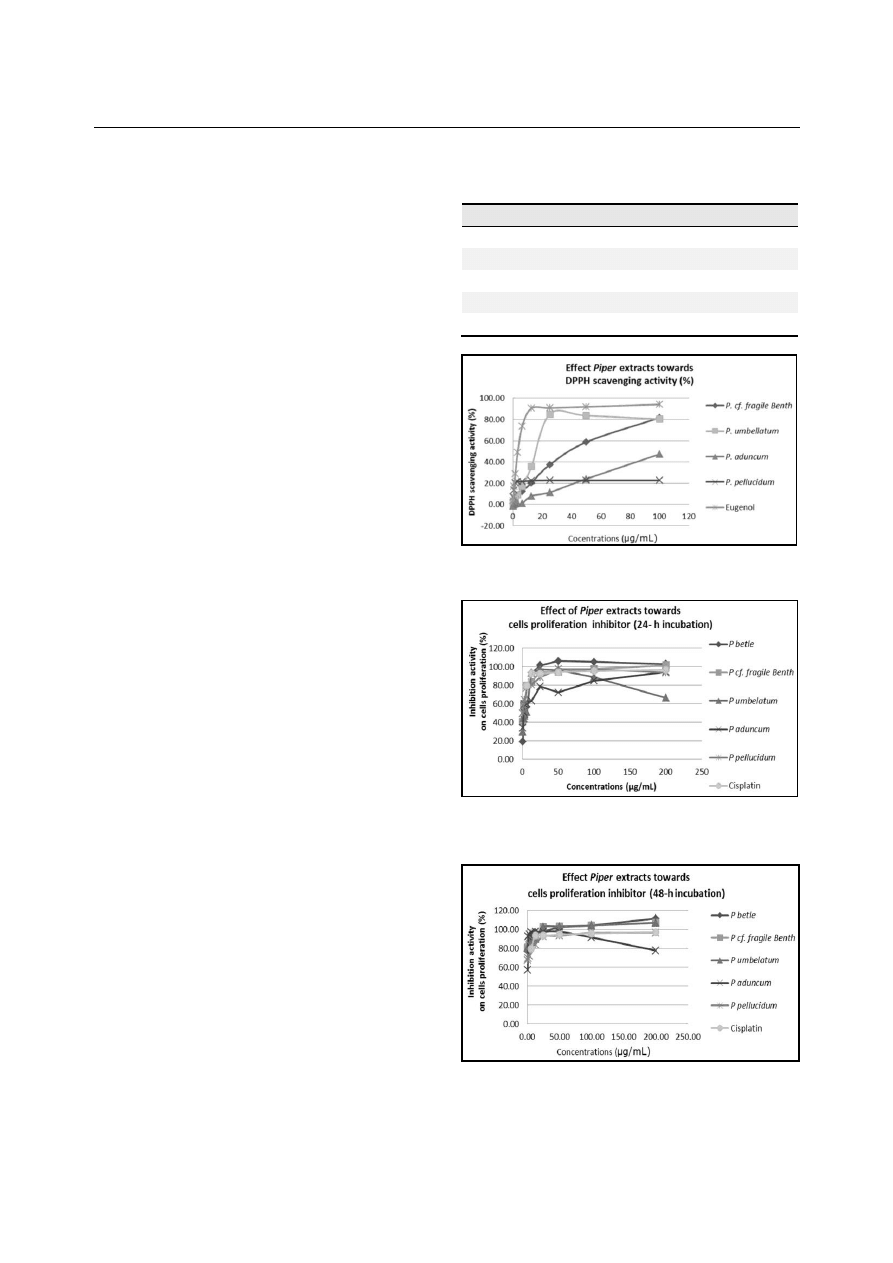
Antioxidant activities of Piper extracts
The antioxidant activities of Piper extracts were
examined in the context of DPPH scavenging and SOD
activities. The DPPH free radical scavenging activity of
P.fragile, P.umbellatum, P.aduncum, and P.pellucidum
extracts and eugenol as a control was measured as a
representative of antioxidant activity. The IC
50
is the
concentration of antioxidant needed to scavenge 50%
of the DPPH free radicals. P.umbellatum, and
P.pellucidum extracts exhibit high levels of DPPH
scavenging activity, and P.fragile and P.aduncum have
low DPPH scavenging activity (Table 1, Fig.1).
The SOD activity of Piper extracts can be seen in
Table 2. SOD activity was found to be concentration-
dependent. The highest SOD activity at 500 µg/ml and
125 µg/ml was shown by the P.betle extract, while
P.umbellatum and P.pellucidum showed the highest
activities at 31.25 µg/ml; the lowest SOD activity at all
concentrations was exhibited by eugenol.
Anticancer activity of Piper extracts
The viability of HeLa cells treated with extracts of
P.betle, P.fragile, P.umbellatum, P.aduncum, and
P.pellucidum decreased in a concentration-dependent
manner; higher extract concentrations exhibited
stronger anticancer activity (Figs.2&3).
Anticancer activity was also found to be concentration-
dependent; higher concentrations more strongly inhibit
proliferation (Figs.2&3). The IC
50
of Piper ethanol
extracts in HeLa cells after 24 h incubation demonstra-
ted that P.fragile extract was the most active and that
Table 1. IC
50
DPPH scavenging activity of Piper extracts.
The DPPH scavenging activity test was measured triplicate
for each extract. [Linear equations, coefficient of regression
(R
2
), and IC
50
were calculated.]
Samples
Linear equation
R
2
IC
50
(µg/ml)
P.fragile
y=0.843x+5.751
0.945
52.49
P.umbellatum
y=3.311x+0.849
0.991
15.36
P.aduncum
y=0.482x+0.429
0.995
102.84
P.pellucidum
y=4.675x+7.94
0.785
9
Eugenol
y=11.443x+6.5
0.965
3.8
Figure 1. DPPH scavenging activity of Piper extracts diluted in
methanol to 100, 50, 25, 12.5, 6.25, 3.125, 1.563, 0.781, 0.391, and
0.195 µg/ml.
Figure 2. Anticancer activity of Piper ethanol extracts diluted in
DMSO to 200, 100, 50, 25, 12.5, 6.25, 3.125, 1.563, and 0.781 µg/ml
and incubated for 24 h. Inhibition of cell proliferation was interpreted
as anticancer activity.
Figure 3. Anticancer activity of Piper ethanol extracts diluted in
DMSO to 200, 100, 50, 25, 12.5, 6.25, 3.125, 1.563, and 0.781 µg/ml
and incubated for 48 h. Inhibition of cell proliferation was interpreted
as anticancer activity.

Widowati et al: Antioxidant, anticancer and apoptotic activity of Piper extracts
228
DOI 10.5455/jeim.160513.or.074
cisplatin was more active than all the extracts. At 48 h
of incubation, the P.fragile extract was more active
than cisplatin and all other extracts (Table 3).
Apoptosis- inducing effect of Piper extracts
P.betle, P.fragile, P.umbellatum., P.aduncum, and
P.pellucidum ethanol extracts induced apoptosis in
Table 2. Mean and Tukey’s HSD post hoc test of SOD
activity of Piper extracts. SOD activity was measured in
triplicate for each extract. [Linear equation, coefficient of
regression (R
2
) of SOD standard and SOD activity of Piper
extracts and eugenol were calculated.]
Samples
Concentrations (μg/ml)
500
125
31.25
P. betle
5.21 ± 0.49
c
4.47 ± 0.17
d
0.64 ± 0.14
b
P.fragile
2.19 ± 0.41
b
0.63 ± 0.15
a
0.09 ± 0.06
a
P.umbelatum
2.72 ± 0.32
b
2.44 ± 0.04
c
1.89 ± 0.11
c
P.aduncum
2.58 ± 0.25
b
1.82 ± 0.19
b
0.95 ± 0.09
b
P.pellucidum
2.56 ± 0.12
b
2.21 ± 0.1
c
1.68 ± 0.12
c
Eugenol
0.87 ± 0.05
a
0.67 ± 0.15
a
0.2 ± 0.02
a
Data are presented as mean ± standard deviation. Different letters in
the same column (among extracts) are significant at P < 0.05
(Tukey’s HSD post hoc test).
Table 3. The IC
50
of Piper ethanol extracts in HeLa cells
after 24 and 48 h incubation. [Each extract was measured in
triplicate and growth inhibition was analyzed using probit.]
Samples
IC
50
(µg/ml)
24 hours
48 hours
P.betle extract
7.13
0.136
P.fragile extract
2.93
0.005
P.umbellatumextract
6.71
0.439
P.aduncum extract
3.91
0.53
P.pellucidum extract
2.85
0.12
Cisplatin
0.07
0.01
HeLa cells after 24 and 48 h incubation (Table 4);
increased incubation of contact between the cancer
cells and anticancer agent increased apoptotic
induction. The strongest apoptosis inducers after 24 h
incubation were P.betle at 12.5 µg/ml (80.9%),
P.umbellatum at 25 µg/ml (85.41%), P.aduncum at
100 µg/ml (80.72%), and cisplatin at 100 µg/ml
(75.21%). The strongest apoptosis inducers at 48 h
incubation were P.betle at 12.5 and 25 µg/ml (95.35
and 95.87%, respectively), P.fragile at 50 µg/ml
(87.17%), P.aduncum at 100 µg/ml (81.52%), and
cisplatin at 100 µg/ml (95.53%).
DISCUSSION
The data in Table 1 shows that P.pellucidum extract
and eugenol, a component of P.betle [11, 12], exhibited
the most active DPPH scavenging activity, consistent
with previous indications that the essential oil of
P.betle is a strong antioxidant [13] and that the ethanol
extract of P.betle exhibits good DPPH scavenging
activity [14]. Essential oil, methanol and aqueous
extracts of P.betle exhibit antioxidant activities,
including DPPH scavenging, iron chelation and
reducing power [11]. This result is consistent with
previous findings that P.betle extract exhibits
antioxidant activity [15]. In the present study, P.betle
extract exhibited the strongest SOD activity compared
to other samples (Table 2). Eugenol was the poorest
antioxidant among the tested Piper extracts. These data
were not consistent with the DPPH scavenging activity
(Table 1) and also with previous reports that eugenol
can improve the antioxidant status of the rat intestine
after short- and long-term (15 days and 90 days,
respectively) oral administration of 1000 mg/kg, a
dosage reported to be highly hepatoprotective; thus,
eugenol seem to be nontoxic and protective [16]. In
another study, however, eugenol exhibited potential
benefits in the management of isoproterenol-induced
cardiac hypertrophy in rats [17].
Table 4. Effect of various Piper extracts in HeLa cells by SubG1 (%) after 24 and 48 h incubation. [The apoptosis assay was
performed with a flow cytometer. The apoptotic cells were determined on the basis of the SubG1 area from cell cycle analysis
and are presented as a percentage of all cells.]
µg/ml
Cisplatin
P.betle
P.fragile
P.umbelatum
P.aduncum
P.pellucidum
24 h
48 h
24 h
48 h
24 h
48 h
24 h
48 h
24 h
48 h
24 h
48 h
1.56
19.45
34.62
11.03
36.81
6.77
10.79
10.96
10.8
5.91
9.99
7.32
10.8
3.12
20.65
29.84
16.72
16.06
7.9
12.92
7.43
8.94
8.57
8.86
7.26
8.94
6.25
28.54
39.19
33.77
32.75
8.79
10.95
13.14
10.59
11.3
5.89
8.62
10.59
12.5
38.57
64.58
80.9
95.87
10.81
10.99
16.1
15.07
12.82
7.65
8.74
15.07
25
51.33
79.2
16.82
95.35
33.43
35.59
85.41
70.9
12.09
7.54
14.46
70.9
50
59.89
86.44
13.89
8.37
63.55
87.17
50.17
55.11
19.66
11.94
40.85
55.11
100
75.21
95.53
17.41
15.5
23.64
30.38
47.04
36.39
80.72
81.52
27.51
36.39

Journal of Experimental and Integrative Medicine 2013; 3(3):225-230
http://www.jeim.org
229
All ethanol extracts of Piper exhibited potential
anticancer activities (Figs.2&3, Table 3), consistent
with a previous study in which the aqueous extract of
P.betle exhibited anticancer activity in cancerous oral
epidermal lesions [18]. This result is also consistent
with a previous study in which P.betle extract inhibited
T47D cell (human ductal breast epithelial tumor cell
line) proliferation [15]. Ethanol extract of P.betle
leaves exhibit cytotoxic activity against larvae of
Artemia salina Leach. Therefore, based on the brine
shrimp lethality test (BLT), the ethanol extract of
P.betle exhibits anticancer activity [19]. The aqueous
extract of P.betle leaves exhibits cytotoxicity in Hep-2
cells
in
microculture
tetrazolium
assays
and
sulforhodamine B (SRB) assays [20]. The anticancer
activity of Piper extracts varies by its content; for
example, eugenol exhibits dose-dependent cytotoxicity
in
U2OS
(human
osteosarcoma)
cells
[21].
Allylpyrocatechol exhibited anti-inflammatory effects
in an animal model of inflammation, and mechanistic
studies suggest that allylpyrocatechol targets the
inflammatory response of macrophages via inhibition
of
inducible
nitric
oxide
synthase
(iNOS),
cyclooxygenase (COX)-2, and interleukin (IL)-12 p40
through downregulation of the nuclear factor (NF)-κB
pathway [22, 23]. Hydroxychavicol is a component of
P.betle leaves that possesses antioxidant and anti-
inflammatory activities [23, 24], inhibits ATCC 25175
(carcinogenic bacteria), and has anticancer properties
[24].
Piper extracts were able to induce apoptosis in HeLa
cells after 24 and 48 h incubation (Table 4). The most
active apoptosis inducer was P.betle extract. These data
are consistent with those from a previous study in
which an alcoholic extract of betel leaves induced
apoptosis of chronic myelogenous leukemia (CML)
cells expressing wild-type and mutated Bcr-Abl, with
imatinib resistant phenotype (STI571 or Gleevec) [25],
induced apoptosis in imatinib-resistant cells [25, 26],
and exhibited activity against T315I tumor xenografts
[25, 26]. The plant extract NPB001-05 from P.betle
exhibited
anti-tumor
activity
in
T315I
tumor
xenografts, where imatinib failed to exhibit antitumor
activity [27]. Hydroxychavicol induces apoptosis in KB
(human oral carcinoma) cells through induction of
reactive oxygen species (ROS) [28].
The inhibitory effects of Piper extracts as anticancer
agents and apoptosis inducers are associated with
antioxidant glutathione (GSH) levels [21]. The
antioxidant property is correlated with anticancer
properties, since free radicals are involved in all
diseases that involve carcinogenesis [20]. DNA is
highly susceptible to free radical attacks. Free radicals
can react with cell membrane fatty acids and form lipid
peroxides, accumulation of which leads to production
of
carcinogenic
agents
such
as
MDA
[29].
Carcinogenesis may be mediated by ROS and reactive
nitrogen
species
(RNS)
directly
by
chronic
inflammation
(oxidation,
nitration
of
nuclear
DNA/RNA or lipids) or indirectly by the products of
ROS/RNS, proteins, lipids, and carbohydrates that are
capable of forming DNA adducts [29, 30]. Chronic
inflammation leads to excessive production of free
radicals and reduces antioxidant levels [29]. Tumor
cells have higher levels of intracellular ROS than
normal cells and ROS is associated with cell
proliferation
[25, 31].
Hydroxychavicol
induces
apoptosis in CML cells expressing wild-type and
mutated Bcr-Abl, including the untreatable T315I
mutation, and acts through the JNK pathway in a ROS-
dependent manner, which in turn activates endothelial
nitric oxide synthase (eNOS) to kill CML cells [25].
In conclusion, P.betle extract has the highest
antioxidant activities as demonstrated by DPPH
scavenging and SOD activities, and is the strongest
inducer of apoptosis at 24 and 48 h incubation in HeLa
cells. P.betle extract has low anticancer activity in
HeLa cells; however, the strongest anticancer activity
was observed with P.fragile and P.pellucidum extracts.
Piper extracts have great therapeutic potential due to
their antioxidant, anticancer, and apoptosis inducing
activities.
ACKNOWLEDGMENTS
We gratefully acknowledge the financial support of
Directorate General for Higher Education, National Ministry
of Republic Indonesia for research grant of Hibah Bersaing
2010-2011 (No DIPA 0561/023-04.2.01/12/2011).

Widowati et al: Antioxidant, anticancer and apoptotic activity of Piper extracts
230
DOI 10.5455/jeim.160513.or.074
REFERENCES
1. Souri E, Amin G, Farsam H, Jalalizadeh H, Barezi S. Screening
of thirteen medicinal plant extracts for antioxidant activity.
Iranian J Pharm Res 2008; 7:149-54.
2. Hussain SP, Hofseth LJ, Harris CC. Radical causes of cancer.
Nat Rev Cancer 2003; 3:276-86.
3. Cerutti PA, Trump BF. Inflammation and oxidative stress in
carcinogenesis. Cancer Cells 1991; 3:1-7.
4. Whiteman M, Hooper DC, Scott GS, Koprowski H, Halliwel B.
Inhibition of hypochlorus acid-induced cellular toxicity by
nitrite. Proc Natl Acad Sci USA 2002; 99:12061-6.
5. Sharafati-Chaleshtori R, Rafieian-Kopaei M, Mortezaei S,
Sharafati-Chaleshtori A, Amini E. Antioxidant and antibacterial
activity of the extracts of Echinophora platyloba D.C. Afr J
Pharm Pharmacol 2012; 6:2692-5.
6. Tan ML, Sulaiman SF, Najimuddin N, Samian MR, Muhammad
TST. Methanolic extract of Pereskia bloe (Kunth) DC.
(Cactaceae) induces apoptosis in breast carcinoma, T47D cell
line. J Ethnopharmacol 2005; 96:287-94.
7. Mooney LM, Al-Sakkaf KA, Brown BL, Dobson PRM.
Apoptotic mechanisms in T47D and MCF-7 human breast cancer
cells. Br J Cancer 2002; 87:909-17.
8. Malich G, Markovic B, Winder C. The sensitivity and specificity
of the MTS tetrazolium assay for detecting the in vitro
cytotoxicity of 20 chemicals using human cell lines. Toxicology
1997; 124:179-92.
9. Chang HY, Ho YL, Sheu MJ, Lin YH, Tseng MC, Wu SH,
Huang GJ, Chang YS. Antioxidant and free radical scavenging
activities of Phellinus merrillii extracts. Botanical Studies 2007;
38:407-17.
10. Mauier CM, Chan PH. Role of superoxide dismutases in
oxidative
damage
and
neurodegenerative
disorders.
Neuroscientist 2002; 8:323-34.
11. Row LC M, Ho JC. The Antimicrobial activity, mosquito
larvicidal activity, antioxidant, property and tyrosinase inhibition
of Piper betle. J Chin Chem Soc 2009; 56:653-8.
12. Vasuki K, Senthamarai R, Kirubha TSV, Balasubramanian P,
Selvadurai S. Pharmacognostical studies on leaf of Piper betle.
Der Pharmacia Lettre 2011; 5:232-5.
13. Prakash B, Shukla R, Singh P, Kumar A, Miswhra PK, Dubey
NK. Efficacy of chemically characterized Piper betle L. essential
oil against fungal and aflatoxin contamination of some edible
commodities and its antioxidant activity. Int J Food Microbiol
2010; 142:114-9.
14. Pin KY, Chuah AL, Rashih AA, mazura MP, Fadzureena J,
Vimala S, Rasadah M. Antioxidant and anti-inflammatory
activities of extracts of betel leaves (Piper betle) from solvents
with different polarities. J Tropic Forest Sci 2010; 22:448-55.
15. Widowati W, Tjandrawati M, Risdian C, Ratnawati H, Tjahjani
S, Sandra F. The comparison of antioxidative and proliferation
inhibitor properties of Piper betle L., Catharanthus roseus [L]
G.Don, Dendrophtoe petandra L., Curcuma mangga Val.
extracts on T47D cancer cell line. Int Res J Biochem Bioinform
2011; 1:22-8.
16. Vidhya N, Devaraj SN. Antioxidant effect of eugenol in rat
intestine. Indian J Exp Biol 1999; 37:1192-5.
17. Choudary R, Mishra KP, Subramanyam C. Prevention of
isoproterenol-induced cardiac hypertrophy by eugenol, an
antioxidant. Indian J Clin Biochem 2006; 21:107-13.
18. Fathilah AR, Sujata R, Norhanom AW, Adenan MI.
Antiproliferative activity of aqueous extract of Piper betle L. and
Psidium guajava L. on KB and HeLa cell lines. J Med Plant Res
2010; 4:987-90.
19. Srisadono A, Sunoko HR. The early screening of ethanol extract
of sirih leaf (Piper betle Linn) as anticancer using brine shrimp
lethality test (BLT) method. Scientific Article, Faculty of
Medicine, Diponegoro University, Semarang, 2008.
20. Chaurasia S, Kulkarni GT, Shetty LN. Phytochemical studies
and in vitro cytotoxicity screening of Piper betle leaf (PBL)
extract. Middle-East J Sci Res 2010; 6:532-6.
21. Ho YC, Huang FM, Chang YC. Mechanisms of cytotoxicity of
eugenol in human osteoblastic cells in vitro. Int Endod J 2006;
39:389-93.
22. Sarkar D, Saha P, Gamre S, Bhattacharjee S, Hariharan C,
Ganguly S, Sen R, Mandal G, Chattopadhyay S, Majumdar S,
Chatterjee M. Anti-inflammatory effect of allylpyrocatechol in
LPS-induced macrophages is mediated by suppression of iNOS
and COX-2 via the NF-kappaB pathway. Int Immunopharmacol
2008; 8:1264-71.
23. Rai M, Thilackand KR, Palatty PL, Rao P, Rao S, Bhat HP,
Baliga MS. Piper betel linn (betel vine), the maligned Southeast
Asian medicinal plant possesses cancer preventive effects: time
to reconsider the wronged opinion. Asian Pacific J Cancer Prev
2011; 12:2149-56.
24. Sharma S, Khan IA, Ali I. Ali F, Kumar M, Kumar A, Johri RK,
Abdullah ST, Bani S, Pandey A, Suri KA, Gupta BD, Satti NK,
Dutt P, Qazi GN. Evaluation of the antimicrobial, antioxidant,
and anti-inflammatory activities of hydroxychavicol for its
potential use as an oral care agent. Antimicrob Agents
Chemother 2009; 53:216-22.
25. Chakraborty JB, Mahato SK, Joshi K, Shinde V, Rakshit S,
Biswas N, Mukherjee IC, Mandal L, Ganguly D, Chowdhury
AA, Chaudhuri J, Paul K, Pal BC, Vinayagam J, Pal C, Manna
A, Jaisankar P, Chaudhuri U, Konar A, Roy S, Bandyopadhyay
S. Hydroxychavicol, a Piper betle leaf component, induces
apoptosis of CML cells through mitochondrial reactive oxygen
species-dependent JNK and endothelial nitric oxide synthase
activation and overrides imatinib resistance. Cancer Sci 2012;
103:88-99.
26. Wagh V, Chile S, Monahar S, Pal BC, Bandyopadhyay S,
Sharma S, Joshi K. NPB001-05 inhibits Bcr-Abl kinase leading
to apoptosis of imatinib-resistant cells. Front Biosci 2011;
3:1273-88.
27. Wagh V, Mishra P, Thakkar A, Shinde V, Sharma S, Padigaru
M, Joshi K. Antitumor activity of NPB001-05, an orally active
inhibitor of Bcr-Abl tyrosine kinase. Front Biosci 2011; 3:1349-
64.
28. Chang MC, Uang BJ, Wu HL, Lee JJ, Hahn LJ, Jeng JH.
Inducing the cell cycle arrest and apoptosis of oral KB carcinoma
cells by hydroxychavicol: roles of glutathione and reactive
oxygen species. Br J Pharmacol. 2002; 135:619-30.
29. Khansari N, Shakiba Y, Mahmoudi M. Chronic inflammation
and oxidative stress as a major cause of age-related diseases and
cancer. Recent Pat Inflamm Allergy Drug Discov 2009; 3:73-80.
30. Jabs T. Reactive oxygen intermediates as mediators of
programmed cell death in plants and animals. Biochem
Pharmacol 1999; 57:231-45.
31. Trachootham D, Alexandre J, Huang P. Targeting cancer cells by
ROS mediated mechanisms: a radical therapeutic approach? Nat
Rev Drug Discov 2009; 8:579-91.
This is an open access article licensed under the terms of the Creative Commons Attribution Non-Commercial License which permits
unrestricted, non-commercial use, distribution and reproduction in any medium, provided that the work is properly cited.
Wyszukiwarka
Podobne podstrony:
Notch and Mean Stress Effect in Fatigue as Phenomena of Elasto Plastic Inherent Multiaxiality
Evaluation of antioxidant properities and anti fatigue effect of green tea polyphenols
Cytolytic Effects and Apoptosis Induction
Effect of Water Deficit Stress on Germination and Early Seedling Growth in Sugar
Inflammation associated enterotypes, host genotype, cage and inter individual effects drive gut micr
effect of high fiber vegetable fruit diet on the activity of liver damage and serum iron level in po
Guide to the properties and uses of detergents in biology and biochemistry
Basic setting for caustics effect in C4D
Gender and Racial Ethnic Differences in the Affirmative Action Attitudes of U S College(1)
Nutritional composition, antioxidant activity and phenolic compounds
American Polonia and the School Strike in Wrzesnia
improvment of chain saw and changes of symptoms in the operators
Combined Radiant and Conductive Vacuum Drying in a Vibrated Bed (Shek Atiqure Rahman, Arun Mujumdar)
Issue of Gun Control and Violence As Seen in the U S and
[41]Hormesis and synergy pathways and mechanisms of quercetin in cancer prevention and management
13 International meteorological and magnetic co operations in polar regions
22 289 298 Carbide Distribution Effect in Cold Work Steel
więcej podobnych podstron