
SCIENCE CHINA
Life Sciences
© The Author(s) 2012. This article is published with open access at Springerlink.com life.scichina.com www.springer.com/scp
*Corresponding author (email: houzao@gmail.com; liudp@pumc.edu.cn)
•
RESEARCH
PAPER
•
January 2013 Vol.56 No.1: 19–25
doi: 10.1007/s11427-012-4407-7
SIRT1 suppresses PMA and ionomycin-induced ICAM-1
expression in endothelial cells
JIA YuYan, GAO Peng, CHEN HouZao
*
, WAN YanZhen, ZHANG Ran, ZHANG ZhuQin,
YANG RuiFeng, WANG Xu, XU Jing & LIU DePei
*
State Key Laboratory of Medical Molecular Biology, Department of Biochemistry and Molecular Biology, Institute of Basic Medical Sciences,
Chinese Academy of Medical Sciences & Peking Union Medical College, Beijing, 100005, People’s Republic of China
Received August 30, 2012; accepted November 1, 2012; published online December 7, 2012
Intercellular adhesion molecule-1 (ICAM-1) plays an important role in the recruitment of leukocytes to the endothelium, which
causes inflammation and initiation of atherosclerosis. We have previously shown that endothelium-specific over-expression of
class III deacetylase SIRT1 decreases atherosclerosis. We therefore addressed the hypothesis that SIRT1 suppresses ICAM-1
expression in the endothelial cells. Here, we found that expression of SIRT1 and ICAM-1 was significantly induced by PMA
and ionomycin (PMA/Io) in human umbilical vein endothelial cells (HUVECs). Adenovirus-mediated over-expression of
SIRT1 significantly inhibited PMA/Io-induced ICAM-1 expression in HUVECs. Knockdown of SIRT1 by RNA interference
(RNAi) resulted in increased expression of ICAM-1 in HUVECs. Luciferase report assay showed that over-expression of
SIRT1 suppressed ICAM-1 promoter activity both in basic and in PMA/Io-induced conditions. We further found that SIRT1
was involved in transcription complex binding on the ICAM-1 promoter by chromatin immunoprecipitation (ChIP) assays.
Furthermore, SIRT1 RNAi increased NF-κB p65 binding ability to the ICAM-1 promoter by ChIP assays. Overall, these data
suggests that SIRT1 inhibits ICAM-1 expression in endothelial cells, which may contribute to its anti-atherosclerosis effect.
SIRT1, ICAM-1, PMA and ionomycin
Citation: Jia Y Y, Gao P, Chen H Z, et al. SIRT1 suppresses PMA and ionomycin-induced ICAM-1 expression in endothelial cells . Sci China Life Sci, 2013,
56: 19–25, doi: 10.1007/s11427-012-4407-7
Leukocyte recruitment and expression of pro-inflammatory
cytokines characterize early atherogenesis and malfunction
of inflammatory mediators mutes atheroma formation in
mice [1]. As the first barrier of blood vessels, the endothe-
lium plays an extremely important role in maintaining ves-
sel homeostasis [2]. Various leukocyte adhesion molecules
increase accompanied by inflammatory activation in endo-
thelial cells, which contribute to leukocyte recruitment in
atherosclerosis-susceptible mice [3]. Thus, inhibition of
leukocyte recruitment by decreasing the expression of adhe-
sion molecules is important for preventing initiation of ath-
erosclerosis.
The expression of adhesion molecules such as intercellu-
lar adhesion molecule-1 (ICAM-1) is increased in athero-
sclerotic lesions [4]. Gene deletion of ICAM-1 resulted in
significant reduction in monocyte recruitment to athero-
sclerotic lesions in ApoE-deficient mice [3]. ICAM-1 is
upregulated in response to inflammatory stimuli such as
interleukin-1 beta (IL-1), tumor necrosis factor-alpha
(TNF-), LPS and PMA [5]. The divalent cation calcium
ionophore ionomycin (Io) interacts synergistically with
PMA but not with cytokines or LPS in upregulating
ICAM-1 [5]. The combination of PMA and Io (PMA/Io) is
routinely used as a T-cell receptor (TCR)-independent
model to study T-cell activation and proliferation. PMA/Io
mimics the phospholipase C-driven activation of protein

20
Jia Y Y, et al. Sci China Life Sci January (2013) Vol.56 No.1
kinase C and cytosolic Ca
2+
increase and induces activation
of NF-B, NFAT and AP-1. NF-B is of special interest in
endothelial cells since it drives the expression of important
adhesion molecules, such as ICAM-1, which recruit blood
monocytes to atherosclerotic lesions [6]. NFAT cooperated
with NF-B to regulate thrombin-induced ICAM-1 gene
expression by binding to the intronic NF-κB site in the
ICAM-1 gene [7].
SIRT1, a well-known mammalian sirtuin, is involved in
aging and age-related diseases [8]. SIRT1 has been de-
scribed as a “guardian at the gates of adipose tissue inflam-
mation”, playing an important role in anti-inflammatory
processes [9]. SIRT1 inhibits inflammatory pathways in
macrophages and modulates insulin sensitivity [10]. SIRT1
acts to protect the heart from hypertrophy, metabolic
dysregulation and inflammation [11]. Moreover, SIRT1
inhibits NF-B and AP-1 transcriptional activity and the
expression of downstream inflammatory genes [12–15]. We
have previously shown that transgenic mice that over-
express SIRT1 in the vascular endothelium have better en-
dothelium-dependent vasodilation and fewer atherosclerotic
lesions when fed a high-fat diet [16]. Recently, expression
of ICAM-1 was increased in atherosclerotic plaques of Ap-
oE
/
SIRT1
+/
compared with ApoE
/
SIRT1
+/+
mice [17].
However, the mechanism by which SIRT1 regulates
ICAM-1 expression remains unknown.
In the present study, we found that over-expression of
SIRT1 significantly decreased PMA/Io-induced ICAM-1
expression in human umbilical vein endothelial cells (HU-
VECs). Knockdown of SIRT1 by RNA interference (RNAi)
remarkably up-regulated ICAM-1 expression in HUVECs.
Moreover, we found that SIRT1 significantly inhibited both
basic and PMA/Io-induced ICAM-1 promoter activity.
SIRT1 was involved in transcription complex binding on
the ICAM-1 promoter by chromatin immunoprecipitation
(ChIP) assays. Furthermore, SIRT1 RNAi increased NF-B
p65 binding ability to the ICAM-1 promoter by ChIP assays.
These data suggests that SIRT1 inhibits ICAM-1 expression
in endothelial cells, which may contribute to its an-
ti-atherosclerosis effect.
1 Materials and methods
1.1 Drugs
PMA (Cat #P1585), Ionomycin (Cat #I0634), Cyclosporin
A (Cat #C1832), resveratrol (Cat #R5010) and Sirtinol (Cat
#S7942) were purchased from Sigma-Aldrich.
1.2 Plasmids, HUVECs culture and adenovirus gener-
ation
SIRT1 expression vector is a gift from Dr. Ishikawa [18].
Endothelial cells were freshly isolated from human umbili-
cal cord veins as described previously [16] and cultured in
M200 medium (Cascade Biologics Inc., Portland, Oregon,
USA). The growth medium was changed every two days
until >80% cells reached confluence. Cells between the 3rd
and the 6th passages were grown in mono-layers in a hu-
midified atmosphere of 5% CO
2
at 37°C, and used for ex-
periments at >80% confluence. Replication-defective ad-
enoviral vectors expressing SIRT1 (Ad-SIRT1) or a green
fluorescent protein control (Ad-GFP) were prepared using
the AdEasy vector kit (Quantum Biotechnologies) as de-
scribed in the supplier’s protocol. The adenovirus-
mediated knockdown of SIRT1 (Ad-SIRT1 RNAi) and a
control vector (Ad-U6) were generated using the same
system. HUVECs were infected with the above adenovirus
for 24 h and were cultured in fresh M200 medium for fur-
ther treatment.
1.3 Reverse transcription and real-time PCR
Total RNA was extracted from cells using Trizol (Invitro-
gen) according to the manufacturer’s instructions. Two mi-
crograms of total RNA were used to synthesize first-strand
cDNA with M-MuLV reverse transcriptase (New England
BioLabs) using random primers. Real-time PCR was per-
formed using the BioRad iCycler iQ5 Real-Time PCR De-
tection System with the Quantitect SYBR Green One-Step
RT-PCR Kit (QIAGEN). Fluorescence curves were ana-
lyzed with iCycler iQ5 Optical System Software (version
2.0). Primer sequences for specific genes are presented in
Table S1.
1.4 Western blotting analysis
HUVEC protein was extracted with Radioimmunoprecipita-
tion Assay (RIPA) buffer (25 mmol L
1
Tris-HCl pH 7.6,
150 mmol L
1
NaCl, 1% NP-40, 1% sodium deoxycholate
and 0.1% SDS). After lysis on an ice for 0.5 h, samples
were sonicated and centrifuged at 4°C at 12000 r min
1
for
0.5 h. The supernatants were transferred into fresh tubes and
protein concentrations were determined by the bicincho-
ninic acid (BCA) method. Equal amounts of protein (20
g/lane) were separated by SDS-PAGE and transferred onto
polyvinylidene difluoride membranes (Millipore). After
being blocked, the filters were incubated with the following
primary antibodies: anti-SIRT1 (Santa Cruz Biotechnology,
Cat #sc-15404), anti-ICAM-1 (Cell Signal Technology, Cat
#4915). After being washed and incubated with the appro-
priate horseradish peroxidase-conjugated secondary anti-
body (Santa Cruz Biotechnology), the immune complexes
were visualized with a chemiluminescence reagent. Western
blotting was quantified densitometrically with Quantity One
software (Bio-Rad), and the intensity values were normal-
ized to anti-GAPDH.
Jia Y Y, et al. Sci China Life Sci January (2013) Vol.56 No.1
21
1.5 Transfection and luciferase assays
HEK293 cells were grown as recommended by ATCC and
were transfected with Lipofectamine 2000 (Invitrogen) ac-
cording to the manufacturer’s instructions. Luciferase as-
says were performed using a dual luciferase reporter assay
system (Promega). Luciferase activity was normalized by
transfection efficiency using pRL-CMV reporter (Promega)
as an internal control. The results are expressed as percent-
ages of relative luciferase activity of the control group.
1.6 ChIP
Chromatin immunoprecipitation (ChIP) assays were per-
formed in HUVECs as described [19]. A rabbit polyclonal
antibody for SIRT1 (Santa Cruz Biotechnology, Cat
#sc-15404) was used in ChIP assay. Control immunoprecip-
itations were carried out with nonimmune normal rabbit IgG
(Santa Cruz Biotechnology, Cat #sc-2027).
1.7 Statistical analysis
Data are expressed as mean±SD. Statistical analyses were
performed using the two-tailed unpaired Student’s t-test to
determine statistical significances between the groups. A
P-value less than 0.05 was considered significant.
2 Results
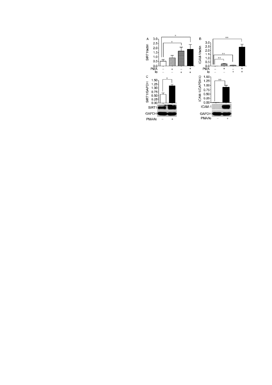
2.1 SIRT1 and ICAM-1 expression is induced by
PMA/Io in HUVECs
Considering that the ICAM-1 promoter has binding sites for
NF-B, NFAT and AP-1, we treated HUVECs with PMA/Io,
which is known to induce activation of transcription factors
NF-B, NFAT and AP-1. Previous studies have shown that
ionomycin (Io) interacted synergistically with PMA in up-
regulating ICAM-1 expression [5]. Here we repeated the
results and found that ICAM-1 mRNA expression level was
induced higher by PMA than by Io (Figure 1B). In contrast,
SIRT1 mRNA level was significantly induced about to
3-fold by Io, but was not significantly changed by PMA
(Figure1A). The results showed that an Io-activated caci-
um-calcineurin-NFAT signal pathway played an important
role in inducing SIRT1 expression. Io also interacted syner-
gistically with PMA in inducing SIRT1 mRNA expression
level about 3.6-fold (Figure 1A). Moreover, SIRT1 and
ICAM-1 protein expression was also induced by PMA/Io in
HUVECs (Figure 1C and D).
2.2 Over-expression of SIRT1 suppresses ICAM-1 ex-
pression in PMA/Io-treated HUVECs
To study the effect of increased expression of SIRT1 on
Figure 1 SIRT1 and ICAM-1 expression is significantly induced in
HUVECs treated with PMA/Io. A and B, HUVECs were treated with PMA
(10 ng mL
1
), Io (0.25 mol L
1
) or PMA/Io (10 ng mL
1
PMA plus 0.25
mol L
1
Io), vehicle DMSO for 3 h. Total RNA was isolated and mRNA
expression level for SIRT1 (A) and ICAM-1 (B) was analyzed by real-time
PCR analysis. Data shown represent the mean±SD of triplicate samples of
one representative of total three independent experiments. Relative mRNA
unit is calculated by using actin as a reference gene. *, P<0.05; **, P<0.01.
C and D, HUVECs were treated with PMA/Io (10 ng mL
1
PMA plus 0.25
mol L
1
Io) for 3 h. Protein expression was analyzed by Western blotting.
Bar graphs show densitometric analysis of immunoblots of SIRT1 (C) and
ICAM-1(D) protein. Data are presented as mean±SD of SIRT1/GAPDH
and ICAM-1/ GAPDH expression ratio (n=3). Immunoblots of SIRT1,
ICAM-1 and GAPDH are representative of three independent experiments.
*, P<0.05; **, P<0.01.
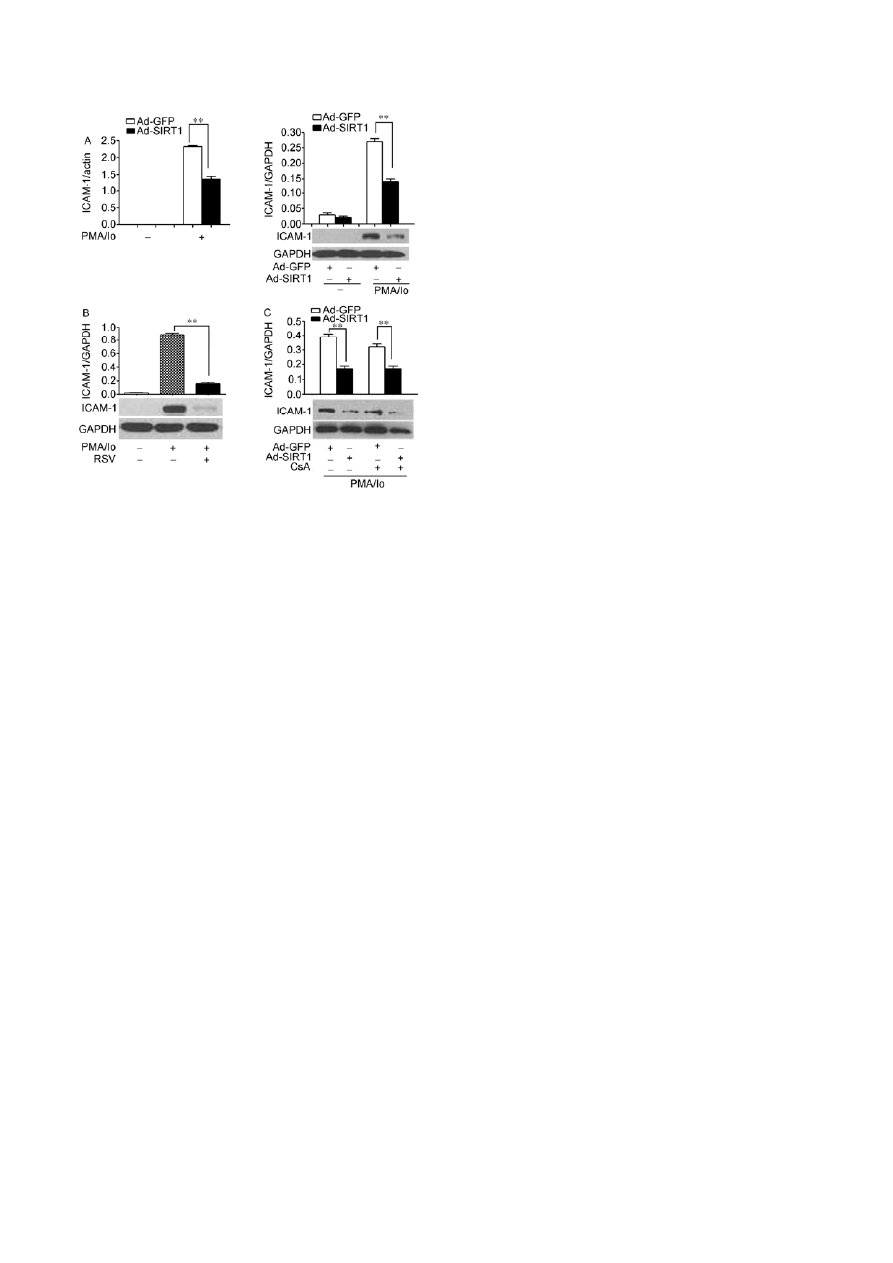
ICAM-1 expression, we over-expressed SIRT1 in PMA/Io-
treated HUVECs. We found that over-expression of SIRT1
suppressed PMA/Io-induced expression of both ICAM-1
mRNA and protein in HUVECs to 60% and 50%, respec-
tively (Figure 2A). Similarly, SIRT1 activator resveratrol
(RSV) suppressed ICAM-1 protein expression to 18% (Fig-
ure 2B).
To detect whether NFAT inhibition influences the sup-
pressive effect of SIRT1 on ICAM-1 expression, we
over-expressed SIRT1 in PMA/Io-treated HUVECs both in
the presence and absence of NFAT inhibitor Cyclosporin A
(CsA). We found that over-expression of SIRT1 suppressed
ICAM-1 protein expression in HUVECs induced by
PMA/Io either in presence or absence of CsA to 44% and
53%, respectively (Figure 2C). The results showed that
there was no significant effect of CsA on inhibition of
ICAM-1 expression by SIRT1.
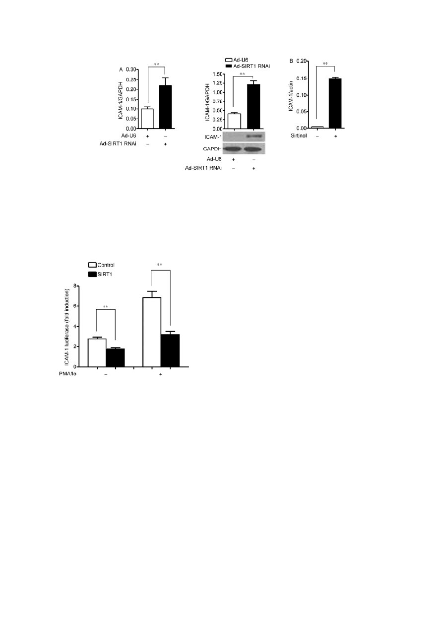
2.3 Knockdown of SIRT1 upregulates ICAM-1 expres-
sion in HUVECs
To examine the effect of SIRT1 knockdown on ICAM-1
expression, we infected HUVECs with adenoviral vectors
encoding SIRT1 RNAi (Ad-SIRT1 RNAi) or control (Ad-
U6). Knockdown of SIRT1 by RNA interference (RNAi)
22
Jia Y Y, et al. Sci China Life Sci January (2013) Vol.56 No.1
Figure 2 Over-expression of SIRT1 suppresses ICAM-1 expression in
HUVECs treated with PMA/Io. A, HUVECs infected with adenoviral
vectors encoding SIRT1 (Ad-SIRT1) or control Ad-GFP were treated with
PMA/Io (10 ng mL
1
PMA plus 0.25 mol L
1
Io) for 3 h. Total RNA was
isolated and mRNA level for ICAM-1 was analyzed by real-time PCR
analysis (left). Relative mRNA unit is calculated by using actin as a refer-
ence gene. Protein expression was analyzed by Western blotting (right).
Bar graphs show densitometric analysis of immunoblots of ICAM-1 pro-
tein. Data are presented as the mean±SD of ICAM-1/GAPDH expression
ratio (n=3). Immunoblots of ICAM-1 and GAPDH are representative of
three independent experiments. **, P<0.01. B, HUVECs were pretreated
with resveratrol (RSV) (30 mol L
1
) or vehicle DMSO for 1 h, then treat-
ed with PMA/Io (10 ng mL
1
PMA plus 0.25 mol L
1
Io) for another 3 h.
Protein expression was analyzed by Western blotting. Bar graphs show
densitometric analysis of immunoblots of ICAM-1 protein. Data are pre-
sented as the mean±SD of ICAM-1/GAPDH expression ratio (n=3). Im-
munoblots of ICAM-1 and GAPDH are representative of three independent
experiments. **, P<0.01. C, HUVECs infected with adenoviral vectors
encoding SIRT1 (Ad-SIRT1) or control Ad-GFP for 24 h, then pretreated
with CsA (1 mol L
1
) or vehicle DMSO for 1 h and treated with PMA/Io
(10 ng mL
1
PMA plus 0.25 mol L
1
Io) for another 3 h. Protein expres-
sion was analyzed by Western blotting. Bar graphs show densitometric
analysis of immunoblots of ICAM-1 protein. Data are presented as the
mean±SD of ICAM-1/GAPDH expression ratio (n=3). Immunoblots of
ICAM-1 and GAPDH are representative of three independent experiments.
**, P<0.01.
upregulated ICAM-1 expression at both the mRNA and pro-
tein levels in HUVECs to 2-fold and 3-fold, respectively
(Figure 3A). Similarly, SIRT1 inhibitor Sirtinol significantly
upregulated ICAM-1 expression in HUVECs to 37.5-fold
(Figure 3B).
2.4 SIRT1 inhibits ICAM-1 promoter activity induced
by PMA/Io
To further examine whether SIRT1 inhibits ICAM-1 pro-
moter activity, we performed assays using luciferase report-
er containing the ICAM-1 promoter (ICAM-1-Luc). Lucif-
erase report assay showed that over-expression of SIRT1
suppressed ICAM-1 promoter activity both in basic and
PMA/Io-induced conditions (Figure 4).
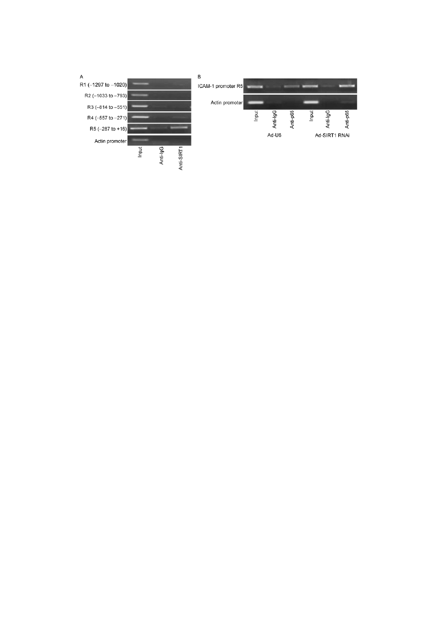
2.5 SIRT1 is involved in transcription complex binding
on the ICAM-1 promoter in HUVECs
The first 1.3 kb upstream of the ICAM-1 transcription start
site are required for ICAM-1basal expression and regulation
by inflammatory stimuli [20]. To explore whether SIRT1is
involved in transcription complex binding on the ICAM-1
promoter, we performed ChIP assays in HUVECs on a 1.3
kb region of the ICAM-1 promoter upstream of the tran-
scription start site, using semiquantitative PCR (qPCR) with
primers for regions (R) named 1 (1297 to 1020 bp); 2
(1033 to 793 bp); 3 (814 to 551 bp); 4 (577 to 271
bp); and 5 (287 to +16 bp). This showed that an increase in
the amount of SIRT1 bound to R5, compared with the oth-
ers regions (Figure 5A). These data indicated that SIRT1
was involved in transcription complex binding on the
ICAM-1 promoter in a region detected by the R5 primers
(287 to +16 bp).
NF-B is deacetylated and transcriptionally suppressed
by SIRT1 and binds at site 187 to 178 bp of the ICAM-1
promoter [12,21]. To examine whether NF-κB is involved
in the effect of SIRT1 on regulating expression of ICAM-1,
we detected NF-B p65 binding ability on ICAM-1 pro-
moter region 5 (287 to +16 bp) by ChIP analysis. As
shown in Figure 5, SIRT1 RNAi increases NF-B p65
binding ability on ICAM-1 promoter region 5 (Figure 5B).
3 Discussion
Here, we found that expression of SIRT1 and ICAM-1 was
significantly increased by PMA/Io in HUVECs. Over-
expression of SIRT1 significantly inhibited PMA/Io- in-
duced ICAM-1 expression in HUVECs. Moreover, SIRT1
suppressed ICAM-1 promoter activity both in basic and
PMA/Io stimulated conditions. We also found that SIRT1
was involved in transcription complex binding on the
ICAM-1 promoter by ChIP assays. Furthermore, SIRT1
RNAi increased NF-B p65 binding ability to the ICAM-1
promoter by ChIP assays.
SIRT1 has been pointed as a key regulator of vascular
endothelial homeostasis controlling angiogenesis, vascular
tone and endothelial dysfunction [22]. More evidence has
pointed out that SIRT1 is a key inducible factor in response
to inflammatory stimulation. For example, we have previ-
ously shown that oxLDL and H
2
O
2
treatment increased
SIRT1 protein levels in HUVECs [16,18], and TNF--
induced SIRT1 expression in vascular smooth muscle cells
Jia Y Y, et al. Sci China Life Sci January (2013) Vol.56 No.1
23
Figure 3 Knockdown of SIRT1 upregulates ICAM-1 expression in HUVECs. A, HUVECs were infected with adenoviral vectors SIRT1 RNAi (Ad-SIRT1
RNAi) or control Ad-U6. Total RNA was isolated and mRNA level for ICAM-1 was analyzed by real-time PCR analysis (left). Data shown represent the
mean±SD of triplicate samples of one representative of total three independent experiments. Relative mRNA unit is calculated by using GAPDH as a refer-
ence gene. Protein expression was analyzed by Western blotting (right). Bar graphs show densitometric analysis of immunoblots of ICAM-1 protein. Data
are presented as the mean±SD of ICAM-1/GAPDH expression ratio (n=3). Immunoblots of ICAM-1 and GAPDH are representative of three independent
experiments. **, P<0.01. B, HUVECs were treated with Sirtinol (25 mol L
1
) or vehicle DMSO for 1 h. Total RNA was isolated and mRNA level for
ICAM-1 was analyzed by real-time PCR analysis. Data shown represent the mean±SD of triplicate samples of one representative of total three independent
experiments. Relative mRNA unit is calculated by using actin as a reference gene. **, P<0.01.
Figure 4 Over-expression of SIRT1 suppresses ICAM-1 promoter activ-
ity. HEK293 cells were transiently transfected with 0.1 g ICAM-1 lucif-
erase reporter (ICAM-1-Luc), 30 ng pRL-CMV, and 0.3 g
SIRT1 expres-
sion vectors or control (pcDNA3.1) for 24 h, then treated with PMA/Io (10
ng mL
1
PMA plus 0.25 mol L
1
Io) or vehicle DMSO for 3 h. The relative
luciferase activities are presented as mean±SD of triplicate samples and are
representative of three independent experiments. **, P<0.01.
(VSMCs) [23]. Here, we found that the SIRT1 level was
induced by PMA/Io in HUVECs. It suggests that SIRT1 has
a compensatory upregulation in endothelial cells in response
to inflammatory factors. Moreover, expression of ICAM-1
was found to be increased in atherosclerotic plaques of
ApoE
/
SIRT1
+/
mice compared with ApoE
/
SIRT1
+/+
mice [17]. Furthermore, we demonstrated that SIRT1 sig-
nificantly inhibited PMA/Io-induced ICAM-1 expression in
HUVECs to 50%. Overall, these data suggests that SIRT1
inhibits ICAM-1 expression in the endothelial cells, which
may contribute to its anti-atherosclerosis effect.
The combination of PMA and Io activated these signals
that mimic the phospholipase C-driven activation of protein
kinase C and increase in cytosolic Ca
2+
and induce the tran-
scription factors NF-B, NFAT and AP-1. Our previous
work demonstrated SIRT1 suppresses the transcriptional
activity of AP-1 [15]. Moreover, SIRT1 inhibits NF-B
transcriptional activity [12]. NF-B plays essential roles in
transcriptional regulation of ICAM-1 in endothelial cells
and binds at site 187 to 178 bp of the ICAM-1 promoter
[21]. In addition, ERK, JNK, AP-1 and NF-κB are all in-
volved in interleukin-1-beta-induced ICAM-1 expression
enhancing leukocyte adhesion in human rheumatoid arthritis
synovial fibroblasts [24]. We found that SIRT1 bound the
ICAM-1 promoter at the R5 region (287 to +16 bp) and
NF-B was included in the region. Moreover, SIRT1 inhib-
its PMA/Io induced NF-B transcriptional activity and
ICAM-1 promoter activity. SIRT1 is involved in transcrip-
tion complex binding on the ICAM-1 promoter and SIRT1
RNAi increases NF-B p65 binding ability to the ICAM-1
promoter. The data indicated that suppressive effect of
SIRT1 on ICAM-1 expression was at least partly mediated
by NF-B. In addition, NFAT cooperates with NF-B by
binding to the intronic NF-B site on the ICAM-1 gene [7].
Our findings demonstrated that over-expression of SIRT1
inhibited ICAM-1 expression in PMA/Io-treated HUVECs
both in the presence and absence of NFAT inhibitor CsA,
although the extent of inhibition was not of statistical sig-
nificance (44% and 53%, respectively). This indicated that
NFAT inhibition may not influence the effect of SIRT1 on
inhibition of ICAM-1 expression.
24
Jia Y Y, et al. Sci China Life Sci January (2013) Vol.56 No.1
Figure 5 SIRT1 is involved in transcription complex binding on the ICAM-1 promoter. A, ChIP assays were performed with chromatin prepared from
HUVECs. Chromatin was immunoprecipitated with normal rabbit IgG or antibody against SIRT1, and precipitated genomic DNA was an analyzed by semi-
quantitative PCR using primers for the specific ICAM-1 promoter region, respectively. B, HUVECs were infected with adenoviral vectors encoding SIRT1
RNAi (Ad-SIRT1 RNAi) or control Ad-U6 for 24 h. ChIP assays were performed with chromatin prepared from HUVECs. Chromatin was immunoprecipi-
tated with normal rabbit IgG, antibody against p65, and precipitated genomic DNA was analyzed by semiquantitative PCR using primers for the specific
ICAM-1 promoter region 5 (287 to +16 bp).
In conclusion, our data provide evidence that SIRT1 sig-
nificantly inhibited PMA/Io-induced ICAM-1 expression in
endothelial cells. Therefore, inhibition of ICAM-1 expres-
sion by activation of SIRT1 may be a potential therapeutic
strategy for inflammation in atherosclerosis.
This work was supported by National Natural Science Foundation of China
(31271227, 31028005, 31021091) and National Basic Research Program
of China (2011CB503902, 2012BAI39B03).
1 Libby P. Inflammation in atherosclerosis. Nature, 2002, 420: 868–
874
2 Libby P, Ridker P M, Hansson G K. Progress and challenges in
translating the biology of atherosclerosis. Nature, 2011, 473: 317–
325
3 Collins R G, Velji R, Guevara N V, et al. P-selectin or intercellular
adhesion molecule (ICAM)-1 deficiency substantially protects against
atherosclerosis in apolipoprotein E-deficient mice. J Exp Med, 2000,
191: 189–194
4 Poston R N, Haskard D O, Coucher J R, et al. Expression of intercel-
lular adhesion molecule-1 in atherosclerotic plaques. Am J Pathol,
1992, 140: 665–673
5 Myers C L, Desai S N, Schembri-King J, et al. Discriminatory effects
of protein kinase inhibitors and calcium ionophore on endothelial
ICAM-1 induction. Am J Physiol, 1992, 262: C365–373
6 Kim I, Moon S O, Kim S H, et al. Vascular endothelial growth factor
expression of intercellular adhesion molecule 1 (ICAM-1), vascular
cell adhesion molecule 1 (VCAM-1), and E-selectin through nuclear
factor-kappa B activation in endothelial cells. J Biol Chem, 2001, 276:
7614–7620
7 Xue J, Thippegowda P B, Hu G, et al. NF-kappaB regulates throm-
bin-induced ICAM-1 gene expression in cooperation with NFAT by
binding to the intronic NF-kappaB site in the ICAM-1 gene. Physiol
Genomics, 2009, 38: 42–53
8 Blander G, Guarente L. The Sir2 family of protein deacetylases. Annu
Rev Biochem, 2004, 73: 417–435
9 Cho K W, Lumeng C N. SirT1: a guardian at the gates of adipose
tissue inflammation. Diabetes, 2011, 60: 3100–3102
10 Yoshizaki T, Schenk S, Imamura T, et al. SIRT1 inhibits inflamma-
tory pathways in macrophages and modulates insulin sensitivity. Am
J Physiol Endocrinol Metab, 2010, 298: E419–428
11 Planavila A, Iglesias R, Giralt M, et al. Sirt1 acts in association with
PPARalpha to protect the heart from hypertrophy, metabolic dysreg-
ulation, and inflammation. Cardiovasc Res, 2011, 90: 276–284
12 Yeung F, Hoberg J E, Ramsey C S, et al. Modulation of NF-kappaB-
dependent transcription and cell survival by the SIRT1 deacetylase.
EMBO J, 2004, 23: 2369–2380
13 Lagouge M, Argmann C, Gerhart-Hines Z, et al. Resveratrol im-
proves mitochondrial function and protects against metabolic disease
by activating SIRT1 and PGC-1alpha. Cell, 2006, 127: 1109–1122
14 Li X, Zhang S, Blander G, et al. SIRT1 deacetylates and positively
regulates the nuclear receptor LXR. Mol Cell, 2007, 28: 91–106
15 Zhang R, Chen H Z, Liu J J, et al. SIRT1 suppresses activator pro-
tein-1 transcriptional activity and cyclooxygenase-2 expression in
macrophages. J Biol Chem, 2010, 285: 7097–7110
16 Zhang Q J, Wang Z, Chen H Z, et al. Endothelium-specific over-
expression of class III deacetylase SIRT1 decreases atherosclerosis
in apolipoprotein E-deficient mice. Cardiovasc Res, 2008, 80: 191–
199
17 Stein S, Schafer N, Breitenstein A, et al. SIRT1 reduces endothelial
activation without affecting vascular function in ApoE
/
mice. Aging
(Albany NY), 2010, 2: 353–360
18 Takata T, Ishikawa F. Human Sir2-related protein SIRT1 associates
with the bHLH repressors HES1 and HEY2 and is involved in HES1-
and HEY2-mediated transcriptional repression. Biochem Biophys
Res Commun, 2003, 301: 250–257
19 Zhou S, Chen H Z, Wan Y Z, et al. Repression of P66Shc expression
by SIRT1 contributes to the prevention of hyperglycemia-induced
endothelial dysfunction. Circ Res, 2011, 109: 639–648
20 Voraberger G, Schafer R, Stratowa C. Cloning of the human gene for
intercellular adhesion molecule 1 and analysis of its 5′-regulatory re-
gion. Induction by cytokines and phorbol ester. J Immunol, 1991, 147:
2777–2786
21 Ledebur H C, Parks T P. Transcriptional regulation of the intercellu-
lar adhesion molecule-1 gene by inflammatory cytokines in human
endothelial cells. Essential roles of a variant NF-kappa B site and p65
homodimers. J Biol Chem, 1995, 270: 933–943
22 Potente M, Dimmeler S. Emerging roles of SIRT1 in vascular endo-
thelial homeostasis. Cell Cycle, 2008, 7: 2117–2122

Jia Y Y, et al. Sci China Life Sci January (2013) Vol.56 No.1
25
23 Zhang H N, Li L, Gao P, Chen H Z, et al. Involvement of the
p65/RelA subunit of NF-kappaB in TNF-alpha-induced SIRT1 ex-
pression in vascular smooth muscle cells. Biochem Biophys Res
Commun, 2010, 397: 569–575
24 Yang C M, Luo S F, Hsieh H L, et al. Interleukin-1beta induces
ICAM-1 expression enhancing leukocyte adhesion in human rheu-
matoid arthritis synovial fibroblasts: involvement of ERK, JNK,
AP-1, and NF-kappaB. J Cell Physiol, 2010, 224: 516–526
Open Access This article is distributed under the terms of the Creative Commons Attribution License which permits any use, distribution, and reproduction
in any medium, provided the original author(s) and source are credited.
Supporting Information
Figure S1 Over-expression of SIRT1 suppresses PMA/Io induced NF-κB transcriptional activity. HEK293 cells were transiently transfected with 0.1 g
NF-B luciferase reporter (NF-B-Luc), 30 ng pRL-CMV and 0.3 g of
SIRT1 expression vectors or control (pcDNA3.1) for 24 h, then treated with
PMA/Io (10 ng/ml PMA plus 0.25 M Io) or vehicle DMSO for 3 h. The relative luciferase activities are presented as the mean ± SD of triplicate samples
and are representative of three independent experiments. **, P<0.01.
Table S1 Oligonucleotide sequences used in this study
The supporting information is available online at life.scichina.com and www.springerlink.com. The supporting materials
are published as submitted, without typesetting or editing. The responsibility for scientific accuracy and content remains en-
tirely with the authors.
Wyszukiwarka
Podobne podstrony:
art 10 1007 s00482 013 1385 z
art 10 1007 s11096 013 9846 0
art 10 1007 s00044 011 9581 9 i Nieznany (2)
art 10 1007 BF02980046 id 69338 Nieznany (2)
art 10 1007 BF02853186 id 69336 Nieznany
art 10 1007 s00482 013 1385 z
art 10 1007 s11908 000 0055 3
art 10 1007 BF02853186
535 0a56c Art 10 orto 04 08 czamara
Nielaty, ART 10 UPN, Postanowienie z dnia 25 listopada 2010 r
art 10 1617 s11527 006 9205 x
art. 10 konkordatu, lokal
Wykład, art 10, Wizje końca stulecia
Prawo wekslowe, ART 10 PR. WEKSL, 1970
kk, ART 10 KK, Wyrok z dnia 29 września 2009 r
535 0a56c Art 10 orto 04 08 czamara
więcej podobnych podstron