
J
OURNAL OF
B
ACTERIOLOGY
,
0021-9193/98/$04.00
10
Oct. 1998, p. 5020–5029
Vol. 180, No. 19
Copyright © 1998, American Society for Microbiology. All Rights Reserved.
Isolation of Candida glabrata Homologs of the Saccharomyces
cerevisiae KRE9 and KNH1 Genes and Their Involvement
in Cell Wall
b-1,6-Glucan Synthesis
SHIGEHISA NAGAHASHI,† MARC LUSSIER,
AND
HOWARD BUSSEY*
Department of Biology, McGill University, Montre´al, Quebec, Canada H3A 1B1
Received 18 May 1998/Accepted 28 July 1998
The Candida glabrata KRE9 (CgKRE9) and KNH1 (CgKNH1) genes have been isolated as multicopy suppres-
sors of the tetracycline-sensitive growth of a Saccharomyces cerevisiae mutant with the disrupted KNH1 locus
and the KRE9 gene placed under the control of a tetracycline-responsive promoter. Although a cgknh1
D mutant
showed no phenotype beyond slightly increased sensitivity to the K1 killer toxin, disruption of CgKRE9 resulted
in several phenotypes similar to those of the S. cerevisiae kre9
D null mutant: a severe growth defect on glucose
medium, resistance to the K1 killer toxin, a 50% reduction of
b-1,6-glucan, and the presence of aggregates of
cells with abnormal morphology on glucose medium. Replacement in C. glabrata of the cognate CgKRE9 promot-
er with the tetracycline-responsive promoter in a cgknh1
D background rendered cell growth tetracycline sensi-
tive on media containing glucose or galactose. cgkre9
D cells were shown to be sensitive to calcofluor white spe-
cifically on glucose medium. In cgkre9 mutants grown on glucose medium, cellular chitin levels were massively
increased.
Candida (Torulopsis) glabrata, an imperfect fungus, is a hap-
loid yeast of the genus Candida and has been demonstrated to
be a pathogen of opportunistic yeast infections (1). There are
increasing concerns over C. glabrata, because it causes not only
mucocutaneous but also systemic infections in transplant and
immunosuppressed patients (21, 58, 59). Moreover, the exten-
sive use of topical and systemic antifungal drugs has resulted in
the appearance of azole-resistant infections with Candida spe-
cies, including C. glabrata (41, 59). Thus, there is a need to
develop new antifungal drugs with novel modes of action and
broad spectra.
Fungal cell wall biosynthesis is one possible target for new
antifungal drugs, since it is essential for fungal viability and
does not occur in mammals (18, 19). Fungal cell wall biosyn-
thesis has been studied quite extensively in Saccharomyces
cerevisiae (11, 14, 30) and Candida albicans (5, 11, 19, 36, 37)
but not in C. glabrata. However, in addition to the advantage of
its haploidy in genetic manipulation, recent progress on the
molecular biology of C. glabrata, including development of
host-vector systems (28, 29, 60), a controllable gene expression
system (40), and the isolation of several structural sequences
(17, 28, 35, 44), provides us with an opportunity to study cell
wall biosynthesis in this organism.
b-1,6-Glucan is a component of fungal cell walls, where it
occurs as a polymer covalently attached to glycoproteins (26,
38) and to other cell wall structural polymers such as
b-1,3-
glucan and chitin (14, 30). In S. cerevisiae, many genes involved
in
b-1,6-glucan synthesis were isolated through mutations (kre
[killer resistant] mutations) that confer resistance to the K1
killer toxin, which kills sensitive yeast cells following binding to
this
b-1,6-glucan polymer (4, 6, 8, 15, 34, 48, 49). While other
genetic studies have identified additional genes affecting cel-
lular levels of
b-1,6-glucan (24, 25, 46, 55), it still remains
unclear how these genes, including the KRE genes, are con-
cerned in
b-1,6-glucan biosynthesis. Among them, KRE9 and
its homolog KNH1, genes encoding cell surface O glycopro-
teins, are required for
b-1,6-glucan synthesis in S. cerevisiae (6,
8, 15). The S. cerevisiae kre9
D null mutant shows several phe-
notypes: resistance to K1 killer toxin; slow growth, especially
on glucose media; an 80% reduction of alkali-insoluble
b-1,6-
glucan; and defects in cell separation. Overexpression of KNH1
can partially suppress these phenotypes of a kre9
D null mutant
(15). Although a knh1
D null mutant showed no obvious phe-
notype, disruption of both KRE9 and KNH1 was synthetically
lethal (15). Further, the SKN7 gene encoding a yeast homolog
of bacterial two-component regulators has also been isolated
as a multicopy suppressor of the slow-growth phenotype of the
kre9
D null mutant (7, 9). Recently, a homolog of the KRE9
gene has been isolated from C. albicans (33).
Here we report isolation of the KRE9 and KNH1 homologs
in C. glabrata and several lines of evidence, including the first
analysis of cell wall components in C. glabrata, suggesting evo-
lutionary conservation of these molecules as essential compo-
nents of
b-1,6-glucan synthesis.
MATERIALS AND METHODS
Strains, growth media, and procedures.
The S. cerevisiae and C. glabrata
strains used in this study are listed in Table 1. YPD and YPGal are complex yeast
media with 2% glucose and 2% galactose, respectively, and YNB is a synthetic
medium with either 2% glucose or 2% galactose and supplemented for auxotro-
phic requirements. Yeast transformations were carried out by the modified
lithium acetate method (20, 23) and the one-step transformation method (12).
Tetracycline assays were carried out as previously described (39). Seeded-plate
assays for killer toxin sensitivity were performed as previously described (8).
Spotting assays were performed as previously described (31). 5-Fluoro-orotic
acid, G418 (Geneticin), and calcofluor white (CFW) were purchased from PCR
Inc. (Gainesville, Fla.), GIBCO BRL (Grand Island, N.Y.), and Polysciences Inc.
(Warrington, Pa.), respectively. Plasmid DNA was propagated in Escherichia coli
XL-1-blue cells (Stratagene, La Jolla, Calif.).
Manipulation of DNA.
Techniques for manipulation of DNA were performed
as previously described (52). Yeast genomic DNA was prepared as previously
described (51). Southern blots were performed by using nylon membranes (Hy-
bond N; Amersham Canada Limited, Oakville, Ontario, Canada) and following
* Corresponding author. Mailing address: Department of Biology,
McGill University, 1205 Dr. Penfield Ave., Montre´al, Quebec, Canada
H3A 1B1. Phone: (514) 398-6439. Fax: (514) 398-2595. E-mail: hbussey
@monod.biol.mcgill.ca.
† Present address: Department of Mycology, Nippon Roche Re-
search Center, Kamakura, Kanagawa 247-8530, Japan.
5020
the instructions of the manufacturer. A PCR fragment harboring the entire
coding sequence for S. cerevisiae KRE9 was used as a probe. DNA sequencing
was performed by the dideoxy method (53) on an ABI 373A sequencer with
Bluescript universal and reverse primers and synthetic oligonucleotides comple-
mentary to specific regions of CgKRE9 and CgKNH1.
Plasmids.
A 0.7-kbp HindIII fragment harboring the tetO-HOP1 chimeric
promoter and a 1.4-kbp NotI fragment harboring the kanamycin resistance gene
(Kan
r
) were excised from p97t (39) and pKanMX2 (57), respectively, and sys-
tematically cloned into Bluescript SKII
1 (Stratagene) to generate p97tKan. A
0.4-kbp SpeI-SacII fragment of pMPY-ZAP (54), harboring the hisG sequence,
was blunted with T4 DNA polymerase (GIBCO BRL) and cloned into the
EcoRV site of Bluescript SKII
1 to construct phisG1 and phisG2. The latter
plasmids have their hisG sequences in opposite orientations. A 0.4-kbp SmaI-
EcoRV fragment of phisG
1, a 1.1-kbp SmaI-HindIII fragment of pMPY-ZAP
(harboring the S. cerevisiae URA3 gene), and a 0.4-kbp HindIII-SmaI frag-
ment of phisG
2 were systematically cloned into Bluescript SKII1 to generate
pSNZAP3, harboring a modified hisG-URA3-hisG module.
A 1.0-kbp EcoRI-HindIII fragment harboring the entire CgKRE9 sequence
was generated by PCR from C. glabrata genomic DNA with a pair of primers
(5
9-AAAGAATTCGGATCCAACACGCCTGTTGTG-39, 59-TTTCTCAAGCT
TTTGGAAGATGGGAGGAC-3
9), cloned into pUC118, and subjected to re-
placement of the region between KpnI and SalI sites with the C. glabrata TRP1
(CgTRP1) sequence (28) to generate pCGK9
DT (Fig. 1A). The 59 portion of the
CgKNH1 sequence was generated by PCR with a pair of primers (5
9-ATATGG
TACCAATCAAATGCTCTCG-3
9, 59-CGTTGGGCCCGACACTCTGCGAC
ACTTC-3
9) as a 0.3-kbp KpnI-SmaI fragment. The 39 portion of the CgKNH1
sequence was generated by PCR with a pair of primers (5
9-ATATGGATCCTT
ACGGGGAACAGAACGG-3
9, 59-AAGAGAGCTCAGTAAGTAGAGTGAA
TATAC-3
9) as a 0.4-kbp BamHI-SacI fragment. These two fragments and a 1.0-
kbp XhoI fragment harboring the C. glabrata HIS3 (CgHIS3) gene (28) were
cloned into Bluescript SKII
1 to generate pCGK1DH (Fig. 1B). A portion of the
CgKRE9 sequence including the start codon was generated by PCR with pSB2-1
as a template and a pair of primers (5
9-CCATCGATGAATTCATGCTGCTG
CTGGCTATACTGCTATC-3
9, 59-TTTCTCAAGCTTTTGGAAGATGGGAG
GAC-3
9) as a 0.3-kbp EcoRI-KpnI fragment. This fragment and a 1.4-kbp SacI-
BamHI fragment of pSB2-1 were cloned into p97t (39) to generate pCGK9tetAB
(Fig. 2A).
pRS424 (13) was used to clone fragments for deletional analysis of the inserts
of pSB2-1 and pSBG9-1. A 4.4-kbp PstI fragment of pSB2-1 (Fig. 3A) and a 3.2-
kbp SacI-EcoRI fragment of PstI fragment-deleted pSBG9-1 (Fig. 3B) were used
for construction of plasmids derived from pRS316, pRS416 (56), pCgACT-14,
and pCgACH-3 (29).
Construction of tetracycline-sensitive mutants of S. cerevisiae KRE9 (Tet
s
KRE9).
Replacement of the cognate KRE9 promoter with the tetracycline-re-
sponsive promoter, 97t (39), was achieved by the one-step gene replacement
method (3, 54) with slight modifications. A DNA fragment was amplified by PCR
using p97tKan as a template and a pair of primers (5
9-GAATAGAACAGGAG
TCTCAAAGCATTCTTGAAGCCAGATTGCAACAGCTATGACCATG-3
9,
5
9-AAAGCACATATGATGGAATTTCTTTGTAAACGCATTATGAATTCT
TTTCTGAGATAAAG-3
9) and subsequently was used for transformation of the
S. cerevisiae strain FAHAP4, which harbors the tetR-HAP4AD fusion activator
gene (39). After selection on G418-containing plates, the correct integration was
confirmed by PCR and the strain was designated SNB50-1. Disruption of the
KNH1 gene in SNB50-1 was achieved by using a DNA fragment amplified by
PCR using pSNZAP3 as a template and a pair of primers (5
9-CTGATAGTAT
TATTCTTAACATTATTTTGTTCGGTAGTGTTCCGTAAAACGACGGCC
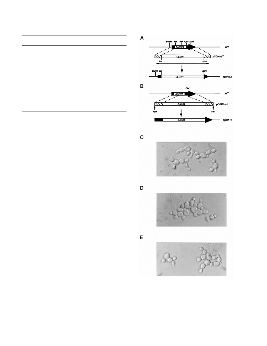
FIG. 1. Disruption of CgKRE9 and CgKNH1 and morphological effects of the
deletions. (A) Disruption of CgKRE9. A PCR-amplified fragment (double-head-
ed arrow) from pCGK9
DT (Materials and Methods) was used for the one-step
gene replacement. (B) Disruption of CgKNH1. A KpnI-SacI fragment of
pCGK1
DH (Materials and Methods) was used for the one-step gene replace-
ment. Homologous recombination between the two regions (hatched boxes)
resulted in disruption of the chromosomal copy. The wild-type strain, 2001HTU
(C), cgkre9
D deletion strain SNBG1-7-7 (D), and cgknh1D deletion strain
SNBG2-26 (E) as viewed by Normarski optics are shown. Cells precultured on
galactose medium were cultured on glucose medium.
TABLE 1. Yeast strains used in this study
Strain
Genotype or description
Source or
reference
S. cerevisiae
SEY6210
MAT
a leu2-3,112 ura3-52 his3-D200 lys2-801
trp1-
D901 suc2-D9
S. D. Emr
HAB813
MAT
a kre9D::HIS3 in SEY6210
6
FAHAP4
MATa ade2-101 his3-
D200 leu2-D1 lys2-801
trp1-
D63::TRP1-tetRHAP4AD ura3-52
39
SNB50-1
MATa Kan
r
-97t-KRE9 in FAHAP4
This work
SNB54-5
MATa knh1
D::hisG in SNB50-1
This work
C. glabrata
2001HTU
cgura3
D cgtrp1D cghis3D
28
ACG22
cgtrp1
D::CgTRP1-TAGAL4 in 2001HTU
40
SNBG1-7-7 cgkre9
D::CgTRP1 in 2001HTU
This work
SNBG2-26
cgknh1
D::CgHIS3 in 2001HTU
This work
SNBG3-10
URA3-97t-CgKRE9 in ACG22
This work
SNBG4-49
cgknh1
D::CgHIS3 in SNBG3-10
This work
SNBG5
cgkre9
D::CgTRP1; suppressor from SNBG1-7-7 This work
V
OL
. 180, 1998
C. GLABRATA HOMOLOGS OF S. CEREVISIAE KRE9 AND KNH1
5021
AGT-3
9, 59-CATTATCTGTGCCTCAAAGCATTAACTTTTCTTGCAGTCA
GAGAAACAGCTATGACCATG-3
9). The correct integration was confirmed
by PCR. The strain was subjected to 5-fluoro-orotic acid selection and finally
designated SNB54-5 after the elimination of the URA3 gene was confirmed by
PCR.
Cloning of C. glabrata KRE9 and KNH1 genes.
SNB54-5 cells were transformed
with a pRS424-based C. glabrata subgenomic bank, harboring EcoRI 4- to 7-kbp
fragments of C. glabrata genomic DNA, and spread onto both YNB-glucose and
YNB-galactose plates containing tetracycline (50
mg/ml). After incubation at
30°C for 3 days, colonies appeared on the plates, cells were collected, and
plasmid DNA was recovered from them.
Disruption of CgKRE9 and CgKNH1 and construction of tetracycline-sensitive
mutants of CgKRE9 (Tet
s
CgKRE9).
Disruption of CgKRE9 in strain 2001HTU
was achieved by using a DNA fragment amplified by PCR using pCGK9
DT as a
template and a pair of primers (5
9-CCATCGATGAATTCATGCTGCTGCT
GGCTATACTGCTATC-3
9, 59-CAACTGGACAAATATCTAAC-39) (Fig. 1).
The correct integration was confirmed by PCR, and the strain was designated
SNBG1-7-7. A KpnI-SacI fragment of pCGK1
DH was used to disrupt CgKNH1
(Fig. 1) in strain 2001HTU. The correct integration was confirmed by PCR, and
the strain was designated SNBG2-26.
A KpnI-ClaI fragment harboring target sequences for CgKRE9 and S. cerevi-
siae URA3 was excised from pCGK9tetAB and used for replacement of the
CgKRE9 promoter region with the tetracycline-responsive promoter, 97t (39, 40),
in the C. glabrata strain ACG22 (40) (Fig. 2A). After the correct integration was
confirmed by PCR, the strain was designated SNBG3-10. To construct SNBG4-
49, a KpnI-SacI fragment of pCGK1
DH was used to disrupt CgKNH1 in SNB3-
10. The correct integration was confirmed by PCR.
Cell wall component analysis.
The levels of cell wall alkali-insoluble
b-glucan
were determined as previously described (15). The alkali-soluble and alkali-
insoluble Zymolyase-resistant cell wall fractions were subjected to a dot blot
FIG. 2. Construction of a tetracycline-sensitive mutant of CgKRE9 (Tet
s
CgKRE9). (A) Scheme for replacement of the cognate CgKRE9 promoter region with the
tetracycline-responsive promoter. A PCR-amplified fragment (double-headed arrow) from pCGK9tetAB (Materials and Methods) was used for the one-step gene
replacement. The solid arrow indicates the ORF of CgKRE9. Open and shaded boxes indicate the S. cerevisiae URA3 gene and the tetracycline-responsive promoter,
97t, respectively. Homologous recombination between the two regions (hatched boxes) resulted in generation of the Tet
s
CgKRE9 mutant. (B) Growth inhibition by
tetracycline on the Tet
s
CgKRE9 mutants. A total of 10
4
cells were inoculated and were cultured on YPD (solid bars) or on YPGal (open bars) for 20 h at 30°C. Growth
of cells with tetracycline (50
mg/ml) is expressed as percent of optical density at 600 nm of cells without tetracycline. As the wild type (WT), strain ACG22 (Table 1)
was used. Error bars, standard deviations.
5022
NAGAHASHI ET AL.
J. B
ACTERIOL
.
analysis by using anti-
b-1,6-glucan antibody as previously described (33) with
standardization by cell wall dry weight. The content of cellular chitin was deter-
mined as previously described (10) with Streptomyces griseus chitinase (Sigma, St.
Louis, Mo.) and standardization by cell dry weight.
Sequence analysis and homology search.
Sequence analysis was performed by
using GeneWorks (Intelligenetics, Mountain View, Calif.) and GeneJockey (Bio-
soft, Cambridge, United Kingdom) software. A homology search for C. glabrata
sequences against S. cerevisiae sequences was performed by using the WU-
BLAST2 program in the Saccharomyces Genome Database (Stanford Universi-
ty).
Nucleotide sequence accession numbers.
The nucleotide sequence data re-
ported in this paper have been submitted to the GenBank database. The acces-
sion numbers of the C. glabrata KRE9 (CgKRE9) and KNH1 (CgKNH1) genes are
AF064251 and AF064252, respectively.
RESULTS
Construction of tetracycline-sensitive mutants of the S. cer-
evisiae KRE9 gene.
To isolate the S. cerevisiae KRE9 homolog
from C. glabrata, we performed complementation screening. As
convenient hosts for the screening, tetracycline-sensitive mu-
tants of the S. cerevisiae KRE9 gene (Tet
s
KRE9) were con-
structed. The KRE9 promoter region was replaced with a tet-
racycline-responsive promoter in a strain, FAHAP4, harboring
the tetR-HAP4AD fusion activator gene for tetracycline-con-
trollable gene expression (39). As shown in Fig. 4, addition of
tetracycline (50
mg/ml) inhibited growth of cells of Tet
s
KRE9
mutant strain SNB50-1 on glucose medium but not on galac-
tose medium. These observations resemble and are consistent
with the finding that an S. cerevisiae kre9
D mutant grows ex-
tremely slowly on glucose medium while growing somewhat
better on galactose medium (15) and suggest that the concen-
tration of tetracycline used in the present study is sufficient to
repress the expression of KRE9 driven by the tetracycline-
responsive promoter. The tetracycline sensitivity of the Tet
s
KRE9 mutant was complemented by introduction of an extra-
genic copy of KRE9 on pRS316 (6) (data not shown). Disrup-
tion of KNH1 in a Tet
s
KRE9 mutant rendered cell growth
tetracycline sensitive on glucose or galactose media (Fig. 4). This
result is consistent with the known synthetic lethality between
kre9
D and knh1D mutations in S. cerevisiae (15). This Tet
s
KRE9
knh1
D mutant strain, SNB54-5, was used for complementation
cloning of a C. glabrata homolog(s).
Cloning of C. glabrata KRE9 and KNH1 genes.
By genomic
Southern hybridization using the S. cerevisiae KRE9 sequence
as a probe, 5- and 6-kbp EcoRI fragments of C. glabrata ge-
nomic DNA were shown to contain sequences hybridizing to
S. cerevisiae KRE9 (data not shown). This result allowed us to
make a subgenomic C. glabrata bank harboring EcoRI frag-
ments ranging from 4 to 7 kbp to assist in their cloning by
functional complementation.
After screening Tet
s
KRE9 knh1
D cells transformed with the
subgenomic bank on plates containing glucose as a carbon
source and tetracycline (50
mg/ml), pSB2-1 harboring the 6-
kbp EcoRI fragment was isolated as a plasmid which allowed
the mutant cells to grow as well as wild-type cells. However,
plasmids harboring the 5-kbp EcoRI fragment, which also gave
a hybridization signal in Southern analysis, were not isolated.
Since the expression of KNH1 is induced by galactose in S. cer-
evisiae (15), we screened a population of transformed cells for
growth on plates containing galactose as a carbon source. In
this way, pSBG9-1, a plasmid harboring the 5-kbp EcoRI frag-
ment was isolated, as well as pSB2-1. As shown in Fig. 4, while
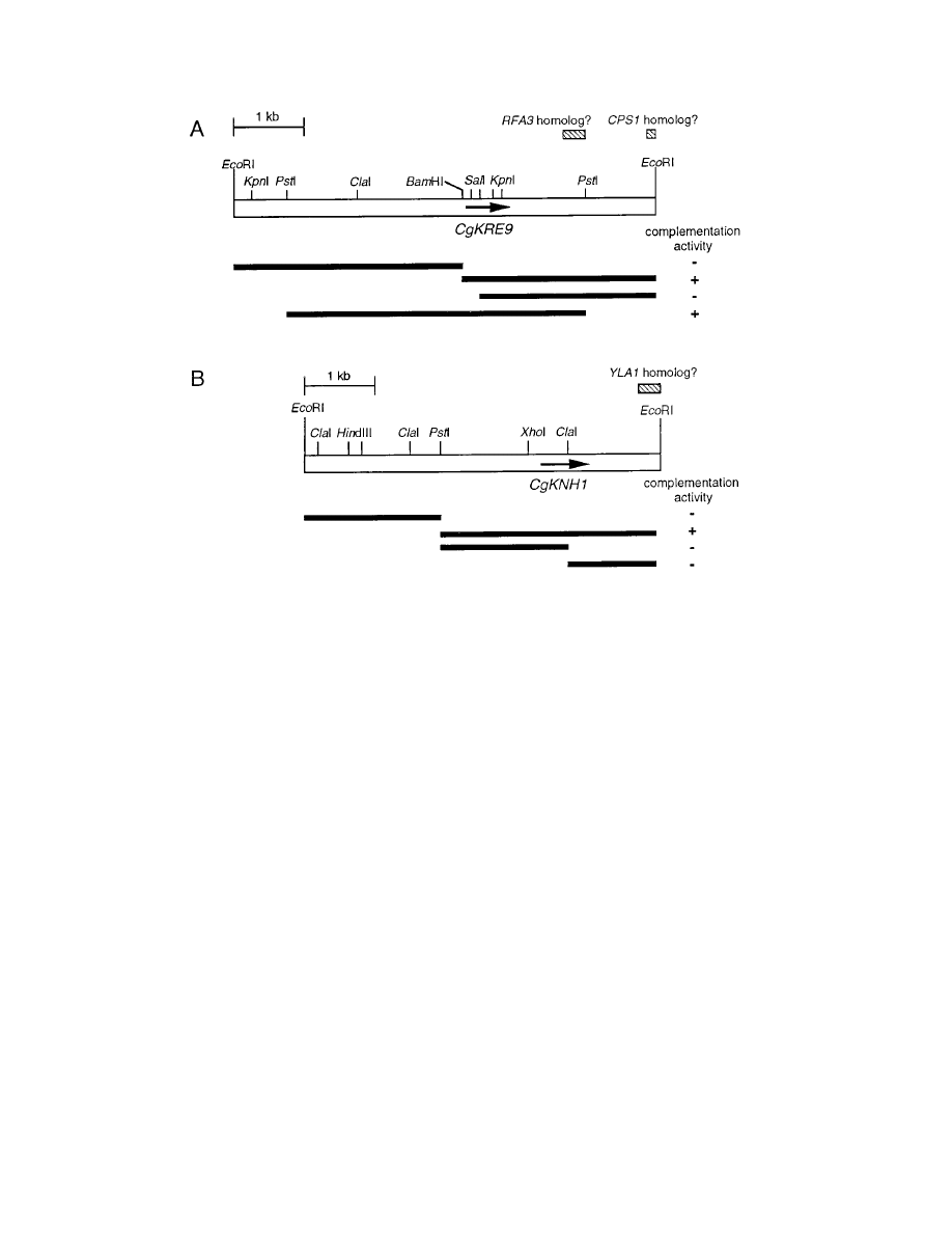
FIG. 3. Restriction maps and deletional analysis of inserts of C. glabrata genomic DNA on pSB2-1 and pSBG9-1. Open bars indicate the inserts on pSB2-1 (A) and
pSBG9-1 (B). Fragments used for deletional analysis are represented by solid bars. The presence and absence of complementation activity in Tet
s
KRE9 knh1
D cells
are indicated as
1 and 2, respectively. Arrows indicate ORFs of CgKRE9 (A) and CgKNH1 (B). Hatched bars indicate regions with homology to the syntenic
S. cerevisiae genes.
V
OL
. 180, 1998
C. GLABRATA HOMOLOGS OF S. CEREVISIAE KRE9 AND KNH1
5023
the tetracycline sensitivity of Tet
s
KRE9 knh1
D cells was com-
plemented by pSBG9-1 partially on glucose medium but com-
pletely on galactose medium, pSB2-1 completely comple-
mented the tetracycline sensitivity of Tet
s
KRE9 knh1
D cells on
both media.
Deletional analysis of the inserts of the two plasmids dem-
onstrated that a 1.4-kbp BamHI-PstI fragment of pSB2-1 and a
3.0-kbp PstI-EcoRI fragment of pSBG9-1 were sufficient for
the complementation activity (Fig. 3). DNA sequencing deter-
mined that the two plasmids harbored distinct open reading
frames (ORFs). The ORF on pSB2-1 was predicted to encode
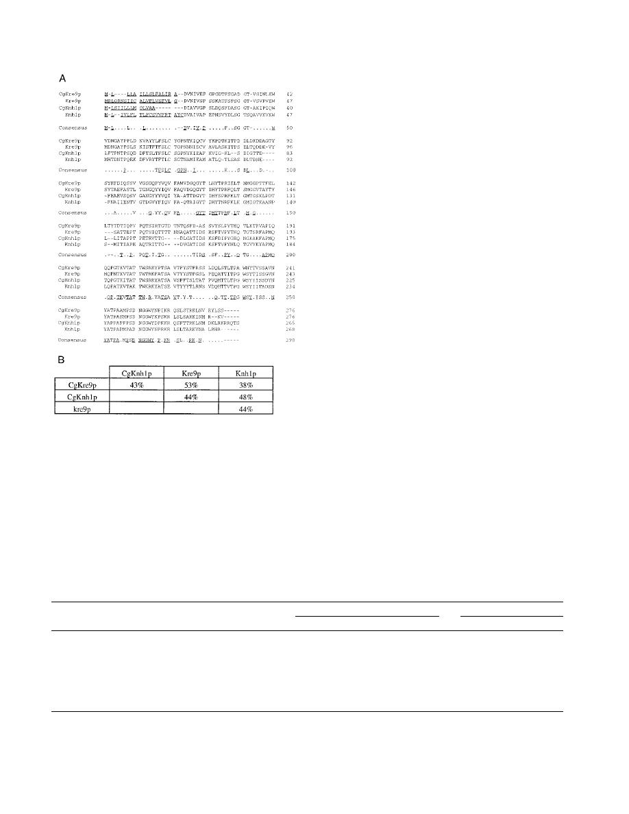
a protein (276 amino acids) similar to S. cerevisiae Kre9p with
53% overall identity, and the protein (265 amino acids) de-
duced from the ORF on pSBG9-1 revealed 48% overall iden-
tity with S. cerevisiae Knh1p (Fig. 5). We designated the genes
on pSB2-1 and pSBG9-1 CgKRE9 and CgKNH1, respectively.
Both predicted gene products showed features characteristic of
their S. cerevisiae counterparts: putative N-terminal signals for
secretion, a high proportion of serine/threonine residues (22%
in both proteins) that could be potential sites for O glycosyla-
tion, and C termini rich in basic amino acid residues (Fig. 5).
Extensive sequencing on 3
9 flanking regions of both CgKRE9
and CgKNH1 identified additional regions similar to the genes
flanking the KRE9 and KNH1 genes in the S. cerevisiae ge-
nome. On pSB2-1, two sequences homologous to the RFA3
and CPS1 genes, respectively, which are located in the 3
9 re-
gion of the KRE9 locus on chromosome X of S. cerevisiae, were
found (Fig. 3A). A sequence homologous to the YLA1 gene,
located in the 3
9 region of the KNH1 locus on chromosome IV,
was found on pSBG9-1 (Fig. 3B).
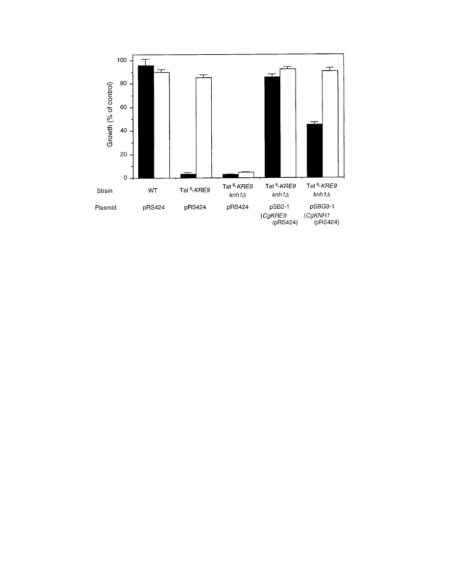
Complementation activity of either CgKRE9 or CgKNH1 on
a yeast centromeric plasmid, pRS416 (56), was also examined
in the Tet
s
KRE9 knh1
D mutant. The tetracycline sensitivity of
the mutant cells on glucose or galactose medium was comple-
mented by introducing a plasmid, CgKRE9-pRS416, whereas
CgKNH1-pRS416 complemented the sensitivity only on galac-
tose medium (Fig. 6), suggesting that expression of CgKNH1 is
induced by galactose in S. cerevisiae.
Complementation of the killer phenotype of the S. cerevisiae
kre9 mutant by CgKRE9 and CgKNH1.
Mutations in KRE9
confer resistance to the K1 killer toxin in S. cerevisiae (6, 8). In
order to test whether multiple copies of CgKRE9 and CgKNH1
could complement this phenotype, pSB2-1 and pSBG9-1 were
transformed into the S. cerevisiae kre9
D null mutant strain
HAB813 (Table 1) and the killer sensitivities of the transfor-
mants were examined by measuring zones of killing in a seed-
ed-plate assay (8). The kre9
D mutant cells are known to show
no killer zone in the assay, since the mutant has an 80% re-
duction of
b-1,6-glucan, which is necessary for the toxin bind-
ing. As shown in Table 2, cells harboring pSB2-1 formed killer
zones when grown on glucose or galactose plates while cells har-
boring pSBG9-1 did so only when grown on galactose plates.
The killer zone sizes, however, were smaller than those of wild-
type strain SEY6210 cells, suggesting that the complementa-
tion was partial. We also examined complementation activity
of either CgKRE9 or CgKNH1 on a single-copy plasmid as
assayed via the killer resistance. Cells harboring CgKRE9-
pRS416 formed killer zones in the seeding assay on glucose or
galactose plates to the same extent as those harboring multiple
copies of CgKRE9 (Table 2), whereas cells harboring CgKNH1-
pRS416 failed to form killer zones (data not shown). To show
that the partial complementation of the killer phenotype of
kre9
D mutant was due to restoration of b-1,6-glucan levels,
alkali-insoluble
b-1,6-glucan levels in the mutant cells harbor-
ing either pSB2-1 or pSBG9-1 were determined. As shown in
Table 2, although cells harboring pSBG9-1 showed no resto-
ration, in cells harboring pSB2-1, the alkali-insoluble
b-1,6-glu-
can level was partially elevated over that of the mutant when
the cells were grown on glucose medium.
Disruption of CgKRE9 and CgKNH1 genes and construction
of tetracycline-sensitive mutants of CgKRE9 (Tet
s
CgKRE9).
To explore the physiological essentialness of CgKRE9 and
FIG. 4. Growth of S. cerevisiae Tet
s
KRE9 knh1
D cells harboring either pSB2-1 or pSBG9-1. About 10
4
cells were inoculated and cultured on YNB-glucose for 20 h (solid
bars) or on YNB-galactose for 40 h (open bars) at 30°C. Cells were grown with or without tetracycline (50
mg/ml), and growth on tetracycline is expressed as the percentage
of optical density at 600 nm of cells grown without tetracycline. The strain FAHAP4 (Table 1) was used as the wild type (WT). Error bars, standard deviations.
5024
NAGAHASHI ET AL.
J. B
ACTERIOL
.
CgKNH1, each gene was disrupted with the C. glabrata TRP1
(CgTRP1) and HIS3 (CgHIS3) genes, respectively (Fig. 1). Trans-
formation for disruption of CgKRE9 was performed on plates
containing either glucose or galactose as a carbon source.
cgkre9
D mutants were obtained from only galactose plates,
whereas cgknh1
D mutants were obtained from glucose plates.
This carbon source dependency on the growth of cgkre9
D mu-
tant was confirmed by spotting cells precultured on galactose
medium onto plates containing either 2% galactose, 2% glu-
cose, or 2% glucose and galactose. Although cgknh1
D cells on
all plates and cgkre9
D cells on the galactose containing plate
grew as well as wild-type cells, the growth of cgkre9
D cells was
severely impaired on plates containing glucose as a carbon
source (data not shown). These results suggest that the pres-
ence of glucose is involved in the slow-growth phenotype of the
cgkre9
D mutant. As shown in Fig. 1D, microscopic examination
of cgkre9
D cells transferred from galactose to glucose medium
revealed the presence of aggregates of cells with abnormal
morphology, which are also observed in the S. cerevisiae kre9
D
null mutant (6). However, cgknh1
D cells showed no morpho-
logical change compared to the wild type (Fig. 1C and E).
To test for a possible synthetic lethality between cgkre9 and
cgknh1 mutations, a C. glabrata tetracycline-controllable gene
expression system (40) was applied to control the expression of
CgKRE9. This system uses the same tetracycline-responsive
promoters and tetR fusion activator as the system for S. cerevi-
siae. As shown in Fig. 2A, a tetracycline-sensitive mutant (Tet
s
CgKRE9) was generated by replacing the cognate CgKRE9
promoter region with the tetracycline-responsive promoter in
C. glabrata ACG22, harboring the tetR-GAL4AD fusion acti-
vator gene (40). Consistent with the growth phenotype of a
cgkre9
D mutant, tetracycline (50 mg/ml) inhibited the growth of
Tet
s
CgKRE9 cells specifically on glucose medium (Fig. 2B).
This glucose-specific tetracycline sensitivity was complemented
by introducing an extragenic copy of CgKRE9 on pCgACH-3
(29), a centromeric plasmid for C. glabrata (data not shown).
When CgKNH1 was disrupted in a Tet
s
CgKRE9 mutant, cells
failed to grow on glucose or galactose media in the presence of
tetracycline (Fig. 2B). This result indicates that the disruption
of both CgKRE9 and CgKNH1 is synthetically lethal in C. gla-
brata.
Killer phenotypes and
b-1,6-glucan levels of cgkre9D and
cgknh1
D mutants. Although cgkre9D cells showed severe growth
defects on glucose medium, spontaneous second-site suppres-
sor mutations partially restoring growth arose when the cells
were cultured by serial passage on glucose medium. Since it is
known in S. cerevisiae that those second-site suppressors have
no effects on the killer phenotypes except for enhanced growth
of the original mutants (4, 8, 34, 48), we used such growth-
suppressed cgkre9
D mutants for further analysis as described
below.
To address the killer phenotypes of cgkre9
D and cgknh1D
FIG. 5. Sequence comparisons of CgKre9p and CgKnh1p with their S. cer-
evisiae counterparts. (A) Alignment of the putative Kre9p and Knh1p amino acid
sequences deduced from the C. glabrata (CgKRE9 and CgKNH1) and S. cerevisiae
(KRE9 and KNH1) nucleotide sequences. The residues with conserved identity in
all proteins are underlined in the consensus sequence. The putative N-terminal
signals for secretion are underlined in each protein. Gaps (shown as dashes) were
introduced to improve alignment. (B) Sequence identities between Kre9p and
Knh1p proteins.
TABLE 2. Killer phenotypes of alkali-insoluble
b-glucan levels of the S. cerevisiae kre9 cells harboring either CgKRE9 or CgKNH1
d
Strain
Allele at
KRE9 locus
Plasmid
Alkali-insoluble glucan(s)
a
Killer zone size
b
(cm) on:
b-1,6-Glucan
b-1,3- and b-1,6-glucan
Glucose
Galactose
SEY6210
KRE9
pRS424
138.13
6 5.15
354.55
6 1.54
1.53
6 0.06
1.25
6 0.05
HAB813
kre9
D::HIS3
pRS424
32.33
6 2.00
301.51
6 17.20
No zone
No zone
HAB813
kre9
D::HIS3
pSB2-1 (CgKRE9/pRS424)
85.29
6 2.24
315.28
6 19.66
1.20
6 0.09
1.02
6 0.03
HAB813
kre9
D::HIS3
pSBG9-1 (CgKNH1/pRS424)
32.86
6 1.31
267.45
6 10.53
No zone
0.70
6 0.00
SEY6210
KRE9
pRS416
ND
c
ND
1.48
6 0.08
1.23
6 0.08
HAB813
kre9
D::HIS3
pRS416
ND
ND
No zone
No zone
HAB813
kre9
D::HIS3
CgKRE9-pRS416
ND
ND
1.10
6 0.05
1.02
6 0.10
a
b-Glucan levels are expressed as micrograms of glucan per milligram (dry weight) of cell wall.
b
Killer zone size (diameter) was determined by seeded-plate assays as previously described (8).
c
ND, not determined.
d
All values are the means of at least three determinations
6 1 standard deviation.
V
OL
. 180, 1998
C. GLABRATA HOMOLOGS OF S. CEREVISIAE KRE9 AND KNH1
5025
mutants, we asked whether C. glabrata was sensitive to the K1
killer toxin. C. glabrata wild-type strain 2001HTU (Table 1) was
found to be sensitive to the toxin on plates containing glucose
or galactose as carbon sources, as measured by killer zones
formed in a seeded-plate assay (Table 3). When mutant cells
were assayed, growth-suppressed cgkre9
D cells clearly formed
smaller killer zones than those of wild-type cells, whereas
cgknh1
D cells formed slightly larger killer zones than those of
wild-type cells (Table 3). We also examined the killer sensitiv-
ity of cgkre9
D cells which had been stored on galactose medium
to prevent second-site suppressor mutations. Although such
mutant cells grew extremely slowly on glucose plates, sizes of
killer zones of the cells were the same as those of growth-
suppressed cgkre9
D cells (data not shown).
To establish that the killer toxin resistance seen in the
growth-suppressed cgkre9
D cells was directly due to decreased
levels in
b-1,6-glucan, we attempted to determine b-1,6-glucan
levels in C. glabrata cells. Following the method used in S. cer-
evisiae, alkali-insoluble cell wall fractions were digested with
Zymolyase, a commercial
b-1,3-glucanase preparation, and re-
sidual polymers were measured as hexose. As shown in Table
3, in growth-suppressed cgkre9
D cells, hexose levels in the
alkali-insoluble Zymolyase-resistant fraction were reduced to
40 and 50% of wild-type levels in cells grown on glucose and
galactose medium, respectively. To verify the presence of
b-
1,6-linkage in these fractions, alkali-soluble and alkali-insolu-
ble Zymolyase-resistant fractions from all three strains grown
on glucose medium were subjected to a dot blot analysis using
affinity-purified anti-
b-1,6-glucan polyclonal antibody (33). In
cgkre9
D cells, the amount of material recognized by the anti-
body in both fractions was estimated at less than 50% of those
of wild-type by comparing signals from serially diluted spotted
samples (data not shown). These results strongly suggest that
disruption of CgKRE9 results in a more than 50% reduction of
cell wall
b-1,6-glucan independent of the carbon source used
for growth.
Sensitivity to CFW and cellular chitin levels in cgkre9 and
cgknh1 mutants.
CFW, a negatively charged fluorescent dye
that preferentially binds to nascent chains of chitin and inter-
feres with cell wall assembly (16, 50), is a useful compound for
surveying a broad range of cell wall defects in S. cerevisiae (32,
46). To test for cell wall defects in cgkre9
D and cgknh1D mu-
tants, CFW sensitivities of both growth-suppressed cgkre9
D
and cgknh1
D cells were determined by a spotting assay (31) on
plates containing glucose or galactose as a carbon source. Al-
though cgknh1
D cells grew as well as wild-type cells even in the
presence of 25-
mg/ml CFW, growth-suppressed cgkre9D failed
to grow at this concentration of CFW when glucose was used
as a carbon source (Table 3).
In S. cerevisiae, kre9
D mutant cells gave strong fluorescence
when stained by CFW (6). This evidence and glucose-specific
CFW sensitivity of growth-suppressed cgkre9
D cells led us to
determine cellular chitin levels in C. glabrata cells. As shown in
Table 3, on glucose medium, more than fourfold more cellular
chitin was detected in growth-suppressed cgkre9
D cells than in
wild-type cells, while cgknh1
D cells had almost the same amount
of chitin as wild-type cells. On galactose medium, no significant
difference was seen in chitin levels among these three strains.
To assess a possible correlation between this chitin increase
and the second-site mutations suppressing the growth defect on
glucose medium, we measured cellular chitin levels in cgkre9
D
cells without such suppressor mutations. For this purpose, two
different strategies were taken. In one, a Tet
s
CgKRE9 mutant
was used. In the other, cgkre9
D cells, which had been stored on
galactose medium, were switched from galactose to glucose me-
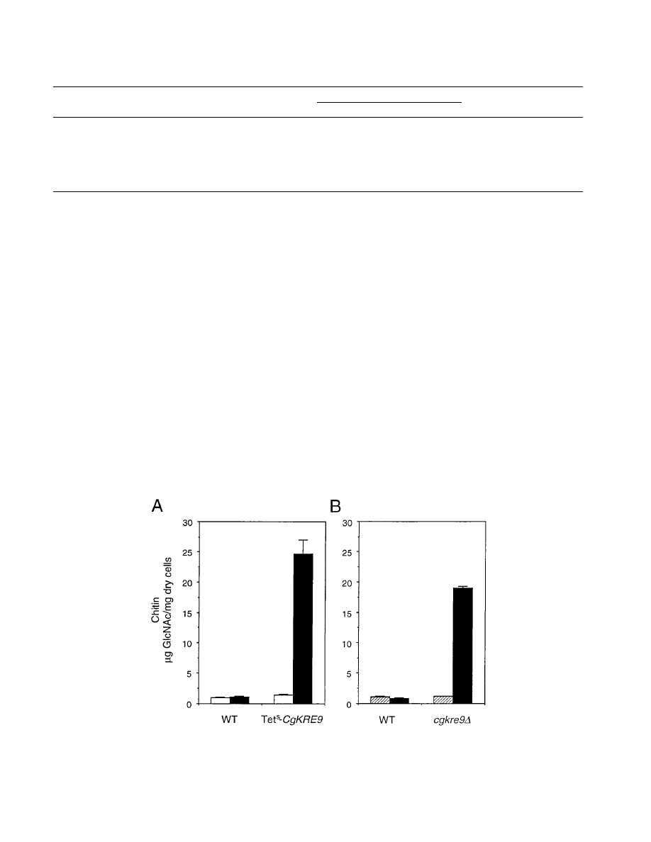
dium. As shown in Fig. 7A, although the repression of CgKRE9
expression is expected to be partial since the inoculum for the
tetracycline assay was increased to permit sufficient cells to be
obtained for the chitin measurement, addition of tetracycline
resulted in an
;17-fold increase of chitin levels in the Tet
s
CgKRE9 mutant cells while there was no obvious change in
cells of the parent strain, ACG22. When cgkre9
D cells were
transferred from galactose to glucose medium, cellular chitin
levels increased by
.15-fold (Fig. 7B). These results suggest
that a considerable amount of chitin is present in cgkre9
D cells
grown in the presence of glucose and that such levels are
unrelated to second-site mutations leading to growth suppres-
sion.
Overexpression of CgKNH1 and S. cerevisiae KRE9 in
cgkre9
D cells. We asked if multiple copies of either CgKNH1
or S. cerevisiae KRE9 could complement the phenotypes of
a cgkre9
D mutant. CgKNH1 was cloned into pRS316 (56),
which is known to be a multicopy plasmid for C. glabrata
(60). CgKNH1-pRS316 and KRE9-pRS316 (6) were trans-
formed into growth-suppressed cgkre9
D cells. As summarized
in Table 4, the killer sensitivities and
b-1,6-glucan levels of the
mutant cells were partially restored by multiple copies of S. cer-
evisiae KRE9 whereas multiple copies of CgKNH1 showed no
effect. Further, multiple copies of either CgKNH1 or S. cerevi-
siae KRE9 allowed growth-suppressed cgkre9
D cells to grow as
well as wild-type cells on plates containing glucose and CFW
(25
mg/ml). In the cells harboring CgKNH1-pRS316, the chitin
increase was slightly suppressed (Table 4).
DISCUSSION
The CgKRE9 and CgKNH1 genes have been identified by
functional screening using an S. cerevisiae Tet
s
KRE9 knh1
D
mutant. Both C. glabrata gene products have significant overall
identity with their S. cerevisiae counterparts (Fig. 5B). Partial
restoration of the killer sensitivity and
b-1,6-glucan levels of
kre9
D mutant cells harboring multiple copies of CgKRE9 (Ta-
ble 2) clearly indicates that CgKRE9 is an ortholog of S. cer-
evisiae KRE9. Furthermore, a single copy of CgKRE9 was suf-
ficient to partially complement the killer phenotype of the
FIG. 6. Growth of S. cerevisiae Tet
s
KRE9 knh1
D cells harboring a single copy
of either CgKRE9 or CgKNH1. About 10
4
cells were inoculated and cultured on
YNB-glucose for 20 h (solid bars) or on YNB-galactose for 40 h (open bars) at
30°C. Growth of cells with tetracycline (50
mg/ml) is expressed as percent of
optical density at 600 nm of cells without tetracycline. FAHAP4 (Table 1) was
used as the wild-type. Error bars, standard deviations.
5026
NAGAHASHI ET AL.
J. B
ACTERIOL
.
kre9
D mutant (Table 2). This result also supports the argument
for the functional similarity between Kre9p and CgKre9p and
implies that the promoter activity of CgKRE9 and the N-ter-
minal signal for secretion of CgKre9p are active in S. cerevisiae.
Disruption of CgKRE9 resulted in cells with phenotypes
similar to that of the S. cerevisiae kre9
D null mutant (6): a
severe growth defect on glucose medium, resistance to the K1
killer toxin, a reduction of
b-1,6-glucan, and the presence of
aggregates of cells with abnormal morphology on glucose me-
dium (Table 3; Fig. 1D). Some of these phenotypes were par-
tially complemented by multiple copies of S. cerevisiae KRE9
(Table 4). Recent cloning of the C. albicans KRE9 (CaKRE9)
gene has demonstrated that CaKre9p is also required for
b-
1,6-glucan synthesis in C. albicans (33). These lines of evidence
indicate that the function of Kre9p as an essential component
for
b-1,6-glucan biosynthesis is conserved at least among S. cer-
evisiae, C. albicans, and C. glabrata.
cgknh1
D mutants, however, had no phenotype beyond a slight-
ly increased sensitivity to the K1 killer toxin. Further, multiple
copies of CgKNH1 failed to restore the killer sensitivity and
alkali-insoluble
b-1,6-glucan levels in cgkre9D cells grown on
glucose medium (Table 4). However, in addition to the syn-
thetic lethality suggested by the tetracycline sensitivity of Tet
s
CgKRE9 cgknh1
D mutant (Fig. 6B), its ability to complement a
range of kre9 defects in S. cerevisiae and C. glabrata implies that
CgKnh1p is related to Kre9p/CgKre9p and is an ortholog of
S. cerevisiae Knh1p. These complementation abilities include
S. cerevisiae kre9 mutant phenotypes (Fig. 4 and Table 2), CFW
sensitivity, and chitin increase of growth-suppressed cgkre9
D
cells (Table 4).
We have demonstrated that cellular chitin levels were sig-
nificantly increased in cgkre9 mutants on glucose medium (Ta-
ble 3 and Fig. 7). It is known that chitin levels are also
increased in several cell wall mutants of S. cerevisiae such as
gas1
D, fks1D, and knr4D mutants (22, 27, 45, 47). Based on
genetic interaction between gas1
D and chs3D mutations and
the sensitivity to nikkomycin Z (a competitive inhibitor of
chitin synthases) of a gas1
D mutant, it has been hypothesized
that such a chitin increase is essential for growth as a compen-
sation mechanism to support the impaired cell wall integrity of
FIG. 7. Cellular chitin levels in cgkre9 mutants of C. glabrata. (A) Effects of addition of tetracycline on cellular chitin levels in Tet
s
CgKRE9 mutants. About 10
6
cells were cultured on YPD with (solid bars) or without (open bars) tetracycline (50
mg/ml) at 30°C for 20 h, and the cellular chitin levels were measured. As the wild
type (WT), strain ACG22 (Table 1) was used. (B) Effect of switching the carbon source on cellular chitin levels in cgkre9
D mutant. Cells precultured on YPGal were
inoculated onto either YPD (solid bars) or YPGal (hatched bars) and cultured at 30°C for 20 h, and the cellular chitin levels were measured. As the wild type (WT),
strain 2001HTU (Table 1) was used. Error bars, standard deviations.
TABLE 3. Alkali-insoluble glucan and cellular chitin levels in C. glabrata cells grown on either glucose or galactose
e
Medium
Strain
Genotype
Killer zone
size
b
(cm)
Alkali-insoluble glucan(s)
a
CFW
sensitivity
c
Chitin
d
b-1,6-Glucan
b-1,3- and b-1,6-glucan
YPD
2001HTU
WT
1.35
6 0.00
52.48
6 0.54
178.44
6 4.54
R
0.88
6 0.07
SNBG5
cgkre9
D::CgTRP1
0.73
6 0.08
20.14
6 1.34
233.56
6 5.75
S
3.87
6 1.10
SNBG2-26
cgknh1
D::CgHIS3
1.55
6 0.00
52.57
6 1.40
179.46
6 4.29
R
0.90
6 0.04
YPGal
2001HTU
WT
1.17
6 0.02
76.14
6 1.07
243.82
6 9.06
R
1.02
6 0.02
SNBG5
cgkre9
D::CgTRP1
0.63
6 0.03
38.66
6 1.62
260.74
6 1.92
R
1.12
6 0.05
SNBG2-26
cgknh1
D::CgHIS3
1.53
6 0.02
84.11
6 2.20
242.65
6 5.46
R
1.08
6 0.03
a
b-Glucan levels are expressed as micrograms of glucan per milligram (dry weight) of cell wall.
b
Killer zone size (diameter) was determined by seeded-plate assays as previously described (8).
c
CFW sensitivity was scored by growth of 10
4
cells on plates containing CFW (25
mg/ml). R, resistant; S, sensitive.
d
Chitin levels are expressed as micrograms of N-acetylglucosamine per milligram of dry cells.
e
All values are the means of at least three determinations
6 1 standard deviation.
V
OL
. 180, 1998
C. GLABRATA HOMOLOGS OF S. CEREVISIAE KRE9 AND KNH1
5027

these mutants (27, 45, 47). However, the increase of chitin in
cgkre9 cannot simply be concluded to be the result of such a
compensation mechanism, since it is correlated with a severe
growth defect on glucose medium and is independent of the
reduction of
b-1,6-glucan. This idea that increased chitin levels
slow the growth of cgkre9 mutants is supported by several ob-
servations in the present study. First, considerable amounts of
cellular chitin were detected in both tetracycline-treated Tet
s
CgKRE9 cells grown on glucose medium (Fig. 7A) and cgkre9
D
cells transferred from galactose to glucose medium (Fig. 7B).
Second, there was no obvious increase in chitin levels in cgkre9
D
cells grown on galactose medium (Table 3 and Fig. 7B), on
which they grew as well as the wild type did, in spite of a 50%
reduction of alkali-insoluble
b-1,6-glucan (Tables 3 and 4).
The mechanism and physiological relevance of the chitin in-
crease in cgkre9 mutants and its apparent glucose dependence
remain to be elucidated. In S. cerevisiae, at least five genes have
been known to be involved in the chitin synthase activity (11,
14). Cloning of these homologs and an enzymatic analysis of
chitin synthesis in C. glabrata will be helpful in addressing this
question. It will be useful to see if a chitin increase is common
to S. cerevisiae kre9 and other kre mutants, since second-site
mutations suppressing growth defects have been isolated in
many kre mutants and act without restoration of killer sensi-
tivity or
b-1,6-glucan levels (4, 8, 34, 48). Glucose-specific
cross-linking changes in the cell wall of cgkre9
D cells may result
in elevated chitin levels and a severe growth defect on glucose
medium.
Extensive sequencing of regions around both the CgKRE9
and CgKNH1 loci show that genomic organization in the 3
9
regions of both homologs is conserved between C. glabrata and
S. cerevisiae (Fig. 3). This synteny in regions of two chromo-
somes further indicates a close evolutionary relationship be-
tween C. glabrata and S. cerevisiae, consistent with the phylo-
genetic trees deduced from comparison of 5S (2) and 18S (43)
rRNA genes. Further, CgKre9p and CgKnh1p have lower over-
all identity between themselves than to their orthologous
S. cerevisiae counterparts (Fig. 5B). This observation implies
that the duplication of the KRE9 and KNH1 genes took place
before the divergence of these two fungi from a common
ancestor. In contrast, no chromosomal conservation between
S. cerevisiae and C. albicans was found in the 8-kbp fragment
containing the CaKRE9 locus (data not shown). This result
supports the idea of a more distant relationship of C. albicans
and S. cerevisiae based on phylogenetic trees deduced from the
distribution of the serine-tRNA gene (42, 43) and comparison
of rRNA genes (2, 43). Although the presence of a KNH1 ho-
molog in C. albicans still remains a possibility, this result sug-
gests that extensive genomic reorganization around the CaKRE9
locus has occurred since its divergence from a common ances-
tor with S. cerevisiae. For example, it is possible that the du-
plication event leading to the KRE9 and KNH1 pair in S. cer-
evisiae and C. glabrata occurred after the divergence of these
yeast lineages from that of C. albicans.
In summary, although the molecular functions of the Kre9p/
Knh1p proteins still remain to be characterized, the evolution-
ary conservation of the essentiality of these proteins supports
the idea that compounds that interfere with their functions
would be new antifungal drugs affecting a broad spectrum of
pathogenic fungi. Our data also indicate that C. glabrata is a
useful model pathogenic fungus for understanding biological
processes, including cell wall biosynthesis.
ACKNOWLEDGMENTS
We thank K. Kitada and H. Nakayama for the C. glabrata strains and
plasmids, P. Philippsen for KanMX2, A. B. Futcher for pMPY-ZAP,
G. P. J. Dijkgraaf and T. Ketela for critical comments throughout this
study, A.-M. Sdicu and S. Veronneau for technical assistance, and
S. Shahinian for anti-
b-1,6-glucan polyclonal antibody and suggestions.
S.N. acknowledges continuous support from Nippon Roche and H.
Yamada-Okabe. This work was supported in part by an operating
grant from the Natural Sciences and Engineering Research Council of
Canada. H.B. is a Canadian Pacific Professor.
REFERENCES
1. Aisner, J., S. C. Schimpff, J. C. Sutherland, V. M. Young, and P. H. Wiernik.
1976. Torulopsis glabrata infections in patients with cancer: increasing inci-
dence and relationship to colonization. Am. J. Med. 61:23–28.
2. Barns, S. M., D. J. Lane, M. L. Sogin, C. Bibeau, and W. G. Weisburg. 1991.
Evolutionary relationships among pathogenic Candida species and relatives.
J. Bacteriol. 173:2250–2255.
3. Baudin, A., K. O. Ozier, A. Denouel, F. Lacroute, and C. Cullin. 1993. A
simple and efficient method for direct gene deletion in Saccharomyces cer-
evisiae. Nucleic Acids Res. 21:3329–3330.
4. Boone, C., S. S. Sommer, A. Hensel, and H. Bussey. 1990. Yeast KRE genes
provide evidence for a pathway of cell wall
b-glucan assembly. J. Cell Biol.
110:
1833–1843.
5. Boone, C., A.-M. Sdicu, M. Laroche, and H. Bussey. 1991. Isolation from
Candida albicans of a functional homolog of the Saccharomyces cerevisiae
KRE1 gene, which is involved in cell wall
b-glucan synthesis. J. Bacteriol.
173:
6859–6864.
6. Brown, J. L., and H. Bussey. 1993. The yeast KRE9 gene encodes an O
glycoprotein involved in cell surface
b-glucan assembly. Mol. Cell. Biol. 13:
6346–6356.
7. Brown, J. L., S. North, and H. Bussey. 1993. SKN7, a yeast multicopy sup-
pressor of a mutation affecting
b-glucan assembly, encodes a product with
domains homologous to prokaryotic two-component regulators and to heat
shock transcription factors. J. Bacteriol. 175:6908–6915.
8. Brown, J. L., Z. Kossaczka, B. Jiang, and H. Bussey. 1993. A mutational
analysis of killer toxin resistance in Saccharomyces cerevisiae identifies new
genes involved in cell wall (1-6)-
b-glucan synthesis. Genetics 133:837–849.
9. Brown, J. L., H. Bussey, and R. C. Stewart. 1994. Yeast Skn7p functions in
a eukaryotic two-component regulatory pathway. EMBO J. 13:5186–5194.
10. Bulawa, C. E., M. Slater, E. Cabib, J. Au-Young, A. Sburlati, W. L. Adair, Jr.,
and P. W. Robbins.
1986. The S. cerevisiae structural gene for chitin synthase
is not required for chitin synthesis in vivo. Cell 48:213–225.
TABLE 4. Effects of multiple copies of either CgKNH1 or S. cerevisiae KRE9 on the phenotypes of growth-suppressed cgkre9
D cells
f
Strain
Allele at
CgKRE9 locus
Plasmid
Alkali-insoluble glucan(s)
a
Killer zone
size
b
(cm)
CFW
sensitivity
c
Chitin
d
b-1,6-Glucan
b-1,3- and b-1,6-glucan
2001HTU
CgKRE9
pRS316
76.05
6 3.40
228.22
6 6.11
1.46
6 0.02
R
0.94
6 0.02
SNBG5
cgkre9
D::CgTRP1
pRS316
35.14
6 1.42
246.14
6 3.79
1.10
6 0.02
S
3.71
6 0.38
SNBG5
cgkre9
D::CgTRP1
CgKNH1-pRS316
32.72
6 0.78
218.78
6 10.09
0.89
6 0.01
R
2.95
6 0.52
SNBG5
cgkre9
D::CgTRP1
KRE9-pRS316
44.97
6 3.01
204.59
6 4.96
1.56
6 0.02
R
ND
e
a
b-Glucan levels are expressed as micrograms of glucan per milligram (dry weight) of cell wall.
b
Killer zone size was determined by seeded-plate assays as previously described (8).
c
CFW sensitivity was scored by growth of 10
3
cells on plates containing CFW (25
mg/ml). R, resistant; S, sensitive.
d
Chitin levels were expressed as micrograms of N-acetylglucosamine per milligram of dry cells.
e
ND, not determined.
f
All values are the means of at least three determinations
6 1 standard deviation.
5028
NAGAHASHI ET AL.
J. B
ACTERIOL
.

11. Bulawa, C. E. 1993. Genetics and molecular biology of chitin synthesis in
fungi. Annu. Rev. Microbiol. 47:505–534.
12. Chen, D. C., B. C. Yang, and T. T. Kuo. 1992. One-step transformation of
yeast in stationary phase. Curr. Genet. 21:83–84.
13. Christianson, T. W., R. S. Sikorski, M. Dante, J. H. Shero, and P. Hieter.
1992. Multifunctional yeast high-copy-number shuttle vectors. Gene 110:
119–122.
14. Cid, V. J., A. Dura´n, F. del Ray, M. P. Snyder, C. Nombela, and M. Sa´nchez.
1995. Molecular basis of cell integrity and morphogenesis in Saccharomyces
cerevisiae. Microbiol. Rev. 59:345–386.
15. Dijkgraaf, G. J. P., J. L. Brown, and H. Bussey. 1996. The KNH1 gene of
Saccharomyces cerevisiae is a functional homolog of KRE9. Yeast 12:683–
692.
16. Elorza, M. V., H. Rico, and R. Sentandreu. 1983. Calcofluor white alters the
assembly of chitin fibrils in Saccharomyces cerevisiae and Candida albicans
cells. J. Gen. Microbiol. 129:1577–1582.
17. Geber, A., C. A. Hitchcock, J. E. Swartz, F. S. Pullen, K. E. Marsden, K. J.
Kwon-Chung, and J. E. Bennett.
1995. Deletion of the Candida glabrata
ERG3 and ERG11 genes: effect on cell viability, cell growth, sterol compo-
sition, and antifungal susceptibility. Antimicrob. Agents Chemother. 39:
2708–2717.
18. Georgopapadakou, N. H., and J. S. Tkacz. 1995. The fungal cell wall as a
drug target. Trends Microbiol. 3:98–104.
19. Georgopapadakou, N. H., and T. J. Walsh. 1996. Antifungal agents: chemo-
therapeutic targets and immunologic strategies. Antimicrob. Agents Che-
mother. 40:279–291.
20. Gietz, R. D., R. H. Schiestl, A. R. Willems, and R. A. Woods. 1995. Studies
on the transformation of intact yeast cells by the LiAc/SS-DNA/PEG pro-
cedure. Yeast 11:355–360.
21. Hickey, W. F., L. H. Sommerville, and F. J. Schoen. 1983. Disseminated
Candida glabrata—report of a uniquely severe infection and a literature
review. Am. J. Clin. Pathol. 80:724–727.
22. Hong, Z., P. Mann, K. J. Shaw, and B. DiDomenico. 1994. Analysis of
b-glucans and chitin in a Saccharomyces cerevisiae cell wall mutant using
high-performance liquid chromatography. Yeast 10:1083–1092.
23. Ito, H., Y. Fukuda, K. Murata, and A. Kimura. 1983. Transformation of
intact yeast cells treated with alkali cations. J. Bacteriol. 153:163–168.
24. Jiang, B., A. F. J. Ram, J. Sheraton, F. M. Klis, and H. Bussey. 1995.
Regulation of cell wall
b-glucan assembly: PTC1 negatively affects PBS2
action in a pathway that includes modulation of EXG1 transcription. Mol.
Gen. Genet. 248:260–269.
25. Jiang, B., J. Sheraton, A. F. J. Ram, G. J. P. Dijkgraaf, F. M. Klis, and H.
Bussey.
1996. CWH41 encodes a novel endoplasmic reticulum membrane
N-glycoprotein involved in
b1,6-glucan assembly. J. Bacteriol. 178:1162–
1171.
26. Kapteyn, J. C., R. C. Montijn, E. Vink, J. De La Cruz, A. Llobelle, J. E.
Douwes, H. Shimoi, P. N. Lipke, and F. M. Klis.
1996. Retention of Sac-
charomyces cerevisiae cell wall proteins through a phosphodiester-linked
b1,3-/b1,6-glucan heteropolymer. Glycobiology 6:337–345.
27. Kapteyn, J. C., A. F. J. Ram, E. M. Groos, R. Kollar, R. C. Montijn, H. Van
Den Ende, A. Llobell, E. Cabib, and F. M. Klis.
1997. Altered extent of
cross-linking of
b1,6-glucosylated mannoproteins to chitin in Saccharomyces
cerevisiae mutants with reduced cell wall
b1,3-glucan content. J. Bacteriol.
179:
6279–6284.
28. Kitada, K., E. Yamaguchi, and M. Arisawa. 1995. Cloning of the Candida
glabrata TRP1 and HIS3 genes, and construction of their disruptant strains by
sequential integrative transformation. Gene 165:203–206.
29. Kitada, K., E. Yamaguchi, and M. Arisawa. 1996. Isolation of a Candida
glabrata centromere and its use in construction of plasmid vectors. Gene 175:
105–108.
30. Klis, F. M. 1994. Review: cell wall assembly in yeast. Yeast 10:851–869.
31. Lussier, M., A.-M. Sdicu, E. Winnett, D. H. Vo, J. Sheraton, A. Du¨sterho¨ft,
R. K. Storms, and H. Bussey.
1997. Completion of the Saccharomyces cer-
evisiae genome sequence allows identification of KTR5, KTR6 and KTR7 and
definition of the nine-membered KRE2/MNT1 mannosyltransferase gene
family in this organism. Yeast 13:267–274.
32. Lussier, M., A.-M. White, J. Sheraton, T. di Paolo, J. Treadwell, S. B.
Southard, C. I. Horenstein, J. Chen-Weiner, A. F. J. Ram, J. C. Kapteyn,
T. W. Roemer, D. H. Vo, D. C. Bondoc, J. Hall, W. W. Zhong, A.-M. Sdicu,
J. Davies, F. M. Klis, P. W. Robbins, and H. Bussey.
1997. Large scale
identification of genes involved in cell surface biosynthesis and architecture
in Saccharomyces cerevisiae. Genetics 147:435–450.
33. Lussier, M., A.-M. Sdicu, S. Shahinian, and H. Bussey. The Candida albi-
cans KRE9 gene is required for cell wall beta-1,6-glucan synthesis and is
essential for growth on glucose. Proc. Natl. Acad. Sci. USA, in press.
34. Meaden, P., K. Hill, J. Wagner, D. Slipetz, S. S. Sommer, and H. Bussey.
1990. The yeast KRE5 gene encodes a probable endoplasmic reticulum
protein required for (136)-
b-
D
-glucan synthesis and normal cell growth.
Mol. Cell. Biol. 10:3013–3019.
35. Mehra, R. K., J. L. Thorvaldsen, I. G. Macreadi, and R. R. Winge. 1992.
Cloning system for Candida glabrata using elements from the metallothio-
nein IIa encoding gene that confer autonomous replication. Gene 113:119–
124.
36. Mio, T., T. Yamada-Okabe, T. Yabe, T. Nakajima, M. Arisawa, and H.
Yamada-Okabe.
1997. Isolation of the Candida albicans homologs of Sac-
charomyces cerevisiae KRE6 and SKN1: expression and physiological func-
tion. J. Bacteriol. 179:2363–2372.
37. Mio, T., M. Adachi-Shimizu, Y. Tachibana, H. Tabuchi, S. B. Inoue, T. Yabe,
T. Yamada-Okabe, M. Arisawa, T. Watanabe, and H. Yamada-Okabe.
1997.
Cloning of the Candida albicans homolog of Saccharomyces cerevisiae GSC/
FKS1 and its involvement in
b-1,3-glucan synthesis. J. Bacteriol. 179:4096–
4105.
38. Montijn, R. C., J. van Rinsum, F. A. van Schagen, and F. M. Klis. 1994.
Glucomannoproteins in the cell wall of Saccharomyces cerevisiae contain a
novel type of carbohydrate side chain. J. Biol. Chem. 269:19338–19342.
39. Nagahashi, S., H. Nakayama, K. Hamada, H. Yang, M. Arisawa, and K.
Kitada.
1997. Regulation by tetracycline of gene expression in Saccharomy-
ces cerevisiae. Mol. Gen. Genet. 255:372–375.
40. Nakayama, H., M. Izuta, S. Nagahashi, E. Yamaguchi-Sihta, Y. Sato, T.
Yamazaki, M. Arisawa, and K. Kitada.
A controllable gene expression sys-
tem in the pathogenic fungus Candida glabrata. Microbiology, in press.
41. Newman, S. L., T. P. Flanigan, A. Fisher, M. G. Rinaldi, M. Stein, and K.
Vigilante.
1994. Clinically significant mucosal candidiasis resistant to flucon-
azole treatment in patients with AIDS. Clin. Infect. Dis. 19:684–686.
42. Ohama, T., T. Suzuki, M. Mori, S. Osawa, T. Ueda, K. Watanabe, and T.
Nakase.
1993. Non-universal decoding of the leucine codon CUG in several
Candida species. Nucleic Acids Res. 21:4039–4045.
43. Pesole, G., M. Lotti, L. Alberghina, and C. Saccone. 1995. Evolutional origin
of nonuniversal CUG
Ser
codon in some Candida species as inferred from a
molecular phylogeny. Genetics 141:903–907.
44. Petter, R., and K. J. Kwon-Chung. 1996. Disruption of the SNF1 gene
abolishes trehalose utilization in the pathogenic yeast Candida glabrata.
Infect. Immun. 64:5269–5273.
45. Popolo, L., D. Gilardelli, P. Bonfante, and M. Vai. 1997. Increase in chitin as
an essential response to defects in assembly of cell wall polymers in the ggp1
D
mutant of Saccharomyces cerevisiae. J. Bacteriol. 179:463–469.
46. Ram, A. F. J., A. Wolters, R. Ten Hoopen, and F. M. Klis. 1994. A new
approach for isolating cell wall mutants in Saccharomyces cerevisiae by
screening for hypersensitivity to calcofluor white. Yeast 10:1019–1030.
47. Ram, A. F. J., J. C. Kapteyn, R. C. Montijn, L. H. P. Caro, J. E. Douwes, W.
Baginsky, P. Mazur, H. Van Den Ende, and F. M. Klis.
1998. Loss of the
plasma membrane-bound protein Gas1p in Saccharomyces cerevisiae results
in the release of
b1,3-glucan into the medium and induces a compensation
mechanism to ensure cell wall integrity. J. Bacteriol. 180:1418–1424.
48. Roemer, T., and H. Bussey. 1991. Yeast
b-glucan synthesis: KRE6 encodes a
predicted type II membrane protein required for glucan synthesis in vivo and
for glucan synthase activity in vitro. Proc. Natl. Acad. Sci. USA 88:11295–
11299.
49. Roemer, T., S. Delaney, and H. Bussey. 1993. SKN1 and KRE6 define a pair
of functional homologs encoding putative membrane proteins involved in
b-glucan synthesis. Mol. Cell. Biol. 13:4039–4048.
50. Roncero, C., and A. Dura´n. 1985. Effect of Calcofluor White and Congo red
on fungal cell wall morphogenesis: in vivo activation of chitin polymerization.
J. Bacteriol. 163:1180–1185.
51. Rose, M. D., F. Winston, and P. Hieter. 1990. Methods in yeast genetics.
Cold Spring Harbor Laboratory, Cold Spring Harbor, N.Y.
52. Sambrook, J., E. F. Fritsch, and T. Maniatis. 1989. Molecular cloning: a
laboratory manual, 2nd ed. Cold Spring Harbor Laboratory, Cold Spring
Harbor, N.Y.
53. Sanger, F., S. Nicklen, and A. R. Coulson. 1977. DNA sequencing with
chain-terminating inhibitors. Proc. Natl. Acad. Sci. USA 74:5463–5467.
54. Schneider, B. L., B. Steiner, W. Seufert, and A. B. Futcher. 1996. pMPY-
ZAP: a reusable polymerase chain reaction-directed gene disruption cassette
for Saccharomyces cerevisiae. Yeast 12:129–134.
55. Shahinian, S., G. J. P. Dijkgraaf, A.-M. Sdicu, D. Y. Thomas, C. A. Jakob, M.
Aebi, and H. Bussey.
1998. Involvement of protein N-glycosyl chain glucosy-
lation and processing in the biosynthesis of cell wall
b-1,6-glucan of Saccha-
romyces cerevisiae. Genetics 149:843–856.
56. Sikorski, R. S., and P. Hieter. 1989. A system of shuttle vectors and yeast
host strains designed for efficient manipulation of DNA in Saccharomyces
cerevisiae. Genetics 122:19–27.
57. Wach, A., A. Brachat, R. Po¨hlmann, and P. Philippsen. 1994. New heterol-
ogous modules for classical or PCR-based gene disruptions in Saccharomyces
cerevisiae. Yeast 10:1793–1808.
58. Wingard, J. R., W. G. Merz, M. G. Rinaldi, C. B. Miller, J. E. Karp, and R.
Saral.
1993. Association of Torulopsis glabrata infections with fluconazole
prophylaxis in neutropenic bone marrow transplant patients. Antimicrob.
Agents Chemother. 37:1847–1849.
59. Wingard, J. R. 1995. Importance of Candida species other than C. albicans
as pathogens in oncology patients. Clin. Infect. Dis. 20:115–125.
60. Zhou, P., M. S. Szczypka, R. Young, and D. J. Thiele. 1994. A system for
gene cloning and manipulation in the yeast Candida glabrata. Gene 142:135–
140.
V
OL
. 180, 1998
C. GLABRATA HOMOLOGS OF S. CEREVISIAE KRE9 AND KNH1
5029
Wyszukiwarka
Podobne podstrony:
Isolated contracture of the rectus femoris muscle
Isolated contracture of the rectus femoris muscle
The law of the European Union
A Behavioral Genetic Study of the Overlap Between Personality and Parenting
Pirates of the Spanish Main Smuggler's Song
Magiczne przygody kubusia puchatka 3 THE SILENTS OF THE LAMBS
An%20Analysis%20of%20the%20Data%20Obtained%20from%20Ventilat
Jacobsson G A Rare Variant of the Name of Smolensk in Old Russian 1964
OBE Gods of the Shroud Free Preview
Posterior Capsular Contracture of the Shoulder
Carol of the Bells
50 Common Birds An Illistrated Guide to 50 of the Most Common North American Birds
A practical grammar of the Latin languag
Cast Coinage of the Ming Rebels
Pathfinder Rise of the Runelords Map Counters
[2001] State of the Art of Variable Speed Wind turbines
Aarts Efficient Tracking of the Cross Correlation Coefficient
więcej podobnych podstron