
311
Staphylococcal Enterotoxin B and Related Toxins
Chapter 14
STAPHYLOCOCCAL ENTEROTOXIN B
AND RELATED TOXINS
RobeRt G. UlRich, P
h
D*; catheRine l. Wilhelmsen, DVm, P
h
D, cbsP
†
;
and
teResa KRaKaUeR, P
h
D
‡
INTRODUCTION
DESCRIPTION OF THE AGENT
PATHOGENESIS
CLINICAL DISEASE
Fever
Respiratory Symptoms
Headache
Nausea and Vomiting
Other Signs and Symptoms
DETECTION AND DIAGNOSIS
MEDICAL MANAGEMENT
IMMUNOTHERAPY
VACCINES
SUMMARY
* Microbiologist, Department of Immunology, US Army Medical Research Institute of Infectious Diseases, 1425 Porter Street, Fort Detrick, Maryland
21702
†
Lieutenant Colonel, Veterinary Corps, US Army (Ret); Biosafety Officer, Office of Safety, Radiation Protection, and Environmental Health, US Army
Medical Research Institute of Infectious Diseases, 1425 Porter Street, Fort Detrick, Maryland 21702; formerly, Chief, Division of Toxinology, US Army
Medical Research Institute of Infectious Diseases, 1425 Porter Street, Fort Detrick, Maryland
‡
Microbiologist, Department of Immunology, US Army Medical Research Institute of Infectious Diseases, 1425 Porter Street, Fort Detrick, Maryland
21702

312
Medical Aspects of Biological Warfare
INTRODUCTION
seb is a prototype enterotoxin and potential bio-
logical threat agent produced by many isolates of
S aureus. During the 1960s, seb was studied exten-
sively as a biological incapacitant in the Us offensive
program. Us scientists had completed studies that
clearly demonstrated the effectiveness of seb as a
biological weapon before the ban on offensive toxin
weapons announced by President nixon in Febru-
ary 1970 (3 months after replicating agent weapons
were banned). seb was exceptionally suitable as a
biological agent because its effect was produced with
much less material than was necessary with synthetic
chemicals, and it presumably had an exceptional
“safety ratio” (calculated by dividing the effective
dose for incapacitation by the dose producing lethal-
ity). however, the safety ratio is misleading because
the coadministration of seb or related toxins with
replicating pathogens may profoundly lower the
lethal dose. available countermeasures and diag-
nostics have focused on seb because of its historical
significance in past biowarfare efforts; however, seb
represents many (perhaps hundreds) of related bio-
logically active superantigens that are readily isolated
and manipulated by recombinant Dna techniques.
all of these superantigens are presumed to have a
similar mode of biological action, but very little data
are available for confirmation.
the gram-positive bacteria Streptococcus pyogenes
and Staphylococcus aureus extensively colonize the hu-
man population and are frequent opportunistic patho-
gens. these bacteria secrete a variety of enzymatic and
nonenzymatic virulence factors that are responsible for
many disease symptoms. among these factors, staphy-
lococcal enterotoxins (ses), toxic shock syndrome toxin
(tsst-1), and streptococcal pyrogenic exotoxins of S
pyogenes share a common three-dimensional protein
fold characteristic of the bacterial products called
“superantigens” because of their profound effects
upon the immune system. most strains of S aureus and
S pyogenes examined harbor genes for superantigens
and are likely to produce at least one of these products.
the staphylococcal enterotoxins are most frequently
associated with food poisoning, yet not all superanti-
gens are enterotoxins, and more severe physiological
consequences, such as a life-threatening toxic shock
syndrome, may result from exposure to any of the
superantigens through a nonenteric route. high dose,
microgram-level exposures to staphylococcal entero-
toxin b (seb) will result in fatalities, and inhalation
exposure to nanogram or lower levels may be severely
incapacitating.
1
in addition, the severe perturbation of
the immune system caused by superantigen exposure
may lower the infectious or lethal dose of replicating
agents such as influenza virus.
2
DESCRIPTION OF THE AGENT
an examination of genes encoding superantigens
of S aureus and S pyogenes indicates a common origin
or perhaps an exchange of genetic elements between
bacterial species. the great diversity of superantigens
and the highly mobile nature of their genetic ele-
ments also suggest an accelerated rate of evolution.
staphylococcal and streptococcal strains that colonize
domestic animals are potential genetic reservoirs for
new toxin genes,
3
and the transfer of these sequences
may contribute to hybrid polypeptides. however, the
many similarities among severe diseases caused by
S aureus and S pyogenes superantigens
4
imply a com-
mon mechanism of pathology. amino acid sequence
comparisons indicate that superantigens can be loosely
compiled into three major subgroups and numerous
sequence variations
5
; whereas genetic analysis shows
that they are all likely derived from common ancestral
genes. Despite significant sequence divergence, with
similarities as low as 14%, overall protein folds are
similar among staphylococcal and streptococcal supe-
rantigens. the toxin genes have evolved by strong se-
lective pressures to maintain receptor-binding surfaces
by preserving three-dimensional protein structure. the
contact surfaces with human leukocyte antigen DR
(hla-DR) receptors involve variations of conserved
structural elements,
6,7
which include a ubiquitous hy-
drophobic surface loop, a polar-binding pocket present
in most superantigens, and one or more zinc-binding
sites found in some toxins. comparison of antibody
recognition among superantigens
8
suggests that anti-
genic variation is maximized while three-dimensional
structures, and hence receptor-binding surfaces, are
conserved. From a practical standpoint, this observa-
tion indicates that a large panel of antibody probes will
be required for proper identification of samples.
molecular details of the biological actions of bacte-
rial superantigens are well established. superantigens
target cells mediating innate and adaptive immunity,
resulting in an intense activation and subsequent
pathology associated with aberrant host immune
responses. class ii molecules of the major histocom-
patibility complex (mhc) are the primary receptors,
and the mhc-bound superantigen in turn stimulates
t cells. most superantigens share a common mode
for binding class ii mhc molecules, with additional
stabilizing interactions that are unique to each one.
9
313
Staphylococcal Enterotoxin B and Related Toxins
a second, zinc-dependent molecular binding mode
for some superantigens increases t-cell signaling and
may impart greater toxicities in some cases. in normal
t-cell responses to peptide antigens, the cD4 mol-
ecule stabilizes interactions between t-cell antigen
receptors and class ii mhc molecules on antigen-
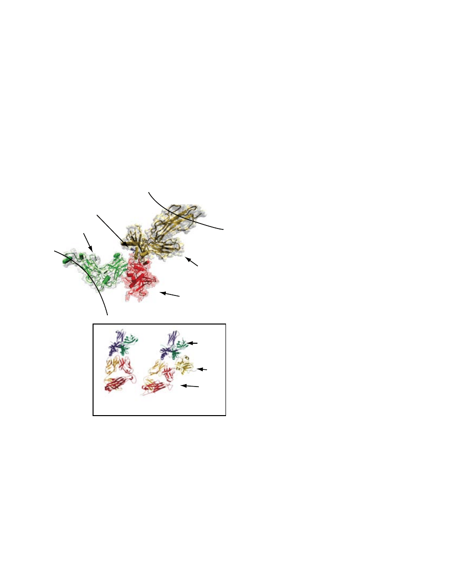
presenting cells (Figure 14-1). superantigens also
cross-link t-cell antigen receptors and class ii mhc
molecules, mimicking the cD4 molecule,
10
and hence
stimulate large numbers of t cells. in addition, each
superantigen preferentially stimulates t cells bearing
distinct subsets of antigen receptors, predominantly
dictated by the specific Vβ chain. an intense and
rapid release of cytokines such as interferon-γ, inter-
leukin-6 and tumor necrosis factor-α is responsible
for the systemic effects of the toxins.
11
in addition to
direct t-cell activation, the gastrointestinal illness
especially prominent after ingestion of staphylococ-
cal enterotoxins is also associated with histamine and
leukotriene release from mast cells.
12
Furthermore,
the cD44 molecule reportedly provides protection
from liver damage in mice caused by seb exposure
through a mechanism linked to activation-induced
apoptosis of immune cells.
13
individuals within the human population may re-
spond differently to superantigen exposure as a result
of mhc polymorphisms, age, and many physiological
factors. each toxin exhibits varying affinities toward
the hla-DR, DQ, and DP isotypes and distinct alleles
of class ii mhc molecules, observed by differences
in t-cell responses in vitro. in addition, primates, in-
cluding humans, are most sensitive to superantigens
compared to other mammals.
14
lethal or incapacitating
doses of toxin may be lowered by coexposure to endo-
toxin from gram-negative bacteria
11
or hepatotoxins,
15
or by infection with replicating agents.
2
Rodents and other domestic animals infected with
strains that produce tsst-1 and se
16,17
are potential
environmental reservoirs. both ovine- and-bovine spe-
cific staphylococcal toxins, which are associated with
mastitis, are almost identical to tsst-1 in amino acid
sequence.
18
toxigenic strains are frequent or universal
in both clinical and nonclinical isolates of S aureus and
S pyogenes, and these strains contribute significantly to
several diseases. approximately 50% of nonmenstrual
toxic shock syndrome (tss) cases are linked to tsst-1,
while the remaining cases are attributable to se, with
seb predominating.
19
Kawasaki’s syndrome and some
forms of arthritis are loosely associated with organisms
producing streptococcal pyrogenic exotoxins (sPes),
sea, and tsst-1.
20
in addition, streptococcal pneumo-
nia with accompanying tss-like symptoms is caused
by sPe-producing bacteria.
21
most of the streptococcal superantigens are encoded
by mobile genetic elements. sPe-a, sPe-c, sea, and
see are all phage-borne, while seD is plasmid-en-
coded. a chromosomal cluster of se and se-like genes
is present in strains of S aureus.
22
because little evi-
dence of genetic drift exists, it has been hypothesized
that the majority of staphylococcal and streptococcal
tss-like bacterial isolates have each descended from
single clones.
23
Production of many ses is dependent
on the phase of cell-growth cycle, environmental ph,
and glucose concentration. transcriptional control of
tsst-1, seb, sec, and seD is mediated through the
accessory gene regulator (agr) locus,
24
whereas sea
expression appears to be independent of agr. strains
that are agr-negative are generally low toxin producers.
antigen-presenting cell
T lymphocyte
T cell antigen
receptor
peptide
SEB
HLA-DR
SEB
HLA-DR
TCR
TCR-[HLA-DR]
TCR-[HLA-DR]-SEB
Fig. 14-1. molecular model of receptor binding. staphylococ-
cal enterotoxins and other bacterial superantigens target the
multireceptor communication between t cells and antigen-
presenting cells that is fundamental to initiating pathogen-
specific immune clearance. the superantigen inserts itself
between the antigen receptor of t cells and the class ii major
histocompatibility complex molecule displaying peptides
from potential pathogens. toxin exposure results in hy-
peractivation of the immune system, and the pathology is
mediated by tumor necrosis factor-α, interferon-γ, and other
cytokines.
hla-DR: human leukocyte antigen DR
seb: staphylococcal enterotoxin b
tcR: t cell receptor

314
Medical Aspects of Biological Warfare
however, there are also considerable differences in
production levels among agr-positive isolates. in ad-
dition, a feedback-mediated regulatory mechanism for
increasing expression of seb and tsst-1 and suppress-
ing all other exotoxins has been demonstrated.
25
at the cellular level, the interaction of superantigens
with receptors on antigen-presenting cells and t cells
leads to intracellular signaling.
26
high concentrations
of seb elicit phosphatidyl inositol production and
activation of protein kinase c and protein tyrosine ki-
nase pathways,
26–28
similar to mitogenic activation of t
cells. ses also activate transcription factors nF-κb and
aP-1, resulting in the expression of proinflammatory
cytokines, chemokines, and adhesion molecules. both
interleukin-1 and tumor necrosis factor-α can directly
activate the transcription factor nF-κb in many cell
types, including epithelial cells and endothelial cells,
perpetuating the inflammatory response. another
mediator, interferon-γ, produced by activated t cells
and natural killer cells, synergizes with tumor ne-
crosis factor-α and interleukin-1 to enhance immune
reactions and promote tissue injury. the substances
induced directly by seb and other superantigens—
chemokines, interleukin-8, monocyte chemoattractant
protein-1, macrophage inflammatory protein-1α, and
macrophage inflammatory protein-1β—can selectively
chemoattract and activate leukocytes. thus, cellular
activation by seb and other superantigens leads to
severe inflammation, hypotension, and shock. addi-
tional mediators contributing to seb-induced shock
include prostanoids, leukotrienes, and tissue factor
from monocytes; superoxide and proteolytic enzymes
from neutrophils; tissue factor; and chemokines from
endothelial cells. activation of coagulation via tissue
factor leads to disseminated intravascular coagulation,
tissue injury, and multiorgan failure. se-induced tss
thus presents a spectrum and progression of clinical
symptoms, including fever, tachycardia, hypotension,
multiorgan failure, disseminated intravascular coagu-
lation, and shock.
Given the complex pathophysiology of toxic shock,
the understanding of the cellular receptors and signal-
ing pathways used by staphylococcal superantigens,
and the biological mediators they induce, has provided
insights to selecting appropriate therapeutic targets.
Potential targets to prevent the toxic effects of ses
include (a) blocking the interaction of ses with the
mhc, tcRs,
26
or other costimulatory molecules
29–32
;
(b) inhibition of signal transduction pathways used
by ses
26
; (c) inhibition of cytokine and chemokine
production
33,34
; and (d) inhibition of the downstream
signaling pathways used by proinflammatory cyto-
kines and chemokines.
most therapeutic strategies in animal models of
seb-induced shock have targeted proinflammatory
mediators. therapeutic regimens include corticoste-
roids and inhibitors of cytokines, caspases, or phos-
phodiesterases. although several clinical trials of
treatment of sepsis with high-dose corticosteroids were
unsuccessful, a multicenter clinical trial using lower
doses of corticosteroids for longer periods reduced
the mortality rate of septic shock.
35
a newer interven-
tion targeting the coagulation pathway by activated
protein c improved the survival of septic patients
with high aPache (acute Physiology and chronic
health evaluation, a system for classifying patients
in the intensive care unit) score.
36
because coagulation
and endothelial dysfunction are important facets of
seb-induced shock, activated protein c may also be
useful in treating tss.
limited therapeutics for treating superantigen-
induced toxic shock are currently available. intrave-
nous immune globulin was effective as a treatment
in humans after the onset of tss. antibody-based
therapy targeting direct neutralization of seb or other
superantigens represents another form of therapeu-
tics, most suitable during the early stages of exposure
before cell activation and the release of proinflamma-
tory cytokines. because some neutralizing antibodies
cross-react among different superantigens,
8
a relatively
small mixture of antibodies might be effective in treat-
ing exposures to a greater variety of superantigens.
Vaccines of seb and sea with altered critical residues
involved in binding class ii mhc molecules were also
used successfully to vaccinate mice and monkeys
against seb-induced disease.
37,38
PATHOGENESIS
Rhesus macaques (Macaca mulatta) have been used
extensively as a model for lethal disease caused by
inhaled seb. Rabbits, endotoxin-primed mice, and ad-
ditional animal models have been developed. because
seb and related toxins primarily affect primates, the
following unpublished rhesus monkey data are highly
relevant for understanding potential human pathol-
ogy. Young and mature adult male and female rhesus
monkeys developed signs of seb intoxication
39
after
being exposed to a lethal dose of aerosolized seb for
10 minutes in a modified henderson head-only aerosol
exposure chamber.
40
these animals demonstrated no
detectable anti-seb antibody before exposure. after
inhalation exposure, microscopic lymphoproliferation
of t-cell–dependent areas of the lymphoid system,
consistent with the potent stimulatory effect of seb
315
Staphylococcal Enterotoxin B and Related Toxins
on the rhesus monkey immune system, was appar-
ent. immunohistochemical analysis, using anti-cD3
antibody, of the large lymphocytes present in the pul-
monary vasculature of the monkeys identified these
lymphocytes as t cells.
41
Generally, the seb-intoxicated rhesus monkeys de-
veloped gastrointestinal distress within 24 hours post-
exposure. clinical signs were mastication, anorexia,
emesis, and diarrhea. after mild, brief, self-limiting
gastrointestinal signs, the monkeys had a variable
period of up to 40 hours of clinical improvement. at
approximately 48 hours postexposure, the monkeys
generally had an abrupt onset of rapidly progressive
lethargy, dyspnea, and facial pallor, culminating in
death or euthanasia within 4 hours of onset.
at necropsy, most of the monkeys had similar gross
pulmonary lesions. the lungs were diffusely heavy
and wet, with multifocal petechial hemorrhages and
areas of atelectasis. clear serous-to-white frothy fluid
often drained freely from the laryngeal orifice. the
small and large intestines frequently had petechial
hemorrhages and mucosal erosions. typically, the
monkeys had mildly swollen lymph nodes, with moist
and bulging cut surfaces.
most of the monkeys also had similar microscopic
pulmonary lesions. the most obvious lesion was
marked multifocal to coalescing interstitial pulmonary
edema involving multiple lung lobes. Peribronchovas-
cular connective tissue spaces were distended by pale,
homogeneous, eosinophilic, proteinaceous material
(edema), variably accompanied by entrapped, beaded
fibrillar strands (fibrin), extravasated erythrocytes,
neutrophils, macrophages, and small and large lym-
phocytes. Perivascular lymphatics were generally
distended by similar eosinophilic material and inflam-
matory cells. most of the monkeys had intravascular
circulating and marginated neutrophils, monocytes,
mononuclear phagocytes, and lymphocytes, including
large lymphocytes with prominent nucleoli (lympho-
blasts), some in mitosis (Figure 14-2). extravascular
extension of these cell types was interpreted as exo-
cytosis/chemotaxis.
loss of airway epithelium was inconsistent. some
monkeys had multifocal, asymmetric denudation of
bronchial epithelium, with near total loss of bronchiolar
epithelium. Former bronchioles were recognized only
by their smooth muscle walls. scant bronchial intralumi-
nal exudate consisted of mucoid material, neutrophils,
macrophages, and sloughed necrotic cells.
a common finding was multifocal alveolar flood-
ing and acute purulent alveolitis. alveolar septa
were distended by congested alveolar capillaries.
alveolar spaces were filled with pale, homogeneous,
eosinophilic material (edema), with deeper embedded
eosinophilic beaded fibrillar strands (fibrin), or with
condensed, curvilinear, eosinophilic deposits hugging
the alveolar septal contours (hyaline membranes). a
variably severe cellular infiltrate of neutrophils, eosino-
phils, small lymphocytes, large lymphocytes (lympho-
blasts), erythrocytes, and alveolar macrophages filled
alveolar spaces. Replicate pulmonary microsections
stained with phosphotungstic-acid–hematoxylin
demonstrated alveolar fibrin deposition. Replicate
microsections stained with Giemsa revealed scarce
sparsely granulated connective-tissue mast cells.
in the upper respiratory tract, the tracheal and
bronchial lamina propria was thickened by clear
space or pale, homogeneous, eosinophilic material
(edema), neutrophils, small and large lymphocytes,
and (possibly preexisting) plasma cells. the edema
and cellular infiltrate extended transtracheally into the
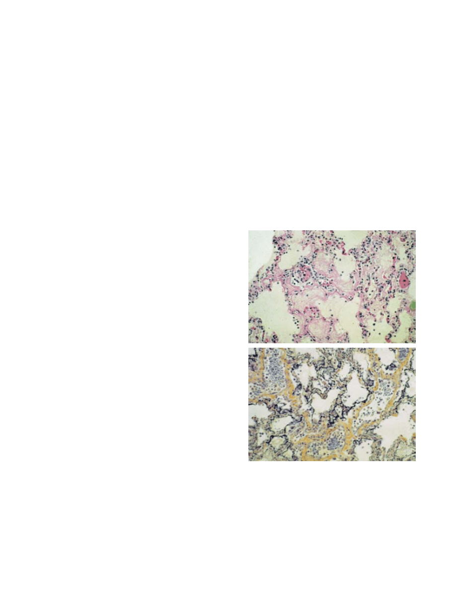
Fig. 14-2. lung of a rhesus monkey that died from inhaled
staphylococcal enterotoxin b. (a) marked perivascular
interstitial edema and focal loss of bronchial epithelium
can be seen (hematoxylin-eosin stain, original magnifica-
tion x 10). (b) the intravascular mononuclear cells include
lymphocytes, lymphoblasts, monocytes, and mononuclear
phagocytes (hematoxylin-eosin stain, original magnifica-
tion x 50).
a
b
316
Medical Aspects of Biological Warfare
mediastinum, with moderate to marked mediastinal
lymphangiectasia.
lymphoid tissues of the respiratory tract had
depletion of b-cell–dependent areas and hyperplasia
of t-cell–dependent areas. the bronchus-associated
lymphoid tissue in some of the monkeys had follicular
lymphocytic depletion. most of the mediastinal lymph
nodes had subcapsular and medullary sinus edema,
histiocytosis, and paracortical lymphoid hyperplasia,
characterized by numerous closely packed small
lymphocytes with interspersed macrophages bearing
tingible bodies and large lymphocytes having promi-
nent nucleoli (lymphoblasts) (Figure 14-3). there were
scattered mitoses, including atypical mitoses. cortical
follicles had small solid centers or hypocellular, hyalin-
ized (depleted) centers.
microscopic changes in lymphoid tissues elsewhere
in the body mirrored changes in the respiratory mu-
cosal lymphoid tissue. mesenteric, axillary, inguinal,
and retropharyngeal lymph nodes had sinus edema
and histiocytosis, paracortical lymphocytic and lym-
phoblastic hyperplasia, and unstimulated or depleted
follicular centers. also depleted were follicular germi-
nal centers of gut-associated lymphoid tissue. splenic
t-cell–dependent periarteriolar sheath zones were
hypercellular, populated by a mix of small and large
lymphocytes and macrophages, whereas b-cell–de-
pendent follicular areas were not recognized. several
monkeys had marked diffuse depletion of cortical
thymocytes, with a “starry sky” appearance attributed
to the presence of numerous thymic macrophages
bearing tingible bodies.
many of the monkeys had a mild erosive enteroco-
litis, with slight, superficial, multifocal mucosal loss
and with numerous lamina proprial macrophages
bearing engulfed cellular debris. crypt enterocytes
had a high nuclear-to-cytoplasmic ratio and numer-
ous mitoses. the crypt epithelium had a conspicuous
population of large mononuclear intraepithelial leu-
kocytes interpreted as lymphoblasts (Figure 14-4). in
the colon of some monkeys, there were many small
crypt abscesses.
Generalized vascular changes in most of the
monkeys were congestion, swollen endothelial cells
with many large intravascular lymphocytes or lym-
phoblasts and inconsistent widening of perivascular
connective tissue spaces (by edema). hepatic lesions
were portal infiltrates of lymphocytes, lymphoblasts,
macrophages, and occasional neutrophils. the choroid
plexus was slightly thickened by edema.
Fig. 14-3. mediastinal lymph node of a rhesus monkey that
died from inhaled staphylococcal enterotoxin b. Paracortical
lymphoproliferation with lymphoblasts can be seen (hema-
toxylin-eosin stain, original magnification x 100).
Fig. 14-4. small intestine of a rhesus monkey that died from
inhaled staphylococcal enterotoxin b. intraepithelial lym-
phoblastic leukocytes can be seen (hematoxylin-eosin stain,
original magnification x 100).
CLINICAL DISEASE
the clinical documentation of tss provides perhaps
the most comprehensive source of information on the
pathology of superantigen (eg, seb) exposure. to meet
the strict centers for Disease control and Prevention
criteria for tss,
42
negative blood (except for S aureus or S
pyogenes), throat, or cerebrospinal fluid cultures, as well
as negative serologic tests for Rocky mountain spotted
fever, leptospirosis, and measles should be obtained.
although tss disease symptoms are well established,
characterized by a rapid drop in blood pressure, elevated
temperature, and multiple organ failure, the respiratory
route of exposure may involve some unique mechanisms.

317
Staphylococcal Enterotoxin B and Related Toxins
the profound hypotension and desquamation of the
palms and soles of the feet that are characteristic of tss
are not observed in exposure by inhalation, and respira-
tory involvement is rapid, unlike in other forms of tss.
Furthermore, the fever prominent after aerosol exposure
is generally not observed in cases of seb ingestion.
Documentation of an accidental laboratory inhala-
tion exposure of nine laboratory workers to seb best
exemplifies the clinical disease, described as a severely
incapacitating illness of rapid onset (3–4 hours) and
modest acute duration (3–4 days).
43
Fever
Fever was prominent in all nine of those exposed.
eight of the individuals experienced at least one
shaking chill that heralded the onset of illness. Using
the morning peak level of seb aerosol generation in
the laboratory as the most likely time of exposure,
onset of fever occurred from 8 to 20 hours post initial
exposure, with a mean time of onset of 12.4 ± 3.9 (sD)
hours. Duration of fever was from 12 to 76 hours after
onset, with a mean duration of 50 ± 22.3 hours. Fever
ranged as high as 106° acutely. myalgias were often
associated with the initial fever. onset of myalgia was
between 8 and 20 hours, with a mean onset of 13 ± 5
hours. Duration was from 4 to 44 hours, and the mean
duration was 16 ± 15 hours.
Respiratory Symptoms
all nine patients were admitted to the hospital with
a generally nonproductive cough. onset was at 10.4 ±
5.4 hours, and duration was 92 ± 41 hours. Five had
inspiratory rales with dyspnea. the three most seriously
compromised patients had dyspnea, moist inspiratory
and expiratory rales, and orthopnea that gradually
cleared. one individual had profound dyspnea for the
first 12 hours that moderated to exertional dyspnea and
rales, which persisted for 10 days. chest radiographs on
admission showed densities compatible with “patches
of pulmonary edema” and Kerley lines suggesting
interstitial edema. During recovery, discoid atelectasis
was noted. moderate compromise of the respiratory
system was often accompanied by radiographic evi-
dence of peribronchial accentuation or “cuffing.” the
mildly ill patients had normal radiographs. one of the
three severely ill patients had severe pulmonary com-
promise and profound dyspnea and received only slight
relief when treated with an aminophylline suppository.
moderately intense chest pain, of a substernal pleuritic
type, occurred in seven individuals. onset of chest pain
was at 12 ± 6.5 hours and lasted for 4 to 84 hours, with
a mean duration of 23 ± 27 hours.
Headache
eight of the nine patients experienced headache.
onset ranged from 4 to 36 hours, and the mean time
of onset was at 13.3 ± 10 hours. Duration ranged from
8 to 60 hours, with a mean duration of 30.6 ± 19 hours.
the headaches ranged from severe to mild, but were
usually mild by the second day of hospitalization.
Five individuals’ headaches responded to Darvon
(propoxyphene hydrochloride; eli lilly & co, india-
napolis, ind) or codeine.
Nausea and Vomiting
Gastrointestinal symptoms occurred in more than
half of the individuals, nausea and anorexia in six,
and vomiting in four. the onset of nausea ranged
from 8 to 24 hours, with a mean onset of 17 ± 6.3
hours. Duration ranged from 4 to 20 hours, with a
mean of 9 ± 5.5 hours. the time to onset of anorexia
ranged from 8 to 24 hours with a mean onset of
18.5 ± 5.6 hours. Duration of anorexia ranged from
4 to 136 hours, and the mean duration was 44.5 ±
45 hours. Vomiting occurred in four patients, some-
times after prolonged paroxysms of coughing. the
range of onset of vomiting was 8 to 20 hours, with
a mean time to onset of 14 ± 5.1 hours. Duration
was not prolonged and usually consisted of one
episode. the patients were successfully treated with
compazine (prochlorperazine; smithKline beecham
Pharmaceuticals, Philadelphia, Pa) and benadryl
(diphenhydramine hydrochloride; Pfizer Pharma-
ceuticals company, new York, nY). only one indi-
vidual demonstrated hepatomegaly and bile in the
urine, although another patient also demonstrated
mildly elevated liver-function tests. no diarrhea was
reported in any of the exposed individuals.
Other Signs and Symptoms
Cardiovascular
all patients who experienced chest pain had nor-
mal electrocardiograms. throughout the illness, all
patients were normotensive. Vomiting was of brief
duration, and no one, including those vomiting, re-
quired intravenous fluid administration. the patients’
pulse rates, when elevated, paralleled temperature
elevation.
Hematology
leukocytosis was observed in most of the patients
12 to 24 hours after exposure to the toxin.

318
Medical Aspects of Biological Warfare
Ocular Effects
none of the patients experienced conjunctivitis,
although one individual later stated he remembered
that his eyes had “burned” during the believed time
of exposure. this contrasts with reports of conjunc-
tivitis resulting from separate accidental laboratory
exposures.
44
DETECTION AND DIAGNOSIS
the staphylococcal enterotoxins are moderately
stable proteins; therefore, immunological evalua-
tion should be possible in field or clinical samples.
a variety of rapid and sensitive detection methods
are available.
45,46
immunoassays can detect picogram
quantities of toxins in environmental samples. Plasma
concentrations of superantigens were measured in
septic patients of an intensive care unit using an en-
zyme-linked immunosorbent assay.
47
in one study,
48
the mean concentration of tsst-1 in human sera from
tss patients was reported to be 440 pg/ml. in con-
trast, anti-tsst-1 antibody titers are often low in tss
patients
49,50
and only recover during convalescence.
Furthermore, most normal human serum samples
contain detectable levels of antibody reacting with sev-
eral different toxins, including seb. therefore, serum
antibody titers are of little diagnostic value. if bacte-
rial sepsis is suspected and cultures can be obtained,
detecting minute quantities of potentially toxigenic
strains is possible by using polymerase chain reaction
amplification and toxin gene-specific oligonucleotide
primers. the results from both polymerase chain reac-
tion and immunoassays are rapid, allowing quantita-
tive or qualitative measurements in less than 24 hours.
Finally, as the best approach to early diagnosis on the
battlefield, toxins may be identifiable in nasal swabs
from individuals exposed to aerosols for at least 12 to
24 hours postexposure.
MEDICAL MANAGEMENT
no specific therapy has been identified or described.
supportive therapy in the nine mild accidental expo-
sure cases described above seemed to provide adequate
care. symptoms of fever, muscle aches, and arthralgias
may respond to cool compresses, fluids, rest, and ju-
dicious use of acetaminophen or aspirin. For nausea,
vomiting, and anorexia, symptomatic therapy should
be considered. antihistamines (eg, diphenhydramine)
and phenothiazine derivatives (eg, prochlorperazine)
have been used parenterally or as suppositories. the
success of these drugs in controlling nausea may have
been augmented by the relatively short duration of
nausea and vomiting induced by aerosolized seb.
because of the brevity of vomiting episodes, fluid re-
placement was not considered or required in the series
discussed. however, replacement may be necessary
in the event of prolonged vomiting resulting in fluid
and electrolyte depletion. Diarrhea was not observed
in human accidental exposure cases, but deposition
of toxin on foodstuffs could produce the syndrome,
which should be treated symptomatically.
initial symptomatic therapy with cough sup-
pressants containing dextromethorphan or codeine
should be routinely employed. Prolonged coughing
unrelieved by codeine might benefit from a semisyn-
thetic centrally acting narcotic antitussive containing
hydrocodone (dihydrocodeinone).
Pulmonary status should be monitored by pulse
oximetry, and when respiratory status is compromised,
prompt evacuation to a site with capacity for intensive
respiratory care by mechanical ventilation should be
considered.
IMMUNOTHERAPY
infusion of intravenous immunoglobulin has been
successfully used
51,52
to treat episodes of Kawasaki’s
syndrome linked to se and tsst-1. an anecdotal
case of tss with elevated tsst-1 and sea levels,
complicated by life-threatening multiorgan dysfunc-
tion, was successfully treated by early introduction
of plasma exchanges.
53
Unpublished studies have
documented the prophylactic and therapeutic value
of human intravenous immunoglobulin in rhesus
monkeys after inhalation of seb, prescribed to the
presence of antibodies to se and tsst-1 in commercial
preparations of intravenous immunoglobulin and
normal human sera. Prior exposure to seb by inhala-
tion does not appear to protect against a subsequent
episode. however, increased antibody titers to seb
are protective, and efforts to devise both passive and
active immunotherapy show promise. because of the
rapidity of receptor binding by these toxins (appar-
ent saturation < 5 min), active immunity should be
considered as the best defense.

319
Staphylococcal Enterotoxin B and Related Toxins
VACCINES
were produced by substitution of active receptor-bind-
ing amino acid side chains that reduced affinities and
consequential t-cell activation,
7,9,37,38
without altering
the three-dimensional structure of the antigen. though
promising, these engineered vaccines are not yet li-
censed or available for general use.
a formalin-treated seb toxoid demonstrated some
degree of efficacy in animal trials, but is not approved
for human use. Vaccines produced by site-specific mu-
tagenesis of the toxins, delivered by intramuscular or
interdermal routes, have also shown promising results
in animal trials. these recombinant subunit vaccines
SUMMARY
seb is representative of a group of bacterial proteins
that exerts profound toxic effects upon the immune
system. many sensitive immunoassays have been
developed for laboratory detection of most of the
staphylococcal and streptococcal superantigen toxins,
but the limit of field detection is unknown. inhalation
exposure to agents such as seb may result in severe but
temporary incapacitation, while high-dose exposures
will result in fatalities. supportive symptomatic ther-
apy is the only known method of treatment. Vaccines
currently under development may afford protection to
individuals but are not yet licensed for human use.
ReFeRences
1. hursh s, mcnally R, Fanzone J Jr, mershon m. Staphylococcal Enterotoxin B Battlefield Challenge Modeling with Medical
and Non-Medical Countermeasures. Joppa, md: science applications international corp; 1995. technical Report mbDRP-
95-2.
2. Zhang WJ, sarawar s, nguyen P. lethal synergism between influenza infection and staphylococcal enterotoxin b in
mice. J Immunol. 1996;157:5049–5060.
3. alber J, el-sayed a, estoepangestie s, lammler c, Zschock m. Dissemination of the superantigen encoding genes
seel, seem, szel and szem in Streptococcus equi subsp. equi and Streptococcus equi subsp. zooepidemicus. Vet Microbiol.
2005;109:135–141.
4. stevens Dl. streptococcal toxic shock syndrome. in: leung DYm, huber bt, schlievert Pm, eds. Superantigens: Mo-
lecular Biology, Immunology, and Relevance to Human Disease. new York, nY: marcel Dekker, inc; 1997: 481–501.
5. Ulrich RG, bavari s, olson m. bacterial superantigens in human diseases: structure, function and diversity. Trends
Microbiol. 1995;3:463–468.
6. Jardetzky ts, brown Jh, Gorga Jc, et al. three-dimensional structure of a human class ii histocompatibility molecule
complexed with superantigen. Nature. 1994;368:711–718.
7. swietnicki W, barnie am, Dyas bK, Ulrich RG. Zinc binding and dimerization of Streptococcus pyogenes pyrogenic
exotoxin c are not essential for t-cell stimulation. J Biol Chem. 2003;278:9885–9895.
8. bavari s, Ulrich RG, leclaire RD. cross-reactive antibodies prevent the lethal effects of Staphylococcus aureus supe-
rantigens. J Infect Dis. 1999;180:1365–1369.
9. Ulrich RG, bavari s, olson m. staphylococcal enterotoxins a and b share a common structural motif for binding class
ii major histocompatibility complex molecules. Nat Struct Biol. 1995;2:554–560.
10. bavari s, Ulrich RG. staphylococcal enterotoxin a and toxic shock syndrome toxin compete with cD4 for human
major histocompatibility complex class ii binding. Infect Immun. 1995;63:423–429.
11. stiles bG, bavari s, Krakauer t, Ulrich RG. toxicity of staphylococcal enterotoxins potentiated by lipopolysaccharide: ma-
jor histocompatibility complex class ii molecule dependency and cytokine release. Infect Immun. 1993;61:5333–5338.

320
Medical Aspects of Biological Warfare
12. scheuber Ph, Denzlinger c, Wilker D, beck G, Keppler D, hammer DK. cysteinyl leukotrienes as mediators of
staphylococcal enterotoxin b in the monkey. Eur J Clin Invest. 1987;17:455–459.
13. mcKallip RJ, Fisher m, Gunthert U, szakal aK, nagarkatti Ps, nagarkatti m. Role of cD44 and its v7 isoform in
staphylococcal enterotoxin b-induced toxic shock: cD44 deficiency on hepatic mononuclear cells leads to reduced
activation-induced apoptosis that results in increased liver damage. Infect Immun. 2005;73:50–61.
14. bavari s, hunt Re, Ulrich RG. Divergence of human and nonhuman primate lymphocyte responses to bacterial su-
perantigens. Clin Immunol Immunopathol. 1995;76:248–254.
15. miethke t, Wahl c, heeg K, echtenacher b, Krammer Ph, Wagner h. t cell-mediated lethal shock triggered in mice
by the superantigen staphylococcal enterotoxin b: critical role of tumor necrosis factor. J Exp Med. 1992;175:91–98.
16. Kenny K, Reiser RF, bastida-corcuera FD, norcross nl. Production of enterotoxins and toxic shock syndrome toxin
by bovine mammary isolates of Staphylococcus aureus. J Clin Microbiol. 1993;31:706–707.
17. ho G, campbell Wh, bergdoll ms, carlson e. Production of a toxic shock syndrome toxin variant by Staphylococcus
aureus strains associated with sheep, goats, and cows. J Clin Microbiol. 1989;27:1946–1948.
18. lee PK, Kreiswirth bn, Deringer JR, et al. nucleotide sequences and biologic properties of toxic shock syndrome toxin
1 from ovine- and bovine-associated Staphylococcus aureus. J Infect Dis. 1992;165:1056–1063.
19. crass ba, bergdoll ms. involvement of staphylococcal enterotoxins in nonmenstrual toxic shock syndrome. J Clin
Microbiol. 1986;23:1138–1139.
20. Freedman JD, beer DJ. expanding perspectives on the toxic shock syndrome. Adv Intern Med. 1991;36:363–397.
21. Reichardt W, muller-alouf h, alouf Je, Kohler W. erythrogenic toxins a, b and c: occurrence of the genes and exotoxin
formation from clinical Streptococcus pyogenes strains associated with streptococcal toxic shock-like syndrome. FEMS
Microbiol Lett. 1992;79:313–322.
22. Jarraud s, Peyrat ma, lim a, et al. eGc, a highly prevalent operon of enterotoxin gene, forms a putative nursery of
superantigens in Staphylococcus aureus. J Immunol. 2001;166:669–677.
23. lee PK, schlievert Pm. molecular genetics of pyrogenic exotoxin “superantigens” of group a streptococci and Staphy-
lococcus aureus. Curr Top Microbiol Immunol. 1991;174:1–19.
24. betley mJ, borst DW, Regassa lb. staphylococcal enterotoxins, toxic shock syndrome toxin and streptococcal pyrogenic
exotoxins: a comparative study of their molecular biology. Chem Immunol. 1992;55:1–35.
25. Vojtov n, Ross hF, novick RP. Global repression of exotoxin synthesis by staphylococcal superantigens. Proc Natl Acad
Sci U S A. 2002;99:10102–10107.
26. chatila t, Geha Rs. signal transduction by microbial superantigens via mhc class ii molecules. Immunol Rev.
1993;131:43–59.
27. chatila t, Wood n, Parsonnet J, Geha Rs. toxic shock syndrome toxin-1 induces inositol phospholipid turnover,
protein kinase c translocation, and calcium mobilization in human t cells. J Immunol. 1988;140:1250–1255.
28. scholl PR, trede n, chatila ta, Geha Rs. Role of protein tyrosine phosphorylation in monokine induction by the
staphylococcal superantigen toxic shock syndrome toxin-1. J Immunol. 1992;148:2237–2241.
29. linsley Ps, ledbetter Ja. the role of the cD28 receptor during t cell responses to antigen. Annu Rev Immunol.
1993;11:191–212.
30. Fraser J, newton m, Weiss a. cD28 and t cell antigen receptor signal transduction coordinately regulate interleukin
2 gene expression in response to superantigen stimulation. J Exp Med. 1992;175:1131–1134.

321
Staphylococcal Enterotoxin B and Related Toxins
31. Krakauer t. costimulatory receptors for the superantigen staphylococcal enterotoxin b on human vascular endothelial
cells and t cells. J Leukoc Biol. 1994;56:458–463.
32. saha b, Jaklic b, harlan Dm, Gray Gs, June ch, abe R. toxic shock syndrome toxin-1-induced death is prevented by
ctla4ig. J Immunol. 1996;157:3869–3875.
33. Krakauer t. immune response to staphylococcal superantigens. Immunol Res. 1999;20:163–173.
34. Krakauer t. inhibition of toxic shock syndrome toxin-1-induced cytokine production and t cell activation by inter-
leukin-10, interleukin-4, and dexamethasone. J Infect Dis. 1995;172:988–992.
35. annane D, sebille V, charpentier c, et al. effect of treatment with low doses of hydrocortisone and fludrocortisone
on mortality in patients with septic shock. JAMA. 2002;288:862–871.
36. bernard GR, Vincent Jl, laterre PF, et al. efficacy and safety of recombinant human activated protein c for severe
sepsis. N Engl J Med. 2001;344:699–709.
37. Ulrich RG, olson m, bavari s. bacterial superantigen vaccines. in: brown F, norrby e, burton D, mekalanos J, eds.
Vaccines 96: Molecular Approaches to the Control of Infectious Diseases. cold spring harbor, nY: cold spring harbor
laboratory Press; 1996: 135–141.
38. bavari s, Dyas b, Ulrich RG. superantigen vaccines: a comparative study of genetically attenuated receptor-binding
mutants of staphylococcal enterotoxin a. J Infect Dis. 1996;174:338–345.
39. Wilhelmsen cl. Unpublished observations, December 1994.
40. henderson DW. an apparatus of the study of airborne infection. J Hyg. 1952;50:53–68.
41. mattix me, hunt Re, Wilhelmsen cl, Johnson aJ, baze Wb. aerosolized staphylococcal enterotoxin b-induced pul-
monary lesions in rhesus monkeys (Macaca mulatta). Toxicol Pathol. 1995;23:262–268.
42. centers for Disease control and Prevention. toxic shock syndrome, United states, 1970–1982. MMWR Morb Mortal
Wkly Rep. 1982;31:201–204.
43. Rusnak Jm. Personal communication, December 2006.
44. Rusnak Jm, Kortepeter m, Ulrich R, Poli m, boudreau e. laboratory exposures to staphylococcal enterotoxin b. Emerg
Infect Dis. 2004;10:1544–1549.
45. alefantis t, Grewal P, ashton J, Khan as, Valdes JJ, Del Vecchio VG. a rapid and sensitive magnetic bead-based
immunoassay for the detection of staphylococcal enterotoxin b for high-throughput screening. Mol Cell Probes.
2004;18:379–382.
46. shriver-lake lc, shubin Ys, ligler Fs. Detection of staphylococcal enterotoxin b in spiked food samples. J Food Prot.
2003;66:1851–1856.
47. azuma K, Koike K, Kobayashi t, mochizuki t, mashiko K, Yamamoto Y. Detection of circulating superantigens in an
intensive care unit population. Int J Infect Dis. 2004;8:292–298.
48. miwa K, Fukuyama m, Kunitomo t, igarashi h. Rapid assay for detection of toxic shock syndrome toxin 1 from hu-
man sera. J Clin Microbiol. 1994;32:539–542.
49. crass ba, bergdoll ms. toxin involvement in toxic shock syndrome. J Infect Dis. 1986;153:918–926.
50. chesney PJ, bergdoll ms, Davis JP, Vergeront Jm. the disease spectrum, epidemiology, and etiology of toxic-shock
syndrome. Annu Rev Microbiol. 1984;38:315–338.

322
Medical Aspects of Biological Warfare
51. takei s, arora YK, Walker sm. intravenous immunoglobulin contains specific antibodies inhibitory to activation of t
cells by staphylococcal toxin superantigens. J Clin Invest. 1993;91:602–607.
52. leung DY, meissner hc, Fulton DR, murray Dl, Kotzin bl, schlievert Pm. toxic shock syndrome toxin-secreting
Staphylococcus aureus in Kawasaki syndrome. Lancet. 1993;342:1385–1388.
53. Kohro s, imaizumi h, Yamakage m, masuda Y, namiki a, asai Y. Reductions in levels of bacterial superantigens/canna-
binoids by plasma exchange in a patient with severe toxic shock syndrome. Anaesth Intensive Care. 2004;32:588–591.
Wyszukiwarka
Podobne podstrony:
Bezp Państwa T 2, W 10, BW
Genomes3e ppt ch14
45 06 BW Hydraulika stosowana
51 07 BW Gospodarka wodna
factoring00wellrich bw
Cersanit wanna, Resources, Budownictwo, BUDOWNICTWO OGÓLNE, Budownictwo Ogólne I i II, Budownictwo o
Akumulator do BOMAG BW BW??L
hartalgebracou0wellrich bw
44 06 BW Budowle wodne
elementarypracti00doddrich bw
bw 1
elementsofalgebr00lillrich bw
BW ch23
IncentiveBee BW
elementarytreati00sherrich bw
temat vi, BW I WSPOL
więcej podobnych podstron