
229
Mol. Nutr. Food Res. 2006, 50, 229 – 234
DOI 10.1002/mnfr.200500156
Review
Green tea catechins as brain-permeable, natural iron
chelators-antioxidants for the treatment of
neurodegenerative disorders
Silvia Mandel, Tamar Amit, Lydia Reznichenko, Orly Weinreb and Moussa B. H. Youdim
Eve Topf and US NPF Centers for Neurodegenerative diseases and Department of Pharmacology, Faculty of
Medicine, Technion, Haifa, Israel
Neurodegeneration in Parkinson’s, Alzheimer’s, or other neurodegenerative diseases appears to be
multifactorial, where a complex set of toxic reactions, including oxidative stress (OS), inflammation,
reduced expression of trophic factors, and accumulation of protein aggregates, lead to the demise of
neurons. One of the prominent pathological features is the abnormal accumulation of iron on top of
the dying neurons and in the surrounding microglia. The capacity of free iron to enhance and promote
the generation of toxic reactive oxygen radicals has been discussed numerous times. The observations
that iron induces aggregation of inert a-synuclein and beta-amyloid peptides to toxic aggregates have
reinforced the critical role of iron in OS-induced pathogenesis of neurodegeneration, supporting the
notion that a combination of iron chelation and antioxidant therapy may be one significant approach
for neuroprotection. Tea flavonoids (catechins) have been reported to possess divalent metal chel-
ating, antioxidant, and anti-inflammatory activities, to penetrate the brain barrier and to protect neu-
ronal death in a wide array of cellular and animal models of neurological diseases. This review aims
to shed light on the multipharmacological neuroprotective activities of green tea catechins with spe-
cial emphasis on their brain-permeable, nontoxic, transitional metal (iron and copper)-chelatable/radi-
cal scavenger properties.
Keywords: ( – )-epigallocatechin-3-gallate / Flavonoid / Hypoxia / Neurodegeneration / Parkinson’s disease
/
Received: September 10, 2005; accepted: October 18, 2005
1 Introduction
The consumption of tea (Camellia sinensis) is believed to
have been initiated five thousands years ago in China and
India. Tea is commonly associated with traditional beverage
rituals and particular lifestyles, especially in Japan, China,
India, and England; nevertheless, nowadays it is considered
as a source of dietary constituents endowed with biological
and pharmacological activities with potential benefits to
human health. Indeed, it is the novel pharmacological activ-
ities that are arousing interest in their possible clinical use
for prevention and therapeutics in several diseases. Several
of these are subject, in the last few years, to intensive investi-
gation in diverse medical disciplines, such as cardiology,
oncology, inflammatory diseases, and neurology [1–3]. The
favorable properties of green tea (GT) extract have been
ascribed to their high content of polyphenolic flavonoids.
Fresh tea leaves contains a high amount of catechins, a group
of flavonoids or flavanols, known to constitute 30–45% of
the solid GT extract [4, 5]. Catechin polyphenols have been
demonstrated to act directly as radical scavengers of oxygen
and nitrogen species and exert indirect antioxidant effects
through activation of transcription factors and antioxidant
enzymes, thus modulating the cellular redox state (see
reviews: [1, 6, 7]. In addition to their radical-scavenging
action, GT catechins possess well-established metal-chelat-
ing properties. Structurally important features defining their
chelating potential are the 39,49-dihydroxyl group in the B
ring [8], as well as the gallate group [9, 10], which may neu-
tralize ferric iron to form redox-inactive iron, thereby pro-
tecting cells against oxidative damage [11].
Correspondence: Dr. Silvia Mandel, Efron St. P.O.B. 9697, Haifa
31096, Israel
E-mail: mandel@tx.technion.ac.il
Fax: +972-4-851-3145
Abbreviations: Ab, amyloid beta; AD, Alzheimer’s disease; APP,
amyloid precursor protein; DFO, desferrioxamine; EGCG, ( – )-epi-
gallocatechin-3-gallate; HIF-1, hypoxia inducible factor-1; IRE, iron
responsive element; IRP, iron regulatory protein; OS, oxidative stress;
PD, Parkinson’s disease; PKC, protein kinase C; sAPPa, soluble APP-
alpha; SN, substantia nigra
i
2006 WILEY-VCH Verlag GmbH & Co. KGaA, Weinheim
www.mnf-journal.com

S. Mandel et al.
Mol. Nutr. Food Res. 2006, 50, 229 – 234
The importance of polyphenolic flavonoids in enhancing
cell resistance to oxidative stress (OS) goes beyond the sim-
ple scavenging activity and is mostly interesting in those
pathologies where OS plays an important role, such as in
neurodegenerative diseases and aging. Aging is character-
ized by decrements in tissue function and accumulation of
mitochondrial DNA mutations, particularly in the brain that
contains postmitotic cells. Many lines of evidence suggest
that iron-mediated OS resulting in reactive oxygen species
(ROS) generation and inflammation plays a pivotal role in
the age-associated cognitive decline and neuronal loss in
neurodegenerative diseases including Alzheimer’s, Parkin-
son’s, and Huntington’s diseases (AD, PD, and HD, respec-
tively). Transitional metal alterations (e.g., iron, copper,
and zinc) have been described in brains of Parkinsonian
patients and of other neurodegenerative diseases, which
may be caused, to a large degree, by endogenous dysregula-
tion of iron uptake, transport, distribution, and storage [12,
13]. The redox-active metals are promoters of membrane-
associated OS including lipid peroxidation and oxidative
modifications of membranes and their coupled proteins,
such as receptors. Dietary metals may influence the risk of
PD, AD, and other neurodegenerative disorders [14], while
persistent iron deprivation has been shown to protect cortex
and hippocampal cells from kainate-induced damage [15].
Furthermore, neurochemical and genomics studies in PD,
and more recently in AD, have provided evidence for the
involvement of supplementary processes, including gluta-
matergic neurotoxicity, nitric oxide elevation, dysfunction
of ubiquitin-proteasome system, and mitochondria, which
may lead to breakdown of energy metabolism and consecu-
tive intraneuronal calcium overload, increased expression
of apoptotic proteins, and loss of tissue reduced glutathione
(GSH, an essential factor for removal of hydrogen perox-
ide) [12, 16–22]. These series of neurotoxic events may act
independently or cooperatively, leading eventually to the
demise of the neurons. Thus, considering the multifactorial
nature of neurodegenerative disorders, drugs directed
against a single target will be ineffective and rather a single
drug or cocktail of drugs with pluripharmacological proper-
ties may be more suitable to be employed.
One innovative therapeutic approach could be the use of
nontoxic, brain-permeable natural plant polyphenol flavo-
noids, reported to possess multifunctional activities, being
iron chelators, radical scavengers, anti-inflammators, and
neuroprotectants [6, 8, 9, 23, 24] as reviewed in [25, 26].
Research from our laboratory has demonstrated that the
antioxidant-iron chelating activity of the major GT poly-
phenol ( – )-epigallocatechin-3-gallate (EGCG) plays a
major role in the prevention of neurodegeneration in a vari-
ety of cellular and animal models of neurodegenerative dis-
eases [27, 28]. Furthermore, collective studies indicated
that beyond this property, catechin flavonoids regulate vari-
ous signaling pathways involved in cellular survival,
growth, and differentiation as protein kinase C (PKC) and
extracellular mitogen-activated protein kinase (MAPK)
[26, 29] and promotion of neurite outgrowth [30]. In addi-
tion, EGCG was shown to down-regulate proapoptotic
genes, such as bad, bax, mdm2, caspase-1, cyclin-depen-
dent kinase inhibitor p21, and TNF-related apoptosis-indu-
cing ligand (TRAIL) [31, 32], and to regulate transcrip-
tional activation [1, 7, 33–35]. These findings suggest that
GT extract may be a source of neuroprotectants, with parti-
cular relevance to neurodegenerative diseases where OS
has also been implicated.
2 GT catechins as brain-permeable,
nontoxic iron chelators to “iron out” iron
from the brain
One of the major pathology of progressive neurodegenera-
tive diseases is the accumulation of iron in the degenerating
neurons [36]. Various metals have been implicated in the
pathophysiology of certain neuropsychiatric diseases –
copper and iron in Wilson’s disease; aluminum, zinc, and
iron in AD; iron in PD, Friedreich’s ataxia, and Hallervor-
den-Spatz-syndrome, just to mention a few [37–39]. Studies
on human and animal brains have shown that the distribu-
tion of brain iron is uneven as compared to other metals.
Thus, iron is present in substantia nigra (SN), globus palli-
dus, and dentate gyrus at a concentration equal to or greater
than that found in the liver. These three brain regions are
known to be associated with neurodegenerative diseases
[40]. Redox-active iron has been observed in the peripheral
halo of Lewy body (LB), the morphological hallmark of PD,
also composed of lipids, aggregated a-synuclein (concen-
trating in the rim of LB), and ubiquitinated, hyperpho-
sphorylated neurofilament proteins [41]. a-synuclein asso-
ciated with presynaptic membrane is not toxic; however, a
number of recent studies [42–44] have shown that it forms
toxic aggregates in the presence of iron and this is consid-
ered to contribute to the formation of LB via OS. In AD,
changes in the levels of iron, ferritin, and transferrin recep-
tor (TfR) have been reported in the hippocampus and cere-
bral cortex [45–47]. Iron promotes both deposition of amy-
loid beta (Ab) peptides and induction of OS, which is asso-
ciated with the cerebral amyloid-containing plaques.
Indeed, it has been demonstrated that amyloid deposits are
enriched with zinc, iron, and copper [39]. Recently, redox-
active iron bound to ribosomes was demonstrated to oxidize
ribosomal RNA in AD [45]. In addition, iron may contribute
to AD via regulation of amyloid precursor protein (APP)
translation, resulting from the existence of an iron-respon-
sive element (IRE-type II) in the 59UTR region of APP
mRNA [48]. This is consistent with biochemical evidence
pointing to APP as a redox-active metalloprotein [49].
The involvement of metals in protein deposition in neurolo-
gical disorders has encouraged the development of iron
230
i
2006 WILEY-VCH Verlag GmbH & Co. KGaA, Weinheim
www.mnf-journal.com
Mol. Nutr. Food Res. 2006, 50, 229 – 234
Green tea catechins as natural iron chelators for neurodegeneration
chelators as a major new therapeutic strategy. Metal chela-
tion has the potential to prevent iron-induced ROS, OS, and
aggregation of a-synuclein and Ab. Indeed, the limited
number of neuroprotective studies that have been carried
out so far indicate that iron-chelation therapy could be a
viable neuroprotective approach for neurodegenerative dis-
orders [50–52]. Animal studies have shown neuroprotective
activity of the prototype iron chelator drug desferrioxamine
(DFO) and the antibiotic iron and copper chelator 5-chloro-
7-iodo-8-hydroxyquinoline (clioquinol) against the neuro-
toxins 6-hydroxydopamine (6-OHDA) and N-methyl-4-
phenyl-1,2,3,6-tetrahydropyridine (MPTP)-induced neuro-
toxicity in mice [53, 54]. However, DFO is a very poor brain
penetrating agent and clioquinol is highly toxic [55]. More
recently, the multifunctional iron chelator-monoamine oxi-
dase (MAO) A and B inhibitor, brain-permeable compound,
M-30, showed neuroprotective activities in neuronal rat
PC12 and P19 cell cultures against serum deprivation, 6-
OHDA [56, 57] and in MPTP-induced parkinsonism [58].
The ability of GT polyphenols to act as metal chelators [9–
11] and to have access to the brain makes them a novel pro-
mising therapeutic approach for treating AD, PD, and amyo-
trophic lateral sclerosis (ALS), in which accumulation of
iron has been found [12, 36, 59]. Ionic iron participates in
Fenton chemistry, generating cytotoxic oxygen radicals, the
most potent being the hydroxyl radical that is particularly
reactive with lipid membranes. Many in vitro studies have
clearly demonstrated the potent peroxyl radical-scavenging
abilities of GT polyphenols in preventing oxidation of lipid
membranes and low-density lipoproteins (LDL). Ingestion
of either black tea or GT extracts protected plasma LDL oxi-
dation in humans [60] and in rats fed with GT extract [61].
GTand black tea extracts were shown to strongly inhibit lipid
peroxidation promoted by iron-ascorbate in homogenates of
brain mitochondrial membranes [62]. A similar effect was
also reported using brain synaptosomes, in which the four
major polyphenol catechines of GT were shown to inhibit
iron-induced lipid peroxidation [9]. In the majority of these
studies, EGCG was shown to be more efficient as a radical
scavenger than its counterparts ECG, EC, and EGC, which
might be attributed to the presence of the trihydroxyl group
on the B ring and the gallate moiety at the 39 position in the C
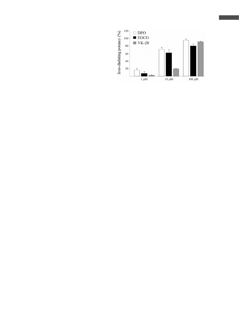
ring. [63]. In our hands, EGCG displayed iron-chelating
potency similar to that of DFO and of our newly developed
nontoxic, lipophilic, brain-permeable iron chelator drug,
VK-28 (Fig. 1). Thus, the cytoprotective effect of tea poly-
phenols against lipid peroxidation may reflect a combination
of a direct scavenging of oxygen, nitrogen, and lipid radicals,
as well as iron chelation.
3 EGCG regulates APP generation/
processing and Ab formation
The capacity of catechins to neutralize excess of free iron
may have a direct implication to AD, which is inherent pri-
marily, to the nature of APP as an iron-regulated protein [48,
64]. APP is post-transcriptionally regulated by iron regula-
tory proteins (IRPs), which are labile iron pool-sensitive
cytosolic RNA proteins, binding specifically to the IREs
located in the 59 or 39 untranslated regions of iron metabo-
lism-associated mRNAs. Thus, reduction of the free-iron
pool by EGCG chelation may lead to suppression of APP
mRNA translation, by targeting the IRE-II sequences in the
APP 59 UTR [48], as was recently shown for DFO and the
bifunctional amyloid-binding/metal-chelating drug XH1
[65] (Fig. 2). In accordance, our recent studies have shown
that prolonged administration of EGCG to mice induced a
reduction in holo-APP levels in the hippocampus [66]. This
is supported by the ability of EGCG to induce a significant
down-regulation of membrane-associated holo-APP levels
in neuroblastoma SH-SY5Y cells (Fig. 3), an effect that was
accompanied by a concomitant decrease in Ab levels, simi-
lar to the novel iron chelator M30, a VK-28 series derivative
(submitted for publication). Furthermore, wine and GT
polyphenols are able to inhibit formation, extension, and
destabilization of Ab fibrils [67], and to protect against Ab-
induced neurotoxicity [66]. Attenuation of APP synthesis
and consequential Ab production by EGCG could be of
therapeutic value for AD therapy, as increased generation of
Ab plays a central role in AD plaque formation [68]. Indeed,
overexpression of mutant human APP gene in transgenic
mice was found to produce excessive Ab, cerebral amyloid
deposition, and an Alzheimer-like pathology [69]. The
increased promotion of holo-APP expression after ische-
mia, hyperglycemia, traumatic brain injury, and cellular
energy depletion have been shown to route the APP metabo-
lism from the nonamyloidogenic to the amyloidogenic path-
way [70–74].
231
i
2006 WILEY-VCH Verlag GmbH & Co. KGaA, Weinheim
www.mnf-journal.com
Figure 1. Iron-chelating activity of EGCG, VK-28, and DFO on
Fe(II). Comparative analysis of the Fe(II) chelating potency of
EGCG, VK-28, and DFO was performed, assessing their abil-
ity to compete with ferrozine for the ferrous ions and further
ferrous ferrozine complexes formation, thereby resulting in a
decrease in the absorbance at 562 nm. Drugs were mixed
with 50 lM ferrozine in 5% ammonium acetate (pH 7) followed
by the addition of 10 lM Fe
2
SO
4
for 2 h. Percentage of the
chelating effect was calculated using the following equation:
[1–(absorbance of sample at 562 nm)/(absorbance of control,
without drugs, at 562 nm)]6100.
S. Mandel et al.
Mol. Nutr. Food Res. 2006, 50, 229 – 234
The other important pharmacological action of EGCG is
related to the recent observation that EGCG promotes the
generation of the soluble N-terminal fragment, soluble
APP-alpha (sAPPa), via PKC-dependent activation of the
enzyme a-secretase, thereby increasing the production of
the nontoxic sAPPa [66] (Fig. 3). This is supported by the
ability of EGCG to up-regulate PKCa and PKCe isoforms
in mice striatum and hippocampus [27, 66]. Since sAPPa
and Ab are formed by two mutually exclusive mechanisms,
stimulation of the secretory processing of sAPPa might pre-
vent the formation of the amyloidogenic Ab. Thus, EGCG
may influence Ab levels, either via translational inhibition
of APP or by stimulating sAPPa secretion (Fig. 4). Clea-
vage of APP within the Ab domain by a-secretases is of
physiological interest, not only because it precludes the for-
mation of Ab, but also because it promotes the generation
of sAPPa that exhibits neuroprotective properties [75, 76].
A number of reports supported the notion that promotion of
a-secretase-mediated APP processing, rather than down-
regulation of Ab production, might offer another approach
to AD treatment [77].
4 GT catechins and induction of iron/
hypoxia-responsive genes
The chelation of iron affects not only the post-transcriptional
regulation of iron homeostasis-related mRNAs (e.g., TfR,
ferritin), but also the induction of genes regulated by the tran-
scription factor hypoxia inducible factor-1 (HIF-1), a master
regulator orchestrating the coordinated induction of an array
of genes sensitive to hypoxia [78]. The target genes of HIF
are especially related to angiogenesis, cell proliferation/sur-
vival, and glucose/iron metabolism [79]. In this context, iron
was recently shown to overcome HIF-1 activation by the GT
catechins, EGCG and epicatechin-3-gallate (ECG), as well
as by DFO [34, 35]. In fact, both HIF-1 and IRP2 share a
common iron-dependent proteasomal degradation pathway,
by the action of key iron and oxygen sensors prolyl hydroxy-
lases, which become inactivated by iron chelation [80, 81].
Thus, the reduction in the free-iron pool by EGCG chelation
may result in the inhibition of prolyl hydroxylases and conse-
quently, in the concerted activation of both HIF and IRP2. As
IRPs and HIF-1 coordinate the expression of a wide array of
genes involved in cellular iron and glucose homeostasis, sur-
vival and proliferation [78, 82], their activation could be of
major importance in neurodegenerative diseases (for a
detailed explanation see Fig. 2).
5 Conclusions
The multifactorial nature of neurodegenerative diseases
makes the use of compounds with polypharmacological
activities or cocktail of drugs, a promising therapeutic
approach for the treatment of these disorders, as practiced
in the management of other diseases such as AIDS, ische-
mia, cancer, and neurotrauma. A wealth of new data sug-
gests that GT catechins may well fulfill the requirements
for a putative neuroprotective drug having diverse pharma-
cological activities. Ordinarily viewed as simple radical
232
i
2006 WILEY-VCH Verlag GmbH & Co. KGaA, Weinheim
www.mnf-journal.com
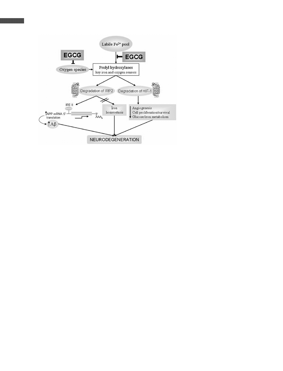
Figure 2. Iron-induced neurodegeneration in
AD via transcriptional activation of APP mRNA
and suppression of hypoxia-inducible genes.
Increase in labile Fe
2+
pool can elevate the pro-
duction of APP via proteasomal-mediated inac-
tivation of IRP2, thereby promoting the transla-
tion of APP mRNA from its 59UTR-typeII).
Increased iron and oxygen species may acti-
vate the prolyl hydroxylase enzymes, which are
key iron and oxygen sensors, leading to protea-
somal-mediated degradation of the transcrip-
tion factor HIF-1 a master regulator orchestrat-
ing the coordinated induction of a wide array of
survival genes. It has been suggested that
IRP2, similar to HIF-1, can be enzymatically
modified by a prolyl hydroxylase, routing it to
proteasomal degradation. Both iron chelation
and oxygen species scavenging by EGCG may
prevent the degradation of IRP2 and HIF-1,
resulting in the promotion of cell survival pro-
cesses such as angiogenesis, glucose metabo-
lism and maintenance of iron homeostasis.
EGCG, IRP, HIF-1. Sharp arrows indicate posi-
tive inputs, whereas blunt arrows are for inhibi-
tory inputs. For a more detailed explanation
read text.
Mol. Nutr. Food Res. 2006, 50, 229 – 234
Green tea catechins as natural iron chelators for neurodegeneration
scavengers, GT catechin polyphenols are considered at pre-
sent to invoke a spectrum of cellular mechanisms of action
related to their neuroprotection/neurorescue activities.
Recently, a new dimension was added to these actions, asso-
ciated with the iron-chelating property of GT catechins and
the impact on neurodegenerative processes, as oxidative
chain breakers and inhibitors of protein aggregation and Ab
plaque formation. Thus, GT catechins may be recognized
as multifunctional, brain-permeable iron chelators that can
prevent or delay neuronal death in the degenerating human
brain [64]. Being of natural origin, they may not exert toxic
side effects inherent to synthetic drugs.
6 References
[1] Higdon, J. V., Frei, B., Crit. Rev. Food Sci. Nutr. 2003, 43,
89 – 143.
[2] Galati, G., O’Brien, P. J., Free Radic. Biol. Med. 2004, 37,
287 – 303.
[3] Wiseman, S., Mulder, T., Rietveld, A., Antioxid. Redox Signal
2001, 3, 1009 – 1021.
[4] Wang, Z. Y., Huang, M. T., Lou, Y. R., Xie, J. G. et al., Cancer
Res. 1994, 54, 3428 – 3455.
[5] Yang, C. S., Wang, Z. Y., J. Natl. Cancer Inst. 1993, 85,
1038 – 1049.
[6] Rice-Evans, C., Curr. Med. Chem. 2001, 8, 797 – 807.
[7] Wiseman, S. A., Balentine, D. A., Frei, B., Crit. Rev. Food
Sci. Nutr. 1997, 37, 705 – 718.
[8] Hider, R. C., Liu, Z. D., Khodr, H. H., Methods Enzymol.
2001, 335, 190 – 203.
[9] Guo, Q., Zhao, B., Li, M., Shen, S. et al., Biochim. Biophys.
Acta 1996, 1304, 210 – 222.
[10] Kumamoto, M., Sonda, T., Nagayama, K., Tabata, M., Biosci.
Biotechnol. Biochem. 2001, 65, 126 – 132.
[11] Grinberg, L. N., Newmark, H., Kitrossky, N., Rahamim, E. et
al., Biochem. Pharmacol. 1997, 54, 973 – 978.
[12] Riederer, P., Sofic, E., Rausch, W. D., Schmidt, B. et al., J.
Neurochem. 1989, 52, 515 – 520.
[13] Connor, J. R., Snyder, B. S., Beard, J. L., Fine, R. E. et al., J.
Neurosci. Res. 1992, 31, 327 – 335.
[14] Mattson, M. P., Ann. N.Y. Acad. Sci. 2004, 1012, 37 – 50.
[15] Shoham, S., Youdim, M. B., Ann. N.Y. Acad. Sci. 2004, 1012,
94 – 114.
[16] Linazasoro, G., Expert. Rev. Neurotherapeutics 2002, 2,
403 – 416.
[17] McNaught, K. S., Belizaire, R., Jenner, P., Olanow, C. W. et
al., Neurosci. Lett. 2002, 326, 155 – 158.
[18] Blum, D., Torch, S., Lambeng, N., Nissou, M. et al., Prog.
Neurobiol. 2001, 65, 135 – 172.
[19] Grunblatt, E., Mandel, S., Jacob-Hirsch, J., Zeligson, S. et al.,
J. Neural. Transm. 2004, 111, 1543 – 1573.
[20] Zhang, Y., James, M., Middleton, F. A., Davis, R. L., Am. J.
Med. Genet. B Neuropsychiatr. Genet. 2005, 137, 5 – 16.
[21] Blalock, E. M., Geddes, J. W., Chen, K. C., Porter, N. M. et
al., Proc. Natl. Acad. Sci. USA 2004, 101, 2173 – 2178.
[22] Poon, H. F., Shepherd, H. M., Reed, T. T., Calabrese, V. et al.,
Neurobiol. Aging 2005.
[23] Morel, I., Lescoat, G., Cogrel, P., Sergent, O. et al., Biochem.
Pharmacol. 1999, 45, 13 – 19.
[24] Joseph, J. A., Shukitt-Hale, B., Casadesus, G., Am. J. Clin.
Nutr. 2005, 81, 313S – 316S.
[25] Mandel, S., Weinreb, O., Amit, T., Youdim, M. B. H., J. Neu-
rochem. 2004, 88, 1555 – 1569.
[26] Mandel, S. A., Avramovich-Tirosh, Y., Reznichenko, L.,
Zheng, H. et al., Neurosignals 2005, 14, 46 – 60.
[27] Mandel, S., Maor, G., Youdim, M. B. H., J. Mol. Neurosci.
2004, 24, 401 – 416.
233
i
2006 WILEY-VCH Verlag GmbH & Co. KGaA, Weinheim
www.mnf-journal.com
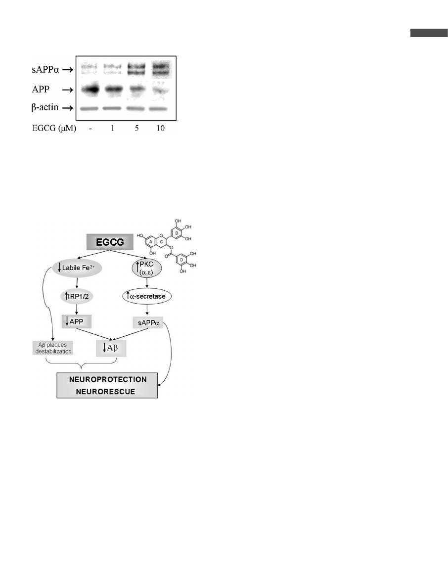
Figure 3. Effect of EGCG on APP protein and sAPPa release.
SH-SY5Y cells were incubated without or with increasing con-
centrations of EGCG (1–10 lM) for 2 days, and then APP and
sAPPa were evaluated in the cell lysate and medium, respec-
tively, by Western blot analysis. mAb against the APP N-termi-
nus (22C11) was used for APP detection, whereas mAb
recognizing an epitope within residues 1–17 of Ab domain of
APP (6E10) was employed for sAPPa determination.
Figure 4. Proposed schematic model for EGCG neuroprotec-
tive/neurorescue effects via regulation of APP processing and
Ab formation. Z, increased levels/activity; z decreased levels/
activity. For full explanation see text.

S. Mandel et al.
Mol. Nutr. Food Res. 2006, 50, 229 – 234
[28] Mandel, S., Youdim, M. B., Free Radic. Biol. Med. 2004, 37,
304 – 317.
[29] Schroeter, H., Boyd, C., Spencer, J. P., Williams, R. J. et al.,
Neurobiol. Aging 2002, 23, 861 – 880.
[30] Reznichenko, L., Amit, T., Youdim, M. B., Mandel, S., J.
Neurochem. 2005, 93, 1157 – 1167.
[31] Levites, Y., Amit, T., Youdim, M. B. H., Mandel, S., J. Biol.
Chem. 2002, 277, 30574 – 30580.
[32] Weinreb, O., Mandel, S., Youdim, M. B. H., FASEB J. 2003,
17, 935 – 937.
[33] Townsend, P. A., Scarabelli, T. M., Davidson, S. M., Knight,
R. A. et al., J. Biol. Chem. 2004, 279, 5811 – 5820.
[34] Zhou, Y. D., Kim, Y. P., Li, X. C., Baerson, S. R. et al., J. Nat.
Prod. 2004, 67, 2063 – 2069.
[35] Thomas, R., Kim, M. H., Biochem. Biophys. Res. Commun.
2005, 334, 543 – 548.
[36] Sofic, E., Paulus, W., Jellinger, K., Riederer, P. et al., J. Neu-
rochem. 1991, 56, 978 – 982.
[37] Moos, T., Morgan, E. H., Ann. N.Y. Acad. Sci. 2004, 1012,
14 – 26.
[38] Richardson, D. R., Ann. N.Y. Acad. Sci. 2004, 1012, 326 –
341.
[39] Atwood, C. S., Obrenovich, M. E., Liu, T., Chan, H. et al.,
Brain Res. Brain Res. Rev. 2003, 43, 1 – 16.
[40] Youdim, M. B. H., Riederer, P. in: Adelman, G., Smith, B.
(eds.), Iron in the Brain, Normal and Pathological, Encyclo-
pedia of Neurosci., Elsevier, Amsterdam 2004.
[41] Jellinger, K. A., Mov. Disord. 2003, 18 Suppl 6, S2 – S12.
[42] Turnbull, S., Tabner, B. J., El-Agnaf, O. M., Moore, S. et al.,
Free Radic. Biol. Med. 2001, 30, 1163 – 1170.
[43] Ostrerova-Golts, N., Petrucelli, L., Hardy, J., Lee, J. M. et al.,
J. Neurosci. 2000, 20, 6048 – 6054.
[44] Ebadi, M., Govitrapong, P., Sharma, S., Muralikrishnan, D. et
al., Biol. Signals Recept. 2001, 10, 224 – 253.
[45] Honda, K., Smith, M. A., Zhu, X., Baus, D. et al., J. Biol.
Chem. 2005, 280, 20978 – 20986.
[46] Beard, J. L., Connor, J. R., Jones, B. C., Nutr. Rev. 1993, 51,
157 – 170.
[47] Sipe, J. C., Lee, P., Beutler, E., Dev. Neurosci. 2002, 24, 188 –
196.
[48] Rogers, J. T., Randall, J. D., Cahill, C. M., Eder, P. S. et al., J.
Biol. Chem. 2002, 277, 45518 – 45528.
[49] Huang, X., Moir, R. D., Tanzi, R. E., Bush, A. I. et al., Ann.
N.Y. Acad. Sci. 2004, 1012, 153 – 163.
[50] Zecca, L., Youdim, M. B., Riederer, P., Connor, J. R. et al.,
Nat. Rev. Neurosci. 2004, 5, 863 – 873.
[51] Youdim, M. B. H., Buccafusco, J. J., Trends Pharmacol. Sci.
2005, 26, 27 – 35.
[52] Rogers, J. T., Lahiri, D. K., Curr. Drug Targets 2004, 5, 535 –
551.
[53] Ben-Shachar, D., Eshel, G., Finberg, J. P., Youdim, M. B., J.
Neurochem. 1991, 56, 1441 – 1444.
[54] Kaur, D., Yantiri, F., Rajagopalan, S., Kumar, J. et al., Neuron
2003, 37, 899 – 909.
[55] Meade, T. W., Br. J. Prev. Soc. Med. 1975, 29, 157 – 169.
[56] Zheng, H., Gal, S., Weiner, L. M., Bar-Am, O. et al., J. Neuro-
chem. 2005, 95, 68 – 78.
[57] Zheng, H., Weiner, L. M., Bar-Am, O., Epsztejn, S. et al.,
Bioorg. Med. Chem. 2005, 13, 773 – 783.
[58] Gal, S., Zheng, H., Fridkin, M., Youdim, M. B., J. Neurochem.
2005, 95, 79 – 88.
[59] Jellinger, K., Paulus, W., Grundke-Iqbal, I., Riederer, P. et al.,
J. Neural Transm. Park. Dis. Dement. Sect. 1990, 2, 327 – 340.
[60] Serafini, M., Ghiselli, A., Ferro-Luzzi, A., Eur. J. Clin. Nutr.
1996, 50, 28 – 32.
[61] Anderson, J. W., Diwadkar, V. A., Bridges, S. R., Proc. Soc.
Exp. Biol. Med. 1998, 218, 376 – 381.
[62] Levites, Y., Youdim, M. B. H., Maor, G., Mandel, S., Bio-
chem. Pharmacol. 2002, 63, 21 – 29.
[63] Nanjo, F., Goto, K., Seto, R., Suzuki, M. et al., Free Radic.
Biol. Med. 1996, 21, 895 – 902.
[64] Mandel, S., Amit, T., Zheng, H., Weinreb, O. et al., in: Luo,
Y., Packer, L. (ed.), Neurodegenerative Disorders, Aging and
Antioxidants, CRC Press, Boca Raton, FL 2006, pp. 277 –
299.
[65] Dedeoglu, A., Cormier, K., Payton, S., Tseitlin, K. A. et al.,
Exp. Gerontol. 2004, 39, 1641 – 1649.
[66] Levites, Y., Amit, T., Mandel, S., Youdim, M. B. H., FASEB J.
2003, 17, 952 – 954.
[67] Ono, K., Yoshiike, Y., Takashima, A., Hasegawa, K. et al., J.
Neurochem. 2003, 87, 172 – 181.
[68] Cuajungco, M. P., Frederickson, C. J., Bush, A. I., Subcell
Biochem. 2005, 38, 235 – 254.
[69] Hsiao, K., Chapman, P., Nilsen, S., Eckman, C. et al., Science
1996, 274, 99 – 102.
[70] Shi, J., Xiang, Y., Simpkins, J. W., Brain Res. 1997, 772,
247 – 251.
[71] Hoyer, A., Bardenheuer, H. J., Martin, E., Plaschke, K., J.
Neural. Transm. 2005, 112, 239 – 253.
[72] Hall, E. D., Oostveen, J. A., Dunn, E., Carter, D. B., Exp. Neu-
rol. 1995, 135, 17 – 27.
[73] Jendroska, K., Poewe, W., Daniel, S. E., Pluess, J. et al., Acta
Neuropathol. (Berl) 1995, 90, 461 – 466.
[74] Murakami, N., Yamaki, T., Iwamoto, Y., Sakakibara, T. et al.,
J. Neurotrauma. 1998, 15, 993 – 1003.
[75] De Strooper, B., Annaert, W., J. Cell Sci. 2000, 113, 1857 –
1870.
[76] Mattson, M. P., Physiol. Rev. 1997, 77, 1081 – 1132.
[77] Esler, W. P., Wolfe, M. S., Science 2001, 293, 1449 – 1454.
[78] Sharp, F. R., Bernaudin, M., Nat. Rev. Neurosci. 2004, 5,
437 – 448.
[79] Lee, J. W., Bae, S. H., Jeong, J. W., Kim, S. H. et al., Exp.
Mol. Med. 2004, 36, 1 – 12.
[80] Hanson, E. S., Rawlins, M. L., Leibold, E. A., J. Biol. Chem.
2003, 278, 40337 – 40342.
[81] Wang, J., Chen, G., Muckenthaler, M., Galy, B. et al., Mol.
Cell Biol. 2004, 24, 954 – 965.
[82] Templeton, D. M., Liu, Y., Biochim. Biophys. Acta 2003,
1619, 113 – 124.
234
i
2006 WILEY-VCH Verlag GmbH & Co. KGaA, Weinheim
www.mnf-journal.com
Wyszukiwarka
Podobne podstrony:
Green tea catechins as a BACE1 inhibitor
(IV)The effect of McKenzie therapy as compared with that of intensive strengthening training for the
Targeting Multiple Neurodegeneratevie Diseases Etiologies with Multimodal Acting Green Tea Catechins
The challenge of developing green tea polyphenols as therapeutic agents
[Pargament & Mahoney] Sacred matters Sanctification as a vital topic for the psychology of religion
ECN, GREEN GAS AS SNG (SYNTHETIC NATURAL GAS) rx04085
Green tea and its polyphenolic catechins medicinal uses in cancer and noncancer applications
Le?nu, J Sheridan Green Tea
Green Tea Press Think Python, How to Think Like a Computer Scientist (2008)
Ground green coffee beans as a functional food
221990530 Green Tea the New Fountain of Youth Myth or Truth
The role of antioxidant versus por oxidant effects of green tea polyphenols in cancer prevention
MOISTURIZING BODY SCRUB CUBES WITH GREEN TEA AND GINGER
CUCUMBER GREEN TEA BODY SPRAY(1)
Evaluation of antioxidant properities and anti fatigue effect of green tea polyphenols
Green tea polyphenols mitgate bone loss of female rats
8 4 1 1 The Internet of Everything Naturally Instructions
więcej podobnych podstron