
AC005639, 30036–28714. Accession numbers for the
other genes are as follows (data are as of 5 January
2000) (complete sequences are available for the first
four and only partial sequences are available for the
remaining genes; LU, location unknown): transcript
GR1F.1,
accession
number
AL035632,
range
7301–8711; GR47F.1, AC005653, 42838–44204;
GR68D.1,
AC006492,
46040–44916;
GR77E.1,
AC006490, 104929–103117; GR28A.1, AC008354,
66711–66973; GR57B.1, AC007837, 102661–103185;
GR65C.1,
AC004251,
23136–24215;
GR93F.1,
AC012873, 35043–35228; GR93F.2, AC012892, 2781–
2650; GR93F.3, AC012892, 4271–4143; GR93F.4,
AC012892, 6482–5559; GR94E.1, AC008200, 72472–
72308;
GR97D.1,
AC007984,
121300–121977;
GR98B.1,
AC007817,
45506–46916;
GR98B.2,
AC007817, 10695–10784; GR98B.3, AC007817,
45189–45284; GR98B.4, AC007817, 39658–39765;
GRLU.1, AC017438, 22141–21398; GRLU.2, AC017138,
10997–11122; GRLU.3, AC015395, 43210–43612;
GRLU.4, BACR28P1-T7, 28–129; GRLU.5, BACR28P1-
T7, 388–734; GRLU.6, BACR06I03-T7, 1028–48; and
GRLU.7, AC012799, 8212–8123.
4. All of the GR proteins were identified as GPCRs when
the algorithm was modified to distinguish previously
described GPCRs from ion channels. The algorithm
was set to positively identify 95% of previously
described GPCRs, with 4.3% false positives. Most ion
channels have six transmembrane domains.
5. R. Falk, N. Bleiser-Avivi, J. Atidia, J. Morphol. 150, 327
(1976).
6. V. Dethier, The Hungry Fly (Harvard Univ. Press, Cam-
bridge, MA, 1976).
7. R. Stocker, Cell Tissue Res. 275, 3 (1994).
8. S. Nayak and R. Singh, Int. J. Insect Morphol. Embryol.
12, 273 (1983).
9. For in situ hybridization to RNA, between 800 bp and
1 kbp of the coding regions of 12 GR transcripts were
subcloned into the pGEM-T Easy vector (Promega).
Digoxygenin-labeled RNA probes were generated and
hydrolyzed according to the manufacturer’s instruc-
tions (Boehringer Mannheim). Initially, hybridization
and detection of probes were performed as was
previously described for the Drosophila odorant re-
ceptors (2), with standard chromogenic detection.
Subsequently, an alternative set of hybridization and
washing conditions was used (21). Both methods
successfully detected expression of the DOR22A.2
gene (2) in the antenna and the pbprp-2 gene (10) in
the labellum, but they did not detect expression of
any of the GR genes, even when many other exper-
imental conditions were varied. Among the variations
tested were the use of increased probe concentra-
tions, nonhydrolyzed probes, combinations of probes,
alternative fixation conditions, and less stringent hy-
bridization and washing conditions. We then tried to
detect expression by adapting an alternative signal
detection method for use on Drosophila cryosections:
tyramide signal amplification in combination with
alkaline-phosphatase–based visualization, described
in (22). This method successfully detected expression
of DOR22A.2 in the antenna but also failed to detect
expression of GR genes.
10. C. Pikielny, G. Hasan, F. Rouyer, M. Rosbash, Neuron
12, 35 (1994).
11. T. Awasaki and K. Kimura, J. Neurobiol. 32, 707
(1997).
12. C. Dambly-Chaudiere et al., Cell 69, 159 (1992).
13. E. Nottebohm et al., Neuron 12, 25 (1994).
14. E. Nottebohm, C. Dambly-Chaudiere, A. Ghysen, Na-
ture 359, 829 (1992).
15. V. Dethier, Q. Rev. Biol. 30, 348 (1955).
16. A. Shiraishi and A. Kuwabara, J. Gen. Physiol. 56, 768
(1970).
17. L. Tompkins, M. Cardosa, F. White, T. Sanders, Proc.
Natl. Acad. Sci. U.S.A. 76, 884 (1979).
18. J. Glendinning and T. Hills, J. Neurophysiol. 78, 734
(1997).
19. R. Chapman, A. Ascoli-Christensen, P. White, J. Exp.
Biol. 158, 241 (1991).
20. J. Carlson, Trends Genet. 12, 175 (1996).
21. L. B. Vosshall, H. Amrein, P. S. Morozov, A. Rzhetsky,
R. Axel, Cell 96, 725 (1999).
22. H. Yang, I. Wanner, S. Roper, N. Chaudhari, J. Histo-
chem. Cytochem. 47, 431 (1999).
23. M. Perin et al., J. Biol. Chem. 266, 615 (1991).
24. Available as supplementary Web material at www.
sciencemag.org/feature/data/1046815.shl
25. Single-letter abbreviations for the amino acid resi-
dues are as follows: A, Ala; C, Cys; D, Asp; E, Glu; F,
Phe; G, Gly; H, His; I, Ile; K, Lys; L, Leu; M, Met; N, Asn;
P, Pro; Q, Gln; R, Arg; S, Ser; T, Thr; V, Val; W, Trp; and
Y, Tyr.
26. The amount of each tissue used to prepare cDNA was
that determined to give approximately the same
signal with a pair of positive control primers, CG-
GATCCCTATGTCAAGGTG and GAAGAGCTTCGTGC-
TGGTCT, representing the Drosophila synaptotagmin
gene (23). Specifically, the amount of tissue used
in each cDNA preparation was as follows: 50 la-
bella, 5 heads from which taste organs (the label-
lum, the LSO, the dorsal cibarial sense organ, and
the ventral cibarial sense organ) had been surgi-
cally removed, 20 thoraces, 20 abdomens, 200 legs,
and 20 anterior wing margins (the portion of the
wing containing chemosensory sensilla). Comple-
mentary DNA preparation and PCR were performed
as in (2). For all genes, primer pairs (24) that span
introns were used to distinguish bands amplified
from cDNA from those amplified from any remain-
ing genomic DNA. All negative results were con-
firmed by testing at least one additional primer
pair.
27. We thank J. Kim for providing candidate transmem-
brane domain sequences and helping to analyze
them, G. Fitzgerald for expert technical assistance, K.
Kimura for the poxn mutant, and J. Nathans for
comments on the manuscript. We are very grateful
to the personnel of the BDGP for their efforts. Sup-
ported by grants from NIH (DC-02174) and the
Human Frontier Science Program to J.R.C.
3 November 1999; accepted 27 January 2000
Correlates of Sleep and Waking
in Drosophila melanogaster
Paul J. Shaw, Chiara Cirelli, Ralph J. Greenspan, Giulio Tononi*
Drosophila exhibits a circadian rest-activity cycle, but it is not known whether
fly rest constitutes sleep or is mere inactivity. It is shown here that, like
mammalian sleep, rest in Drosophila is characterized by an increased arousal
threshold and is homeostatically regulated independently of the circadian clock.
As in mammals, rest is abundant in young flies, is reduced in older flies, and is
modulated by stimulants and hypnotics. Several molecular markers modulated
by sleep and waking in mammals are modulated by rest and activity in Dro-
sophila, including cytochrome oxidase C, the endoplasmic reticulum chaperone
protein BiP, and enzymes implicated in the catabolism of monoamines. Flies
lacking one such enzyme, arylalkylamine N-acetyltransferase, show increased
rest after rest deprivation. These results implicate the catabolism of mono-
amines in the regulation of sleep and waking in the fly and suggest that
Drosophila may serve as a model system for the genetic dissection of sleep.
Sleep is ubiquitous in mammals and birds and
must serve a fundamental biological function
that is as yet unknown (1). Both vertebrates
and invertebrates often display a prominent
circadian organization of rest and activity.
But do invertebrates, such as Drosophila,
sleep? If this were known, powerful genetic
tools could be used to investigate sleep mech-
anisms and functions.
In mammals, sleep is distinguished from
inactivity both behaviorally and electrophysi-
ologically. In invertebrates, the identification of
sleep-like states depends primarily on the be-
havioral analysis of quiescence, increased
arousal threshold, and increased rest after pro-
longed waking (a criterion that indicates that
rest is under homeostatic control) (2). Recently,
molecular screening has revealed that sleep and
waking also differ in the expression of several
neural genes (3). We therefore evaluated
whether Drosophila has sleep-like states by
investigating both behavioral and molecular
characteristics of its rest-activity cycle.
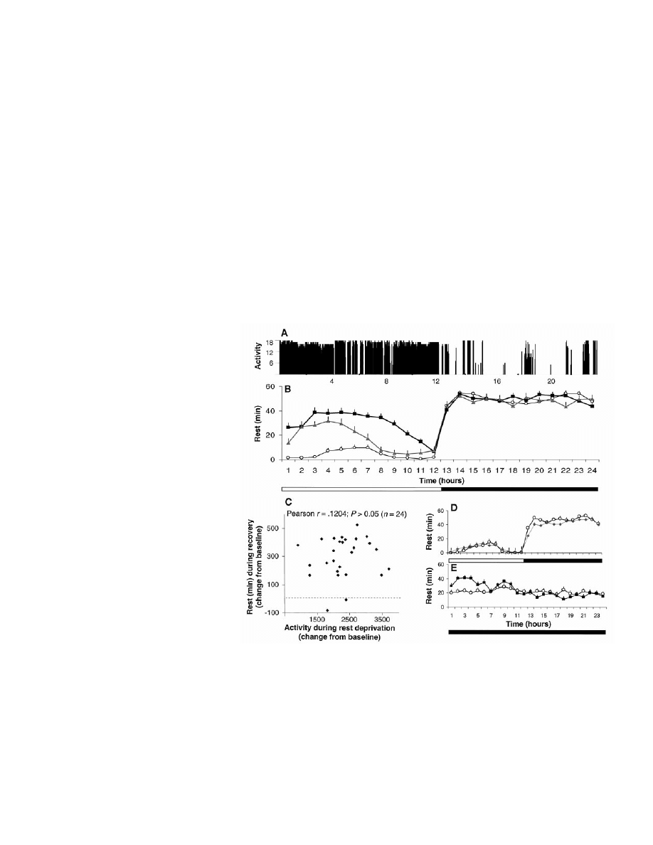
Continuous, high-resolution measurement
of fly behavior (5-day-old virgin females, Can-
ton-S) was achieved with an ultrasound activity
monitoring system (4). This system detects fine
movements of the fly’s head, wings, and limbs,
in good agreement with visual observation (5).
Flies subjected to 12 hour:12 hour light/dark
cycles exhibited sustained periods of activity
and quiescence, with
⬎90% of quiescence
(henceforth referred to as rest) occurring during
the dark period (Fig. 1A) (6). To monitor rest-
activity patterns in large numbers of flies, we
used an infrared activity monitoring system,
which confirmed a robust circadian organiza-
tion of activity and showed good correspon-
dence with the ultrasound system (7).
To determine whether periods of rest are
associated with increased arousal thresholds,
we subjected flies to vibratory stimuli of in-
creasing intensity [0.05g (acceleration), n
⫽ 12;
0.1g, n
⫽ 10; and 6.0g, n ⫽ 8] (8). Flies that
had been behaviorally awake readily responded
to intensities of 0.05g and 0.1g (90% of trials).
Flies that had been behaviorally quiescent for 5
min or longer rarely showed a behavioral re-
sponse to these stimuli (
⬍20% of trials; P ⬍
The Neurosciences Institute, 10640 John Jay Hopkins
Drive, San Diego, CA 92121, USA.
*To whom correspondence should be addressed. E-
mail: tononi@nsi.edu
R
E P O R T S
10 MARCH 2000 VOL 287 SCIENCE www.sciencemag.org
1834
0.001,
2
). However, when the intensity of the
stimulus was increased to 6g, all flies quickly
responded regardless of behavioral state (P
⬎
0.1,
2
). Thus, like sleep in mammals, sustained
periods of quiescence in Drosophila are char-
acterized by increased arousal thresholds.
We next investigated whether the amount
of rest in Drosophila is homeostatically reg-
ulated. Flies were deprived of rest individu-
ally by gentle tapping for 12 hours during the
dark period (i.e., manual rest deprivation).
During the following 12-hour light period, flies
exhibited a large increase in rest compared to
baseline (Fig. 1B). Additionally, an automat-
ed system was used to deprive large numbers
of flies of rest during the 12-hour dark period,
resulting in an increase in rest over baseline
values during the first 6 hours of the follow-
ing light period (Fig. 1B) (8). In the first 24
hours after manual rest deprivation, flies re-
covered 50% of the rest that was lost, a value
comparable to the sleep rebound seen in
mammals after short-term sleep deprivation.
Recordings with the ultrasound system
showed that the rest rebound after deprivation
was characterized by actual immobility, as op-
posed to an increase in stationary waking activ-
ities (such as eating or grooming) that may
result in reduced infrared beam crossing. More-
over, the increase in rest was not accounted for
by levels of prior activity (Fig. 1C). Consistent
with this result, when flies were stimulated in
the apparatus during the 12-hour light period,
rest not only failed to increase, but was actually
reduced by 16
⫾ 4% during the first 6 hours of
recovery (Fig. 1D). Thus, the increase in rest is
not due to physical exhaustion induced by
forced activity (8). To investigate whether the
homeostatic response is separable from circadi-
an factors, we examined per
01
mutants (4),
which are arrhythmic under constant darkness.
In the absence of a circadian rest-activity
rhythm, per
01
flies showed a robust homeostat-
ic response after 12 hours of rest deprivation
(Fig. 1E). This indicates that, as in mammals,
rest is homeostatically regulated and can be
dissociated from circadian control (9).
In mammals, sleep is prominent in the very
young, stabilizes during adolescence and adult-
hood, and declines during old age (10). Rest in
Drosophila follows a similar pattern. On the
first full day after eclosion, the amount of rest
was high but declined steadily until day 3, when
it reached an adult pattern (Fig. 2A). As the flies
aged, the amount of rest during the night de-
clined, and by 33 days of age it was significant-
ly below that found in young adults (Fig. 2B).
Several studies indicate that the homeostatic
regulation of sleep is preserved in older humans
(10). When 33-day-old flies were deprived of
rest, they exhibited a rest rebound similar to
young flies.
Sleep in mammals is modulated by stim-
ulants and hypnotics. For example, caffeine
increases waking and motor activity, whereas
antihistamines reduce sleep latency (11).
Flies given caffeine showed a dose-depen-
dent decrease in rest (Fig. 2C). By contrast,
hydroxyzine, an antagonist of the H1 hista-
mine receptor, increased rest and reduced its
latency (Fig. 2, D and E). Thus, two agents
that modulate waking and sleep in mammals
also modulate vigilance states in Drosophila.
We performed a systematic screening of
gene expression in Drosophila by using mRNA
differential display combined with ribonuclease
protection assays (RPA) (12). RNA was ex-
tracted from whole heads of flies that (i) had
been spontaneously resting for 3 hours during
the dark period, (ii) had been rest-deprived
for 3 hours at the same circadian time, or (iii)
had been spontaneously awake for 3 hours
during the light period, thereby allowing us to
distinguish between changes associated with
behavioral state and those associated with
circadian time (Fig. 3A) (13).
As in the rat (3), only
⬃1% of the tran-
scripts examined in Drosophila were modulat-
ed by behavioral state (14). A transcript whose
expression was higher after periods of rest is
shown in Fig. 3A (“Rest”). As confirmed using
RPA, expression of this mRNA was 45% high-
er during rest than during rest deprivation. None
of the rest-related transcripts matched any pub-
lished sequence. By contrast, several known
genes were expressed at higher levels during
waking than during rest, irrespective of circadi-
an time (Fig. 3A, “Waking”). One, with high
homology to Fatty acid synthase (Fas) (15),
was increased after 3 hours of spontaneous
waking or rest deprivation relative to rest (Fig.
3B). This transcript was localized throughout
the fly brain, including the optic lobes (Fig.
3C), but not in the eye (16). Although the role
of this enzyme in the fly brain is unclear, fatty
acids are modulators of neural activity (17).
Cytochrome P450 (Cyp4e2), a member of a
Fig. 1. (A) Activity record of flies maintained on a 12 hour:12 hour light (horizontal open bar) /dark
(horizontal solid bar) cycle monitored with the ultrasound system. Activity counts indicate the
number of perturbations of the ultrasound standing wave detected over 2-s bins. (B) The
rest-activity cycle monitored with the infrared system (mean
⫾ SEM, n ⫽ 24). Baseline values are
shown in circles. After manual rest deprivation (not shown), flies exhibited a large increase in rest
during the subsequent light period (squares; P
⬍ 0.001; Wilcoxon signed-ranks test). Flies deprived
of rest by the automated system also showed an increase in rest during the subsequent light period
(triangles; n
⫽ 25, P ⬍ 0.001). This finding was replicated in 10 independent experiments (n ⫽ 286).
(C) The amount of rest during the 12-hour recovery period was not correlated with the amount of
activity during rest deprivation. (D) Stimulation of the flies during the light period did not result in a
compensatory increase in rest during recovery (diamonds) with respect to baseline (circles). (E) Under
constant darkness, per
01
flies had the same amount of rest as under light-dark conditions (P
⬎ 0.05),
but this was evenly distributed across the 24 hours (open circles). Twelve hours of automated rest
deprivation resulted in a significant increase in rest during the first 6 hours of recovery (squares)
compared to baseline (circles; n
⫽ 25, P ⬍ 0.001). Because rest is evenly distributed in per
01
flies, rest
deprivation only eliminated
⬃50% of daily rest, compared with 90% in wild-type flies.
R
E P O R T S
www.sciencemag.org SCIENCE VOL 287 10 MARCH 2000
1835
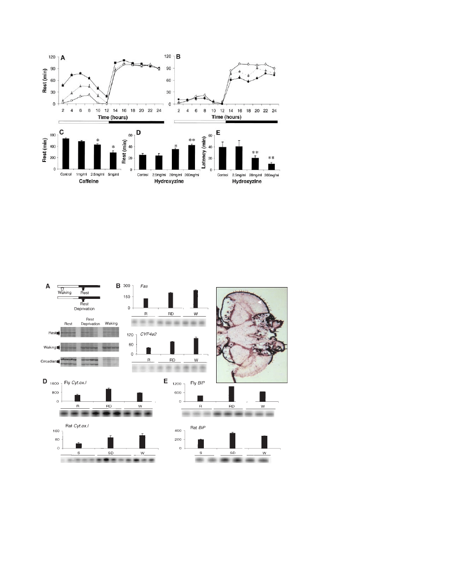
family of detoxifying enzymes, was also in-
creased in waking and rest deprivation relative
to rest in the fly (Fig. 3B) (18).
Several “waking” genes in the fly corre-
sponded to “waking” genes in the rat. For ex-
ample, the mitochondrial gene Cytochrome ox-
idase C, subunit I, showed a rapid increase in
expression during the first few hours of waking
(Fig. 3D), likely a local response of nervous
tissue to the increased metabolic requirements
of waking (3). Another “waking” gene in both
Drosophila and rat is BiP (Hsc70-3), an endo-
plasmic reticulum chaperone protein that may
promote the structural changes necessary for
the establishment of long-term memory (Fig.
3E) (19). Finally, mRNA levels of arylalky-
lamine N-acetyltransferase (Dat), an enzyme
involved in the catabolism of monoamines (20),
were increased by 48% after 2 to 3 hours of
waking relative to rest. In rats, waking is asso-
ciated with a marked increase in brain mRNA
for arylsulfotransferase, another enzyme impli-
cated in the catabolism of monoamines (3).
These findings are of importance because wak-
ing is associated with high central monoamin-
ergic activity, whereas a reduction of such ac-
tivity is a hallmark of sleep (21). This has led to
the suggestion that sleep may serve to counter-
act the effects of continued monoaminergic dis-
charge. According to this hypothesis, an im-
paired catabolism of monoamines should result
in an increased need for sleep (22).
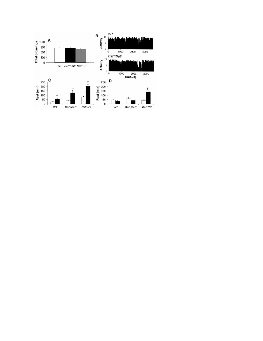
To evaluate this possibility, we examined a
Drosophila mutant in which the transcriptional
level and activity of the Dat enzyme is deficient
(Dat
lo
) (20). By both infrared and ultrasound
measurements, flies homozygous for the Dat
lo
mutation did not differ from wild-type flies in
the percentage and circadian distribution of rest
and waking (Fig. 4A) and showed normal
amounts and patterns of activity (Fig. 4B).
However, after 12 hours of rest deprivation
during the dark period, homozygous Dat
lo
flies
displayed a rest rebound that was greater than in
rest-deprived controls (Fig. 4C). To confirm
that this phenotype maps to the Dat locus and to
assay for gene dosage effects, we crossed Dat
lo
homozygotes with flies carrying a deficiency
(Df ) of the Dat locus, Df(2R)Px1 (20, 23). The
resulting Dat
lo
/Df flies did not differ from wild-
type flies or Dat
lo
homozygotes in the percent-
age and circadian distribution of rest and wak-
ing (Fig. 4A). Dat
lo
/Df flies showed not only an
increased rest rebound during the first 6 hours
of recovery relative to wild-type flies (Fig. 4C),
but also a persistent rebound during the second
6 hours of recovery (Fig. 4D). These results
indicate that the more severely mutant the fly is
at the Dat locus, the greater the rebound. Al-
though the mechanisms responsible for the in-
creased homeostatic response to rest depriva-
tion are currently unclear, these results suggest
a linkage between the catabolism of mono-
amines and the regulation of sleep and waking
in Drosophila.
In conclusion, behavioral, pharmacological,
molecular, and genetic investigations indicate
that Drosophila rest shares many critical fea-
tures with mammalian sleep. The identification
of molecular correlates of sleep and waking that
are conserved across evolution offers a new
Fig. 2. (A) Rest was pronounced during the first full day after eclosion (squares), decreased on day
2 (triangles), and reached adult values by day 3 (circles; P
⬍ 0.001, ANOVA, Tukey post hoc). The
amount of rest remained stable across days 3, 5, and 7 (ANOVA, P
⫽ 0.92). (B) By 16 days of age
(diamonds), rest began to decline during the night and was significantly below day 3 values (open
circles) by 33 days of age (solid circles; P
⬍ 0.001). (C) Flies given caffeine obtained less rest during
the dark period in a dose-dependent fashion (n
⫽ 36 per dose, *P ⬍ 0.0001). Drugs dissolved in
food were continuously available beginning in the final hour of the light period. Hydroxyzine, an H1
antagonist, increased the percentage of rest (D) and decreased its latency (E) during the first hour
of the dark period (n
⫽ 40 per dose; *P ⫽ 0.056, **P ⬍ 0.001). The increase in rest was not
associated with an impairment of fly behavior. The activity per waking minute was unchanged
during the dark period, including the first hour, as was the total amount of activity during the light
period. Responsiveness to arousing stimuli was preserved.
Fig. 3. (A) Examples of transcripts identified with differential display that are expressed differen-
tially depending on behavioral state and circadian time. The waking band corresponds to a gene
with high homology to Fas. (B) RPA confirmed the differential display results. Messenger RNA levels
of Fas and Cyp4e2 are higher during waking (W) and rest deprivation (RD) compared to rest (R)
(P
⬍ 0.01, ANOVA, Tukey post hoc). Densitometric analysis was performed with a PhosphorImager.
(C) In situ hybridization shows that Fas mRNA is present in the central nervous system but not in
the eye (arrow). (D) Cytochrome oxidase C, subunit I, and (E) BiP mRNA levels are higher during
waking in both fly and rat (P
⬍ 0.01, ANOVA, Tukey post hoc).
C
R
E P O R T S
10 MARCH 2000 VOL 287 SCIENCE www.sciencemag.org
1836
approach for studying the phylogeny of sleep.
Most important, the demonstration that a muta-
tion modifies the homeostatic regulation of
sleep-like states opens the way for gene discov-
ery through mutant screening and validates the
use of Drosophila as a model system for eluci-
dating the functions of sleep.
Note added in proof: While this paper was
in review, another group reported that rest in
Drosophila is a sleep-like state (24 ).
References and Notes
1. S. S. Campbell and I. Tobler, Neurosci. Biobehav. Rev.
8, 269 (1984); H. Zepelin and A. Rechtschaffen, Brain
Behav. Evol. 10, 425 (1984); A. Rechtschaffen, Per-
spect. Biol. Med. 41, 359 (1998).
2. I. Tobler, Behav. Brain Res. 8, 351 (1983);
㛬㛬㛬㛬
and
J. Stalder, J. Comp. Physiol. A 163, 227 (1988); I.
Tobler and M. Neuner-Jehle, J. Sleep Res. 1, 231
(1992); W. Kaiser and J. Steiner-Kaiser, Nature 301,
707 (1983); W. Kaiser, J. Comp. Physiol. A 163, 565
(1988).
3. C. Cirelli and G. Tononi, Mol. Brain Res. 56, 293
(1998); C. Cirelli, P. J. Shaw, G. Tononi, Sleep 22
(suppl.), 113 (1999).
4. Flies were cultured at 25°C, 50 to 60% humidity, 12
hour:12 hour light/dark cycle, on yeast, dark corn
syrup, and agar food. We obtained per
01
flies from
J. C. Hall (Brandeis University) and Dat
lo
and
Df(2R)Px1/In(2LR)SM5, al
2
Cy lt
v
sn
2
sp
2
flies from
the Bloomington Drosophila Stock Center. For details
about the ultrasound monitoring system, see Science
Online (www.sciencemag.org/feature/data/1047207.
shl).
5. Five behaviors were visually scored in 2-s bins by an
observer blind to the output of the ultrasound system
on 18 independent trials for a total of 8 hours during
the light period. The correspondence rates were as
follows: locomoting, 99%; inactive, 97%; grooming
anterior limbs, 94%; grooming posterior limbs, 98%;
and eating, 97%.
6. Rest was defined as uninterrupted behavioral quies-
cence lasting for at least 5 min.
7. Drosophila Activity Monitoring System (Trikinetics)
[M. Hamblen et al., J. Neurogenet. 3, 249 (1986)]. The
system was validated by visual observation for 17.75
hours (n
⫽ 7). Flies were awake but did not cross the
infrared beam in 5 of 213 bins (miss rate
⫽ 2.35%).
8. For procedures for arousal thresholds, procedures for
automated rest deprivation, and additional controls
used to validate the infrared system, see Science
Online (www.sciencemag.org/feature/data/1047207.
shl).
9. R. E. Mistlberger, B. M. Bergmann, W. Waldenar, A.
Rechtschaffen, Sleep 6, 217 (1983); I. Tobler, A. A.
Borbely, G. Groos, Neurosci. Lett. 42, 49 (1983); D. M.
Edgar, W. C. Dement, C. A. Fuller, J. Neurosci. 13,
1065 (1993).
10. W. S. Stone, Clin. Geriatr. Med. 5, 363 (1989); D.-J.
Dijk, J. F. Duffy, E. Riel, T. L. Shanahan, C. A. Czeisler,
J. Physiol. 516, 611 (1999).
11. G. Yanik, S. Glaum, M. Radulovacki, Brain Res. 403,
177 (1987).
12. Methods were as in (3), with modifications: 0.5
g of
pooled total RNA (n
⫽ 20) was reverse-transcribed
(two independent pools per condition). Polymerase
chain reactions were performed in duplicate for each
pool (104 primer combinations). For RPA, 1 to 2
g of
total RNA from pooled fly heads (n
⫽ 60) was used.
The amount of sample RNA was normalized using a
riboprobe specific for ribosomal protein rp49.
13. The behavioral state was determined individually for
each fly; only flies that satisfied specific criteria were
selected for analysis. A fly was considered awake if it
was active for at least 90% of the 3-hour light period
and 100% of the hour before killing. A fly was resting
if it was inactive for at least 66% of the 3-hour dark
period and 100% of the hour before killing. Only
about 60 to 70% of the flies examined satisfied these
criteria. Failure to specifically identify rest and wak-
ing results in samples containing a mixture of behav-
ioral states.
14. An estimated
⬃5000 RNA species were screened. For
additional data, see Science Online (www.sciencemag.
org/feature/data/1047207.shl).
15. The sequence matched a Drosophila P1 clone
(AC005554). Analysis using Genescan indicated that
the proposed peptide has a 49% homology with rat
Fas.
16. In situ hybridization was performed as described [K.
Aronstein, V. Auld, R. Ffrench-Constant, Invert. Neu-
rosci. 2, 115 (1996)]. Sense riboprobes gave no spe-
cific hybridization.
17. S. Yehuda et al., Peptides 19, 407 (1998).
18. B. C. Dunkov, R. Rodriguez-Arnaiz, B. Pittendrigh, R. H.
Ffrench-Constant, R. Feyereisen, Mol. Gen. Genet.
251, 290 (1996).
19. D. Kuhl, T. E. Kennedy, A. Barzilai, E. Kandel, J. Cell
Biol. 119, 1069 (1992); D. M. Rubin et al., Gene 128,
155 (1993).
20. D. Brodbeck et al., DNA Cell Biol. 17, 621 (1998).
21. D. J. McGinty and R. M. Harper, Brain Res. 101, 569
(1976); G. Aston-Jones and F. E. Bloom, J. Neurosci. 1,
876 (1981).
22. E. Hartmann, Functions of Sleep (Yale Univ. Press,
New Haven, CT, 1973); J. M. Siegel and M. A. Ro-
gawksi, Brain Res. Rev. 13, 213 (1988).
23. C. B. Bridges, Cytologia Fujii Jubil., 745 (1937).
24. J. Hendricks et al., Neuron 25, 129 (2000).
25. We thank D. F. Robinson, G. A. Davis, M. J. Gallina,
J. M. Salbaum, J. Snook, N. Almassy, and E. Balaban
for his conception of the ultrasound system. The
Neurosciences Institute is supported by the Neuro-
sciences Research Foundation and receives major
support for this program from Novartis. C.C. was a
Joseph Drown Foundation Fellow.
15 November 1999; accepted 8 February 2000
Genetic Suppression of
Polyglutamine Toxicity in
Drosophila
Parsa Kazemi-Esfarjani* and Seymour Benzer
A Drosophila model for Huntington’s and other polyglutamine diseases was
used to screen for genetic factors modifying the degeneration caused by ex-
pression of polyglutamine in the eye. Among 7000 P-element insertions, several
suppressor strains were isolated, two of which led to the discovery of the
suppressor genes described here. The predicted product of one, dHDJ1, is
homologous to human heat shock protein 40/HDJ1. That of the second, dTPR2,
is homologous to the human tetratricopeptide repeat protein 2. Each of these
molecules contains a chaperone-related J domain. Their suppression of poly-
glutamine toxicity was verified in transgenic flies.
Expanded polyCAG tracts in the genes for
Huntington’s disease (HD) and at least seven
other disorders are associated with hereditary
neurodegeneration (1). The polyCAGs are
translated to polyglutamines, which form cy-
toplasmic and/or nuclear aggregates and pro-
duce toxic effects (1, 2). One approach to the
identification of proteins that can modify
polyglutamine aggregation and toxicity is the
isolation of enhancer and suppressor genes.
Fig. 4. (A) The number of infrared beam crossings per day is similar in wild-type, Dat
lo
/Dat
lo
, and
Dat
lo
/Df flies (P
⬎ 0.05, n ⫽ 25). (B) Activity patterns (ultrasound system, units as in Fig. 1A) are
similar in all three Drosophila genotypes (two representative records for 1 hour during the light
period are shown). (C) The amount of rest during the first 6 hours of recovery (solid bars) compared
to baseline (open bars) was higher in Dat
lo
/Dat
lo
and Dat
lo
/Df flies than in wild-type flies (*P
⬍
0.005, Wilcoxon test). (D) In Dat
lo
/Df flies, rest rebound persists into the second 6 hours of
recovery (*P
⬍ 0.005).
R
E P O R T S
www.sciencemag.org SCIENCE VOL 287 10 MARCH 2000
1837
Wyszukiwarka
Podobne podstrony:
Effect of caffeine on fecundity egg laying capacity development time and longevity in Drosophila
Effect of caffeine on fecundity egg laying capacity development time and longevity in Drosophila
1 Application of Joints and Springs in ANSYS
3 T Proton MRS Investigation of Glutamate and Glutamine in Adolescents at High Genetic Risk for Schi
Compare and contrast literature of Whitman and Dickinson in terms of God, man and nature
The Pernicious Blend of Rumination and Fearlessness in NSSI
The Presentation of Self and Other in Nazi Propaganda
Suke Wolton Lord Hailey, the Colonial Office and the Politics of Race and Empire in the Second Worl
RÜDIGER SCHMITT The Problem of Magic and Monotheism in The Book of Leviticus
Wild, Rodden, Grodd, Ruch Neural Correlates of Laughter and H
The Code of Honor or Rules for the Government of Principals and Seconds in Duelling by John Lyde Wil
A contrastive analysis of English and Arabic in relativization
Cases of domestication and foreignization in the
PSYCHIC METHODS OF DIAGNOSIS AND TREATMENT IN ACUPUNCTURE …
Penier, Izabella Re Conceptualization of Race and Agency In Jamaica Kincaid sthe Autobiography of M
The history of translation dates back to the times of Cicero and Horace in first century BCE and St
Elizabeth Coldwell (ed) Sex in London Tales of Pleasure and Perversity in the English Capital [MF]
Fr hlich Stability of Pulegone and Thujone in Ethanolic Solution
więcej podobnych podstron