ARTICLE
Increased osteoclastic activity in acute Charcot
’s
osteoarthopathy: the role of receptor activator of nuclear
factor-kappaB ligand
G. Mabilleau
&
N. L. Petrova
&
M. E. Edmonds
&
A. Sabokbar
Received: 19 December 2007 / Accepted: 22 February 2008 / Published online: 4 April 2008
# Springer-Verlag 2008
Abstract
Aims/hypothesis Our aims were to compare osteoclastic
activity between patients with acute Charcot
’s osteoarthro-
pathy and diabetic and healthy controls, and to determine
the effect of the receptor activator of nuclear factor-kappaB
ligand (RANKL) and its decoy receptor osteoprotegerin
(OPG).
Methods Peripheral blood monocytes isolated from nine
diabetic Charcot patients, eight diabetic control and eight
healthy control participants were cultured in the presence of
macrophage-colony stimulating factor (M-CSF) alone,
M-CSF and RANKL, and also M-CSF and RANKL with
excess concentrations of OPG. Osteoclast formation was
assessed by expression of tartrate-resistant acid phosphatase
on glass coverslips and resorption on dentine slices.
Results In cultures with M-CSF, there was a significant in-
crease in osteoclast formation in Charcot patients compared
with healthy and diabetic control participants (p=0.008). A
significant increase in bone resorption was also seen in the
former, compared with healthy and diabetic control par-
ticipants (p < 0.0001). The addition of RANKL to the
cultures with M-CSF led to marked increase in os-
teoclastic resorption in Charcot (from 0.264 ± 0.06% to
41.6±8.1%, p<0.0001) and diabetic control (0.000±0.00%
to 14.2±16.5%, p<0.0001) patients, and also in healthy
control participants (0.004±0.01% to 10.5±1.9%, p<0.0001).
Although the addition of OPG to cultures with M-CSF and
RANKL led to a marked reduction of resorption in Charcot
patients (41.6±8.1% to 5.9±2.4%, p=0.001), this sup-
pression was not as complete as in diabetic control patients
(14.2±16.5% to 0.45±0.31%, p=0.001) and in healthy
control participants (from 10.5 ±1.9% to 0.00 ±0.00%,
p<0.0001).
Conclusions/interpretation These results indicate that
RANKL-mediated osteoclastic resorption occurs in acute
Charcot
’s osteoarthropathy. However, the incomplete inhi-
bition of RANKL after addition of OPG also suggests the
existence of a RANKL-independent pathway.
Keywords Charcot
’s osteoarthropathy. OPG . Osteoclasts .
Osteolysis . RANKL . Resorption
Abbreviations
LIGHT
homologous to lymphotoxins exhibiting in-
ducible expression and competing with herpes
simplex virus glycoprotein D for herpes virus
entry mediator (HVEM), a receptor expressed
by T lymphocytes
MEM
minimum essential medium
M-CSF
macrophage-colony stimulating factor
OPG
osteoprotegerin
PBMCs
peripheral blood monocytes
RANK
receptor activator of nuclear factor-kappaB
RANKL
receptor activator of nuclear factor-kappaB
ligand
sRANKL
soluble receptor activator of nuclear factor-
kappaB ligand
TRAcP
tartrate-resistant acid phosphatase
Diabetologia (2008) 51:1035
–1040
DOI 10.1007/s00125-008-0992-1
G. Mabilleau
:
A. Sabokbar
Nuffield Department of Orthopaedic Surgery,
Botnar Research Centre, University of Oxford,
Oxford, UK
N. L. Petrova
:
M. E. Edmonds (
*)
Diabetic Foot Clinic,
King
’s College Hospital NHS Foundation Trust,
Denmark Hill,
London SE5 9RS, UK
e-mail: Michael.Edmonds@kch.nhs.uk

Introduction
Although Charcot
’s osteoarthropathy is characterised by
increased local bone resorption [
], the exact cellular
mechanisms contributing to the pathogenesis of this
condition remain unresolved. Osteoclasts have been shown
to be the principal cell type responsible for bone resorption
[
]. These cells originate from the haemopoietic lineage and
are known to undergo various stages of proliferation, fusion
and differentiation before they are fully functionally active,
mature osteoclasts. Recently, receptor activator of nuclear
factor-kappaB (RANK) ligand (RANKL) has been identi-
fied as an essential mediator of osteoclast formation and
activation [
]. RANKL is expressed on a variety of cell
types such as bone forming osteoblasts, T lymphocytes,
dendritic cells, endothelial cells and fibroblasts. RANKL
mediates the process of osteoclastogenesis by binding to its
RANK, which is expressed on mononuclear osteoclast
precursors. The effects of RANKL
–RANK interaction are
physiologically counterbalanced by osteoprotegerin (OPG),
which acts as a soluble receptor decoy for RANKL and
blocks the interaction of RANKL with RANK. The ratio of
RANKL to OPG has been suggested to regulate the extent
of osteoclast formation and resorption. Therefore, any
alteration in the RANKL/OPG ratio could be critical in
the pathogenesis of osteolytic bone disorders [
].
Recently, Jeffcoate hypothesised that the RANK/RANKL/
OPG pathway may play an important role in the osteolysis
seen in acute Charcot
’s osteoarthropathy [
]. Using an in
vitro technique to generate functional human osteoclasts
from peripheral blood monocytes (PBMCs) [
] in the pres-
ence of macrophage-colony stimulating factor (M-CSF) [
and soluble RANKL, it is possible to determine the cellular
mechanisms involved in the process of osteoclast formation
and resorption in physiological and pathological conditions.
To our knowledge, this technique has not yet been studied
in patients with Charcot
’s osteoarthropathy.
The aims of this study were: (1) to generate functional
human osteoclasts in vitro from diabetic patients with acute
Charcot
’s osteoarthropathy and from healthy and diabetic
control participants; (2) to compare the extent of osteoclast
formation and resorption; and (3) to determine the role of
the RANK/RANKL/OPG pathway in osteoclastic activity
in Charcot
’s osteoarthropathy.
Methods
Patients
We studied nine consecutive diabetic patients with recent
onset of acute Charcot
’s osteoarthropathy (five men, four
women; five type 1, four type 2 diabetes), eight diabetic
patients with no previous history of Charcot
’s osteoarthro-
pathy (five men, three women; four type 1, four type 2) and
eight healthy control participants (five men, three women).
Patients with acute Charcot
’s osteoarthropathy were matched
for age and duration of diabetes with the diabetic control
patients and for age with the healthy control participants. The
mean age was similar between patients with Charcot
’s
osteoarthropathy and diabetic control patients (53±2.8 versus
59±2.9 years [mean±SEM], p=0.167) as was the mean age
between the former and healthy control participants (53±2.8
versus 47±2.7 years, p=0.114). The mean duration of diabetes
was similar in both groups with diabetes (31±5.1 [Charcot
patients] versus 27±4.6 years, p=0.606). Diabetes control as
indicated by glycated Hb was also similar in the two diabetes
groups (7.7±0.6 [Charcot
’s] versus 7.8±0.4%, p=0.743).
Diagnosis of Charcot
’s osteoarthropathy was made on
the presentation of a hot swollen foot, with skin foot
temperature 2°C greater than the corresponding site on the
contralateral foot and with typical radiological changes of
subluxation, dislocation or fragmentation of bone on
standard foot radiographs [
]. All patients had intact feet
and no evidence of foot infection or ulceration.
Ethical permission for this study was obtained from the
King
’s College Hospital Research Ethics Committee and all
participants gave written informed consent.
Isolation and culture of monocytes
Peripheral blood mononuclear cells were isolated as
previously described [
]. Briefly, blood was diluted 1:1 in
α-minimum essential medium (MEM; Invitrogen, Paisley,
UK), layered over Histopaque and centrifuged (693 g) for
20 min. The interface layer was resuspended in MEM, then
centrifuged (600 g) for a further 10 min. The resultant cells
were resuspended in MEM with 10% heat-inactivated FCS
and counted in a haemocytometer following lysis of
erythrocytes by a 5% (vol./vol.) acetic acid solution.
To assess the extent of osteoclast formation and resorption,
PBMCs were cultured on glass coverslips and dentine slices.
Initially, 5×10
5
PBMCs were added to 6-mm diameter glass
coverslips and 4-mm diameter dentine slices in MEM
containing 100 UI/ml penicillin, 100
μg/ml streptomycin
and 10% FCS (Gibco, Paisley, UK). After 2 h incubation,
coverslips and dentine slices were vigorously rinsed in
medium to remove non-adherent cells. The cultures were
maintained in MEM/FCS under three different culture
conditions: (1) human M-CSF (R&D Systems Europe,
Abingdon, UK) alone at 25 ng/ml; (2) M-CSF plus 100 ng/ml
human soluble RANKL (sRANKL; Peprotech, London,
UK) (a concentration known to facilitate differentiation of
osteoclast precursors to active bone-resorbing osteoclasts in
vitro); and (3) M-CSF plus sRANKL plus 250 ng/ml human
OPG (R&D Systems Europe).
1036
Diabetologia (2008) 51:1035
–1040

Coverslips and dentine slices were cultured at 37°C in
5% CO
2
for 14 and 21 days respectively.
Osteoclast formation
After 14 days, the coverslips were examined histochemically
for the expression of tartrate-resistant acid phosphatase
(TRAcP), an osteoclast marker. Coverslips with newly formed
osteoclasts were collected and rinsed in PBS buffer, fixed with
formalin (10% [vol./vol.] in PBS buffer) for 10 min and rinsed
in distilled water. TRAcP was histochemically revealed by a
simultaneous coupling reaction using Naphtol AS-BI-phosphate
as substrate and Fast violet B as the diazonium salt. The
coverslips were incubated for 90 min at 37°C in a dark room,
rinsed three times in distilled water and the residual activity
was inhibited by 4% NaF (wt/wt) for 30 min. Coverslips were
then rinsed in distilled water, counterstained with DAPI for
20 min and allowed to dry before mounting, using an aqueous
medium. TRAcP-positive cells with more than three nuclei were
identified as osteoclasts. The number of newly generated osteo-
clasts was assessed using a light microscope examination.
Osteoclast resorption
After 21 days, the dentine slices were removed from the
culture wells, placed in NH
4
OH (1 mol/l) for 30 min and
sonicated for 5 min to remove any adherent cells. They were
then rinsed in distilled water and stained with 0.5% (vol./vol.)
toluidine blue prior to examination by light microscopy. The
surface of each dentine slice was examined for evidence of
lacunar resorption and the extent of eroded surface on each
dentine slice was determined using image analysis and
expressed as the percentage of surface area resorbed.
Statistical analyses
Data were expressed as a mean±SEM. Initially the difference
within the three study groups (Charcot patients, healthy and
diabetic controls) was assessed with the non-parametric
Kruskall
–Wallis test. Then the differences between Charcot
and diabetic patients, and Charcot patients and healthy controls
were assessed by the non-parametric Mann
–Whitney U test. In
each patient group, the differences between the various culture
conditions were also assessed using the Mann
–Whitney U test.
Differences were considered significant at p<0.05.
Results
Osteoclast cultures in the presence of M-CSF
Osteoclast formation The mean number of newly formed
TRAcP-positive multinucleated osteoclasts in the presence
of M-CSF alone was significantly greater in the patients
with acute Charcot
’s osteoarthropathy (48.6±18.2) than in
diabetic (6.8±2.7) and healthy control participants (5.0±0.7)
(p=0.008). The number of TRAcP-positive multinucleated
osteoclasts formed in acute Charcot
’s osteoarthropathy
was 7.2 and 9.7 times greater than those formed in diabetic
(p=0.010) and healthy control groups (p=0.003), respectively.
Osteoclast resorption The newly formed osteoclasts
exhibited increased functional activity as demonstrated by
the extent of resorption on dentine slices, with percentage
area resorption significantly elevated in the patients with
acute Charcot
’s osteoarthropathy (0.264±0.06%) compared
with diabetic (0.000 ±0.00%) and healthy control groups
(0.004 ±0.01) (p<0.0001). The percentage of resorption
was significantly greater in the Charcot patients than in the
diabetic (p=0.001) and healthy control groups (p=0.001).
Osteoclast cultures in the presence of M-CSF and sRANKL
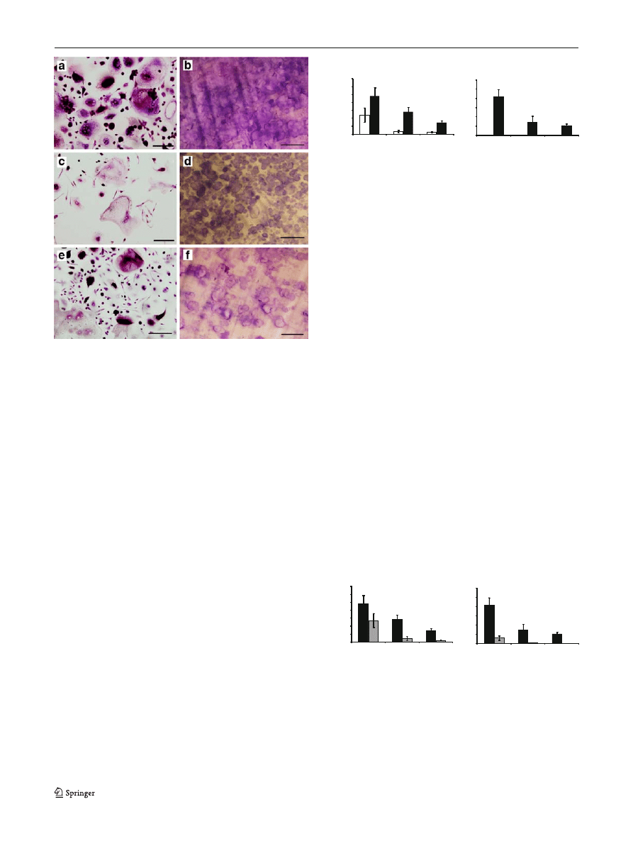
Osteoclast formation The addition of sRANKL led to an
increase in the number of TRAcP-positive multinucleated
osteoclasts in all three groups of patients. The mean number
of these osteoclasts in patients with acute Charcot
’s os-
teoarthropathy was 96.0±21.6, which was significantly
greater than that in the diabetic (56.5±11.5) and healthy
(29.0±5.1) control groups (p=0.010; Fig.
a,c,e). The number
of TRAcP-positive multinucleated osteoclasts in the
patients with acute Charcot
’s osteoarthropathy was 1.7
times higher than in diabetic control patients, but this
finding did not reach significance (p=0.105). However, the
number of these osteoclasts in the acute Charcot group was
3.3 times (and significantly) higher than in the healthy
control group (p=0.005). When the number of cells in the
cultures with M-CSF alone was compared with that after
the addition of sRANKL, there was a significant increase in
the diabetic control patients (from 6.8±2.7 to 56.5±11.5,
p=0.003) and in the healthy participants (from 5.0±0.7 to
29.0±5.1, p=0.002), while the increase in the number of
TRAcP-positive multinucleated osteoclasts in the acute
Charcot group failed to reach significance (increase from
48.6±18.2 to 96.0±21.6, p=0.059; Fig.
a).
Osteoclast resorption The percentage area resorption on
dentine slices with M-CSF and sRANKL was significantly
increased in the acute Charcot group (41.6 ± 8.1%) com-
pared with that in the diabetic (14.2 ± 16.5%) and healthy
control groups (10.5 ±1.9%; p = 0.005). Resorption in the
Charcot patients was 2.9 times higher than in diabetic
control patients (p = 0.008) and four times higher than in
healthy participants (p = 0.005; Fig.
b,d,f). The addition
of sRANKL to the cultures with M-CSF led to the
Diabetologia (2008) 51:1035
–1040
1037
following rises in the percentage area resorption when
compared with M-CSF alone: Charcot
’s 0.264±0.06% to
41.6 ± 8.1%, p < 0.0001; diabetic control 0.000 ± 0.00% to
14.2 ± 16.5%, p < 0.0001; healthy control 0.004 ± 0.01%
to 10.5 ± 1.9%, p < 0.0001 (Fig.
b).
Osteoclast cultures in the presence of M-CSF, sRANKL
and excess concentrations of OPG
Osteoclast formation The addition of excess concentrations
of OPG led to a reduction in the number of TRAcP-positive
multinucleated osteoclasts in the cultures with M-CSF,
sRANKL and OPG in all the three groups of patients.
However, after the addition of OPG, the number of TRAcP-
positive multinucleated osteoclasts was still significantly
increased in the Charcot group (54.4±17.6), as compared
with diabetic (8.8±5.3) and healthy control participants
(4.4±1.2; p=0.003). In the cultures with M-CSF, sRANKL
and OPG, the number of TRAcP-positive multinucleated
osteoclasts was greater in the Charcot patients than in the
diabetic (p=0.005) and healthy control groups (p=0.001).
When OPG was added to the cultures with M-CSF and
sRANKL, the reduction in the number of TRAcP-positive
cells in Charcot patients was not significant (96.0±21.6
versus 54.4 ±17.6, p =0.189). OPG on the other hand
significantly inhibited the number of TRAcP-positive cells
in M-CSF and RANKL-mediated cultures from diabetic
(reduced from 56.5±11.5 to 8.8±5.3, p=0.005) and healthy
control participants (29.0 ± 5.1 to 4.4 ± 1.2, p = 0.003;
Fig.
a).
Osteoclast resorption The addition of OPG led to a marked
reduction of the percentage area resorption on dentine
slices in Charcot patients (from 41.6±8.1% to 5.9±2.4%,
p=0.001) and also in diabetic (14.2±16.5% to 0.45±0.31%,
p = 0.001) and healthy control (from 10.5 ± 1.9% to
0.00±0.00%, p<0.0001) participants (Fig.
b).
However, the percentage area resorption on the dentine
slices was still greater in the cultures with M-CSF, RANKL
and OPG from the patients with acute Charcot
’s osteo-
arthropathy (5.9±2.4%) than in those from diabetic (0.45±
0.31%) and healthy control (0.00 ±0.00%) participants
0
40
80
120
Charcot's
Diabetic
Healthy
a
0
40
20
60
Charcot's
Diabetic
Healthy
b
TRAcP-positive
multinucleated cells (
n)
Total area of
bone resor
ption (%)
Fig. 2 a Quantitative comparison between the number (n) of TRAcP-
positive cells formed in cultures with M-CSF alone (white bars) or with
M-CSF and sRANKL (black bars) in patients with Charcot
’s osteo-
arthropathy and diabetic and healthy control participants. b Quantitative
comparison between the percentage area resorption in the same cultures
and patient groups. Statistical differences between the groups were
determined using the Mann
–Whitney U test, with significance as
follows: a Charcot
’s p=0.059, diabetic control p=0.003, healthy control
p=0.002; b Charcot
’s p<0.0001, diabetic control p<0.0001, healthy
control p<0.0001
TRAcP-posituve
0
40
80
120
Charcot's
Diabetic
Healthy
a
0
20
40
60
Total area of bone
Charcot's
Diabetic
Healthy
b
multinucleat
ed cells (
n)
resorption (%)
Fig. 3 a Comparison between the number (n) of TRAcP-positive cells
formed in cultures with M-CSF and sRANKL (black bars) or with
M-CSF, sRANKL and excess concentrations of OPG (250 ng/ml)
(grey bars) in patients with Charcot
’s osteoarthropathy and diabetic
and healthy control participants. b Comparison between the percent-
age area resorption in the same cultures and patient groups. Statistical
differences between the groups were determined using the Mann
–
Whitney U test, with significance as follows: a Charcot
’s p=0.189,
diabetic control p=0.005, healthy control p=0.003; b Charcot's p=0.001,
diabetic control p=0.001, healthy control p<0.0001
Fig. 1 Multinucleated TRAcP-positive cells were formed on glass
coverslips (a, c, e) capable of lacunar resorption (b, d, f) after 14 and
21 days incubation, respectively, in the presence of 25 ng/ml human
M-CSF and 100 ng/ml sRANKL. Newly formed osteoclasts were
numerous and highly active in Charcot
’s patients (a, b) compared with
diabetic (c, d) and healthy control (e, f) participants. Scale bars, 10
μm
1038
Diabetologia (2008) 51:1035
–1040
(p=0.003). Resorption on the dentine slices was greater in
the Charcot patients than in diabetic (p=0.005) and healthy
control (p=0.003) groups.
Discussion
This study shows that monocytes from patients with acute
Charcot
’s osteoarthropathy cultured in the presence of M-CSF
alone were capable of differentiating into mature osteoclasts
that exhibited increased resorption compared with diabetic
and healthy control participants. Furthermore, osteoclasts
generated after the addition of sRANKL were functionally
more aggressive, exhibiting a considerable increase in the
extent of resorbing activity in patients with acute Charcot
’s
osteoarthropathy. This resorption was partially blocked by the
addition of excess concentrations of OPG, a soluble receptor
decoy for RANKL. This suggests that the increased osteo-
clastic activity in patients with acute Charcot
’s osteoarthrop-
athy is mediated through both a RANKL-dependent and a
RANKL-independent pathway.
Cultures from the patients with Charcot
’s osteoarthrop-
athy showed increased osteoclast formation and resorption
when cultured with M-CSF alone. Although M-CSF is an
essential factor for proliferation, differentiation and survival
of the monocyte-macrophage lineage [
,
], it is not an
osteoclastogenic factor and it is unusual to detect osteoclast
formation and resorption in cultures with M-CSF, as was
seen in the diabetic and healthy controls. This observation
suggests that in acute Charcot
’s osteoarthropathy there may
be increased levels of other circulating pro-inflammatory
factors such as TNF-
α [
], IL-8 [
] and
LIGHT (homologous to lymphotoxins exhibiting inducible
expression and competing with herpes simplex virus
glycoprotein D for herpes virus entry mediator [HVEM],
a receptor expressed by T lymphocytes) [
], which have
been previously shown to stimulate osteoclastogenesis
independently of RANK/RANKL mechanisms. The con-
centrations of these circulating factors in diabetic and
healthy control participants may not be sufficient to induce
the formation and differentiation of active osteoclasts in the
presence of M-CSF alone.
After the addition of sRANKL to M-CSF cultures, the
newly formed osteoclasts exhibited markedly increased
resorption in the patients with Charcot
’s osteoarthropathy,
although the number of osteoclasts did not significantly
increase in these patients compared with cultures with M-CSF
alone. These observations may not be unique to Charcot
’s
osteoarthropathy, and indeed similar observations have been
reported in other conditions associated with increased bone
resorption, such as rheumatoid arthritis where the addition of
sRANKL resulted in a significant increase in lacunar
resorption, but did not lead to a significant increase in the
number of TRAcP-positive cells [
]. Overall, the observed
extensive resorption in acute Charcot patients, in the pres-
ence of M-CSF and sRANKL, as compared with the diabetic
or healthy control groups, may suggest that the osteoclast
precursors circulating in acute Charcot patients are in a
higher activated state and as such are more primed to
becoming osteoclasts (mediated through RANKL) than those
in the control groups.
In order to ascertain that RANKL was a major osteo-
clastic activator in patients with Charcot
’s osteoarthropathy,
excess concentrations of OPG, the soluble receptor decoy
to RANKL, were added to the cultures with M-CSF and
RANKL. The rationale for this approach was that if osteo-
clastogenesis is mediated solely through RANK
–RANKL
interaction, addition of excess concentrations of OPG (as
had been previously determined to be sufficient to block
osteoclastogenesis through RANKL [
]) would complete-
ly abolish the process of osteoclast differentiation and
activation. In the current study, although osteoclast forma-
tion and resorption in the diabetic and healthy control
groups was completely blocked by the addition of OPG, the
latter did not achieve total inhibition of osteoclast formation
and resorption in patients with acute Charcot
’s osteo-
arthropathy. These results suggest that although RANKL-
dependent pathways do play a significant role in the
osteoclastic activity of Charcot
’s osteoarthropathy, an
alternative pathway (other than RANK/RANKL) may also
be involved. Osteoclastogenic mediators other than
RANKL that have been reported to stimulate osteoclast
differentiation independently of the RANKL pathway
include TNF-
α [
], IL-6 [
], IL-8 [
] and LIGHT
]. In acute Charcot
’s osteoarthropathy, it is possible that
one or a combination of these factors may have initiated the
circulating osteoclast precursors to be in a more
‘primed’
condition, a situation which as such could explain the
observed resorption in Charcot monocyte cultures supple-
mented with M-CSF alone, without the exogenous addition
of any osteoclastogenic mediators.
The osteolysis of Charcot
’s osteoarthropathy may be
explained by our observation that osteoclast precursors
from Charcot patients develop into mature osteoclasts that
exhibit increased resorptive activity, especially in response
to RANKL, unlike the increased resorption in response to
bacterial infection, which is not mediated by RANKL [
Increased expression of RANKL has been previously
demonstrated in pathological osteolysis associated with
the development of various bone diseases [
] and a similar
mechanism may contribute to osteolysis of Charcot
’s
osteoarthropathy [
]. Furthermore, patients with Charcot
’s
osteoarthropathy have severe neuropathy, which itself can
also lead to increased expression of RANKL as a result of
the loss of nerve-derived peptides known to antagonise its
effect such as calcitonin gene-related peptide [
]. In
Diabetologia (2008) 51:1035
–1040
1039
addition to the RANKL-dependent pathway, our results
suggest that a RANKL-independent pathway, mediated by
pro-inflammatory cytokines, may also be important. Indeed,
Charcot
’s foot is characterised by excessive inflammation
and proinflammatory cytokines have been implicated in its
pathogenesis [
]. In support of this, a recent immunohis-
tochemical analysis of bone samples isolated from Charcot
’s
osteoarthropathy patients showed excessive osteoclastic
activity in a microenvironment enriched with mediators of
bone resorption (IL-1, IL-6 and TNF-
α) [
]. Thus a
RANKL-independent pathway, which is also known to
play a role in other osteolytic disorders such as rheumatoid
arthritis [
] and aseptic loosening [
], could contribute
also to the pathogenesis of the Charcot
’s osteoarthropathy.
This study has indicated, for the first time that the
RANKL-dependent pathway is important in the pathogen-
esis of Charcot
’s osteoarthropathy, thereby raising the
possibility of the use of RANKL inhibition in the
management of Charcot
’s foot. However, our observations
also suggest that a RANKL-independent pathway may play
a role, but further investigation is required to fully clarify
the mechanism involved. If confirmed, specific pharmaco-
logical agents that counteract the RANKL-independent
pathway, such as anti-TNF strategies, may be useful in the
treatment of Charcot
’s osteoarthropathy. Whatever the
relative importance of either pathway, this in vitro tech-
nique of generating human osteoclasts from PBMCs may
allow specific characterisation of osteoclastic activity in
each patient and could, in the future, lead to individually
tailored anti-osteoclastic treatment for the patient with acute
Charcot
’s osteoarthropathy.
Acknowledgements
G. Mabilleau was supported by Furlong Char-
itable Trust. N. L. Petrova was supported by Diabetes UK Grant:
BDA:05/0003025 and an EFSD/AstraZeneca Clinical Travel Fellowship.
Duality of interest
The authors declare that there is no duality of
interest associated with this manuscript.
References
1. Gough A, Abraha H, Li F et al (1997) Measurement of markers of
osteoclast and osteoblast activity in patients with acute and chronic
diabetic Charcot neuroarthropathy. Diabet Med 14:527
–531
2. Teitelbaum SL (2000) Bone resoption by osteoclasts. Science
289:1504
–1508
3. Yasuda H, Shima N, Nakagawa N et al (1998) Osteoclast
differentiation factor is a ligand for osteoprotegerin/osteoclasto-
genesis-inhibitory factor and is identical to TRANCE/RANKL.
Proc Natl Acad Sci U S A 95:3597
–3602
4. Hofbauer LC, Schoppet M (2004) Clinical implications of the
osteoprotegerin/ RANKL /RANK system for bone and vascular
diseases. JAMA 292:490
–495
5. Jeffcoate W (2004) Vascular calcification and osteolysis in
diabetic neuropathy
—is RANK-L the missing link? Diabetelogia
47:1488
–1492
6. Sabokbar A, Athanasou NS (2003) Generating human osteoclasts
from peripheral blood. Methods Mol Med 80:101
–111
7. Fujikawa Y, Quinn JM, Sabokbar A, McGee JO, Athanasou NA
(1996) The human osteoclast precursors circulate in the monocyte
fraction. Endocrinology 137:4058
–4060
8. Sanders LJ, Frykberg RG (2001) Charcot neuroarthropathy of the
foot. In: Bowker JH, Phiefer MA (eds) Levin & O
’Neal’s the
diabetic foot. 6th edn. Mosby, St Louis, pp 439
–466
9. Flanagan AM, Lader CS (1998) Update on the biologic effects of
monocyte-macrophage colony-stimulating factor. Curr Opin
Hematol 5:181
–185
10. Motoyoshi K (1998) Biological activities and clinical application
of M-CSF. Int J Hematol 67:109
–122
11. Kobayashi K, Takahashi N, Jimi E et al (2000) Tumor necrosis
factor alpha stimulates osteoclast differentiation by mechanism
independent of ODF/RANKL-RANK-interaction. J Exp Med 191:
257
–286
12. Kudo O, Fujikawa Y, Itonaga I, Sabokbar A, Torisu T, Athanasou
NA (2002) Proinflammatory cytokine (TNFalpha/IL-1alpha)
induction of human osteoclast formation. J Pathol 198:220
–227
13. Kudo O, Sabokbar A, Pocock A, Itonaga I, Fujikawa Y, Athanasou
NA (2003) Interleukin-6 and interleukin-11 support human
osteoclast formation by RANKL-independent mechanism. Bone
32:1
–7
14. Bendre MS, Montague DC, Peery T, Akel NS, Gaddy D, Suva LJ
(2003) Interleukin-8 stimulation of osteoclastogenesis and bone
resorption is a mechanism for the increased osteolysis of
metastatic bone disease. Bone 33:28
–37
15. Edwards JR, Sun SG, Locklin R et al (2006) LIGHT (TNFSF14),
a novel mediator of bone resorption, is elevated in rheumatoid
arthritis. Arthritis Rheum 54:1451
–1462
16. Hirayama T, Danks L, Sabokbar A, Athanasou NA (2002)
Osteoclast formation and activity in the pathogenesis of osteopo-
rosis in rheumatoid arthritis. Rheumatology 41:1232
–1239
17. Zou W, Bar-Shavit Z (2002) Dual modulation of osteoclast differen-
tiation by lipopolysaccharide. J Bone Miner Res 17:1211
–1218
18. Grimaud E, Soubigou L, Couillaud S et al (2003) Receptor
activator of nuclear factor kappaB ligand (RANKL)/osteoprote-
gerin (OPG) ratio is increased in severe osteolysis. Am J Pathol
163:2021
–2031
19. Jeffcoate WJ, Game F, Cavanagh P (2005) The role of pro-
inflammatory cytokines in the cause of neuropathic osteoarthrop-
athy (acute Charcot foot) in diabetes. Lancet 366:2058
–2061
20. Baumhauer JF, O'Keefe RJ, Schon LC, Pinzur MS (2006)
Cytokine-induced osteoclastic bone resorption in Charcot arthrop-
athy: an immunohistochemical study. Foot Ankle Int 27:797
–800
21. Adamopoulos IE, Sabokbar A, Wordsworth BP, Carr A, Ferguson
DJ, Athanasou NA (2006) Synovial fluid macrophages are capable
of osteoclast formation and resorption. J Pathol 208:35
–43
22. Sabokbar A, Kudo O, Athanasou NA (2003) Two distinct cellular
mechanisms of osteoclast formation and bone resorption in
periprosthetic osteolysis. J Orthop Res 21:73
–80
1040
Diabetologia (2008) 51:1035
–1040
Document Outline
- Increased...
Wyszukiwarka
Podobne podstrony:
Newell, Shanks On the Role of Recognition in Decision Making
Morimoto, Iida, Sakagami The role of refections from behind the listener in spatial reflection
86 1225 1236 Machinability of Martensitic Steels in Milling and the Role of Hardness
Illiad, The Role of Greek Gods in the Novel
The Role of the Teacher in Methods (1)
THE ROLE OF CATHARSISI IN RENAISSANCE PLAYS - Wstęp do literaturoznastwa, FILOLOGIA ANGIELSKA
The Role of Women in the Church
The Role of the Teacher in Teaching Methods
The?ll of Germany in World War I and the Treaty of Versail
The Role of The Japanese Emperor in the Meiji Restoration
Introduction Blocking stock in warehouse management and the management of ATP
Newell, Shanks On the Role of Recognition in Decision Making
Morimoto, Iida, Sakagami The role of refections from behind the listener in spatial reflection
Aggarwal And Conroy Price Discovery In Initial Public Offerings And The Role Of The Lead Underwriter
The Role of Vitamin A in Prevention and Corrective Treatments
the role of interpersonal trust for enterpreneurial exchange in a trnsition economy
więcej podobnych podstron