
R E S E A R C H A R T I C L E
Open Access
Nature of bacterial colonization influences
transcription of mucin genes in mice during
the first week of life
Anders Bergström
1*
, Matilde B Kristensen
1
, Martin I Bahl
1
, Stine B Metzdorff
2
, Lisbeth N Fink
3
, Hanne Frøkiær
2
and Tine R Licht
1
Abstract
Background: Postnatal regulation of the small intestinal mucus layer is potentially important in the development
of adult gut functionality. We hypothesized that the nature of bacterial colonization affects mucus gene regulation
in early life.
We thus analyzed the influence of the presence of a conventional microbiota as well as two selected
monocolonizing bacterial strains on the transcription of murine genes involved in mucus layer development during
the first week of life.
Mouse pups (N = 8/group) from differently colonized dams: Germ-free (GF), conventional specific pathogen free
(SPF), monocolonized with either Lactobacillus acidophilus NCFM (Lb) or Escherichia coli Nissle (Ec) were analyzed
by qPCR on isolated ileal tissue sections from postnatal days 1 and 6 (PND1, PND6) after birth with respect to:
(i) transcription of specific genes involved in mucus production (Muc1-4, Tff3) and (ii) amounts of 16S rRNA of
Lactobacillus and E. coli. Quantification of 16S rRNA genes was performed to obtain a measure for amounts of
colonized bacteria.
Results: We found a microbiota-independent transcriptional increase of all five mucus genes from PND1 to PND6.
Furthermore, the relative level of transcription of certain mucus genes on PND1 was increased by the presence of
bacteria. This was observed for Tff3 in the SPF, Ec, and Lb groups; for Muc2 in SPF; and for Muc3 and Muc4 in Ec
and Lb, respectively.
Detection of bacterial 16S rRNA genes levels above the qPCR detection level occurred only on PND6 and only for
some of the colonized animals. On PND6, we found significantly lower levels of Muc1, Muc2 and Muc4 gene
transcription for Lb animals with detectable Lactobacillus levels as compared to animals with Lactobacillus levels
below the detection limit.
Conclusions: In summary, our data show that development of the expression of genes encoding secreted
(Muc2/Tff3) and membrane-bound (Muc1/Muc3/Muc4) mucus regulatory proteins, respectively, is distinct and
that the onset of this development may be accelerated by specific groups of bacteria present or absent at the
mucosal site.
Keywords: Germ free mice, Monocolonized, qPCR, LinRegPCR, Postnatal transcription onset, Probiotics,
Lactobacillus acidophilus NCFM, Escherichia coli Nissle, 16S rRNA
* Correspondence:
1
Gut Ecology Group, Department of Food Microbiology, National Food
Institute, Technical University of Denmark, Mørkhøj Bygade 19, Søborg 2860,
Denmark
Full list of author information is available at the end of the article
© 2012 Bergström et al.; licensee BioMed Central Ltd. This is an Open Access article distributed under the terms of the Creative
Commons Attribution License (http://creativecommons.org/licenses/by/2.0), which permits unrestricted use, distribution, and
reproduction in any medium, provided the original work is properly cited.
Bergström
et al. BMC Research Notes 2012, 5:402
http://www.biomedcentral.com/1756-0500/5/402

Background
The interplay between the microbiota of the gut and the
intestinal mucus layer in early life is important in the
development of the epithelial barrier as part of the
innate immune defense [1]. The first weeks and months
after birth are believed to be crucial for establishment of
the gut microbiota and consequently for the health and
integrity of the epithelium throughout life [2,3]. In this
period, a development regulated by endogenous factors
such as hormones, in parallel with gene regulation
caused by the microorganisms present in the gut, takes
place [4,5]. The presence and composition of the micro-
biota has been shown to be directly involved in the
regulation of gene transcription in the intestinal epithe-
lium, including the mucin genes, Muc1-4 and the trefoil
factor Tff3 [4,6].
In the human intestines, MUC1-4 are the most preva-
lent [6] of the different mucin gene transcripts described
to date [1,7,8]. In the gastrointestinal tract, specific
mucins show coordinated expression and localization
with the viscosity regulating trefoil factors (TFF
’s), in
particular TFF3 [1]. Epithelial linings contain both
membrane-bound (MUC1, MUC3, MUC4) and secreted
gel-forming mucins (MUC2) expressing highly specific
oligosaccharide side chains, which are important in rela-
tion to filtering the entry of various moieties e.g. bacteria
and food to the underlying tissue. The membrane-bound
mucins act as cell-surface receptors and sensors, mediat-
ing signals to trigger cell proliferation, apoptosis, differ-
entiation and specific secretions, when relevant [1]. The
four human mucin genes (MUC1-4) all share a fairly
high degree of sequence, distribution and functional
homology to the mouse mucin genes Muc1-4 [9-12].
As facultative anaerobes, lactobacilli and E. coli strains
have been recognized as successful early life colonizers
of the sterile gastro-intestinal tract [13,14]. Strains of
Lactobacillus acidophilus are known to stimulate tran-
scription of mucin genes in vitro [15,16]. Moreover
administrations of probiotic lactobacilli and bifidobac-
teria have been shown to increase ileal gene and protein
levels of Muc3 in adult rats [17] and cell cultures [16],
respectively. Certain E. coli strains have been associated
with increased production of MUC2, MUC3 and MUC4
in human ileal cells [18].
In order to elucidate the role of microbial colonization
in the postnatal regulation of Muc1-4 and Tff3, we inves-
tigated the expression of these genes in mouse ileal seg-
ments isolated at the first day after birth (PND1) and six
days after birth (PND6), respectively, from specific
pathogen free, conventionally bred mice (SPF), mice
monocolonized with either Lactobacillus acidophilus
NCFM (Lb) [19] or E. coli Nissle (Ec) [20,21], and from
germ free (GF) mice [15,22]. Specifically, samples were
collected and analyzed at PND1 and PND6 to examine
the immediate postnatal effects, which are relevant for
immune system priming [22,23]. Quantification of bac-
terial 16S rRNA gene levels was performed to obtain a
measure of bacterial colonization levels in the different
animal groups on PND1 and PND6.
Results and discussion
qPCR
We introduced several new primers in this study, all
scoring successfully on our validation criteria. Lin-
RegPCR [24,25] was utilized for qPCR analysis, as it
enables individual PCR efficiencies to be calculated. The
standard curve assumption, that in all samples the PCR
efficiency/amplicon, based on one
“representative” DNA
sample is constant, is replaced by an assumption-free
method based on linear regression in the exponential
phase of the fluorescence of the actual individual
samples analyzed [24]. Further, by including in the subse-
quent calculation of average efficiency/amplicon, only
successful samples within 5 % of the mean efficiency/
amplicon, contributions from diverging samples to the
final results are excluded.
We tested the choice of reference gene, but interestingly
found no significant difference in the results between beta-
actin [26,27], neuroplastin (Genevestigator recommen-
dation) nor the geometrical mean of them both.
Effect of time and bacterial colonization on regulation
of Muc1-4 and Tff3 transcription
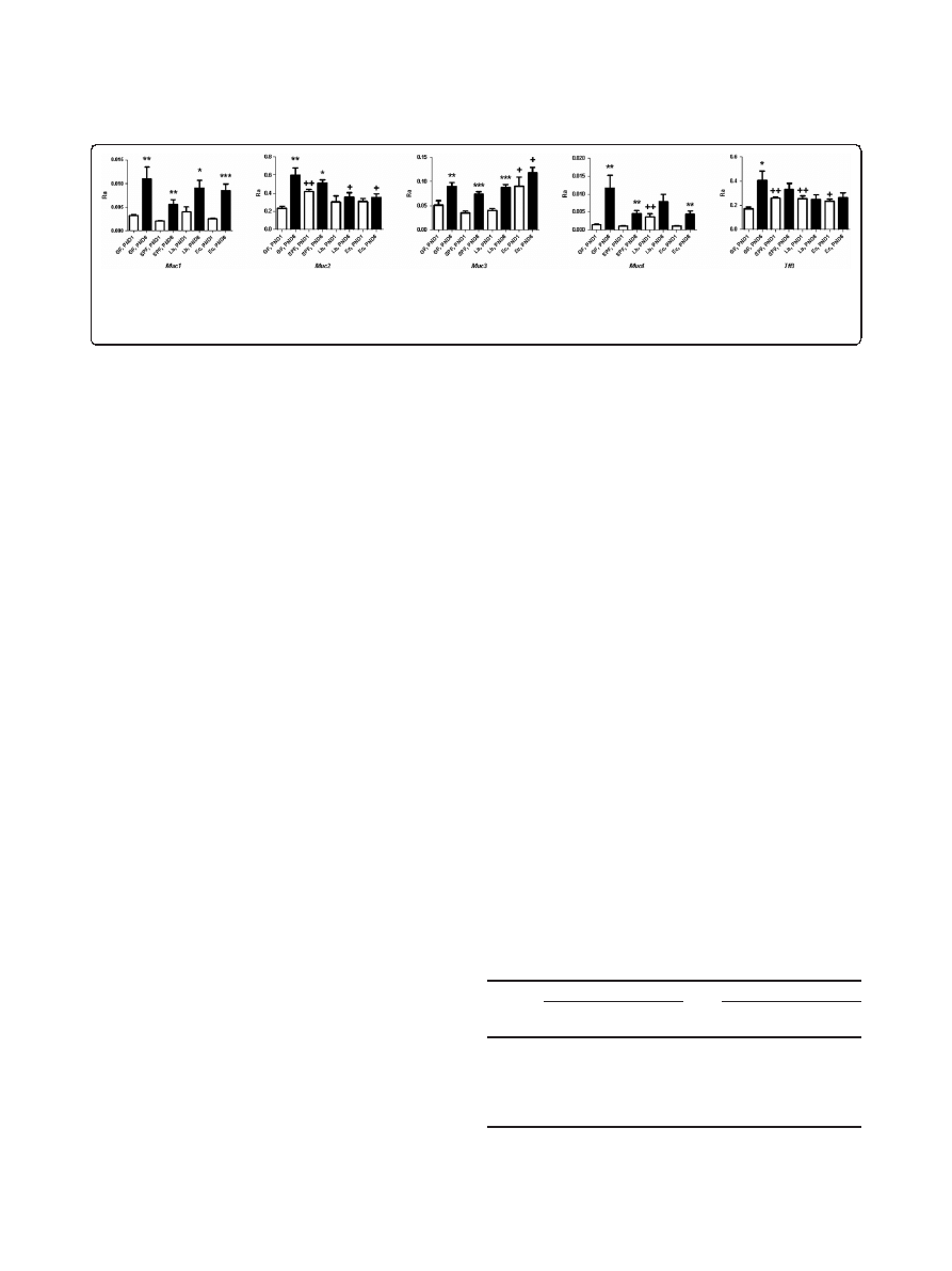
In GF mice, Muc1-4 and Tff3, all showed statistically sig-
nificant increases in transcription from PND1 to PND6,
indicating that this event occurs during the first post-
natal week independently of the presence of microbes
(Figure 1). For certain mucin genes, presence of bacteria
in the colonized animals correlated with an increased
relative abundance of transcripts on PND1 compared to
transcription levels of the same genes in GF mice. This
was particularly evident for the genes Muc2 and Tff3.
Increased transcription on PND1 of Tff3 was observed in
conventional pups (SPF) as well as in pups of dams
’
monocolonized with either Lb or Ec, while for Muc2, this
was observed only in presence of a full microbiota (SPF).
For Muc3 in Ec and Muc4 in Lb, a higher level of tran-
scription was observed on PND1 than in GF pups, indi-
cating that E. coli and Lactobacillus may specifically
stimulate transcription of these genes immediately after
birth (Figure 1).
The higher level of Muc2 and Tff3 transcriptions at
PND1, both encoding secreted proteins with goblet cell
origin [28], indicates that the presence of bacteria affects
gene transcription onset in these exocytotic cells. While
both gene products play protective roles during gut
inflammatory conditions, at sites of epithelial damage
[18,29-34] and during postnatal development [35,36],
Bergström
et al. BMC Research Notes 2012, 5:402
Page 2 of 7
http://www.biomedcentral.com/1756-0500/5/402
Muc2, unlike Tff3, polymerizes into a protective gel-like
structure [1]. Based on the obtained results, it is however
not possible to determine whether there is a connection
between this difference in functionality and the corre-
sponding gene regulation.
Previously, we demonstrated how microbiota affects
ileal gene expression of a number of immune related
genes (specific cytokines and chemokines) during the
first week after birth [23]. As seen for Muc2 in the
present study, and also for a number of Toll-like recep-
tor signaling pathway related genes such as Tlr2/4, Irak1
and the chemokine Cxcl2, encoding MIP-2, the presence
of a full microbiota was required to influence gene
expression on PND1, which was only to a limited degree
affected by monocolonization with either Lactobacilli or
E. coli [23].
Increased transcription of Muc3 and Muc4 on PND1
was observed in Ec and Lb pups, respectively, but not in
SPF (Figure 1). Although specific probiotic bacteria, in-
cluding Lactobacillus acidophilus NCFM [15], Lactoba-
cillus rhamnosus [17], Bifidobacterium bifidum [17],
Lactobacillus plantarum [16,17] as well as two atypical,
enteropathogenic E.coli strains [18], have previously been
shown to stimulate mucin gene expression, this study is
to our knowledge the first to address such effects at a
very early stage of life. Muc1 transcription levels were in
this study apparently not affected by the presence of
bacteria.
Bacterial 16S rRNA abundance on PND1 and PND6
None of the PND1 samples contained Lactobacillus or E.
coli in amounts above the qPCR detection limit (DL), in
any of the four animal groups (Table 1). This was
expected, since only partial bacterial colonization is
achieved so short after birth. On PND6, 5/8 pups in both
the Lb and SPF groups, respectively, were positively
above the Lactobacillus 16S rRNA DL, while 8/8 and 0/8
in the Ec and SPF groups, respectively, were colonized
above the E.coli DL. These observations corresponded to
differences in N
0
values (See Methods) of >300-fold for
Lactobacillus and >160-fold for E.coli. This shows that
bacterial levels in the ileal sections increased between
PND1 and PND6 after birth, although the employed pro-
cedure did not allow detection of bacterial 16S rRNA in
all pups. Culture-based techniques have shown that the
gut mucosal surfaces in newborn mice follow a rather
conserved colonization pattern [37]. In particular, lacto-
bacilli colonize within the first 1
–2 days after birth,
whereas coliforms are normally not quantifiable in the
mucosal layers until approximately 9 days after birth
[14]. These results are thus consistent with findings in
the SPF group in this study. It is however important to
note, that the current analysis was performed on whole
intestinal sections, including both luminal contents and
mucosal surfaces, whereas the other studies referred to
were based on analysis of mucosal surfaces only.
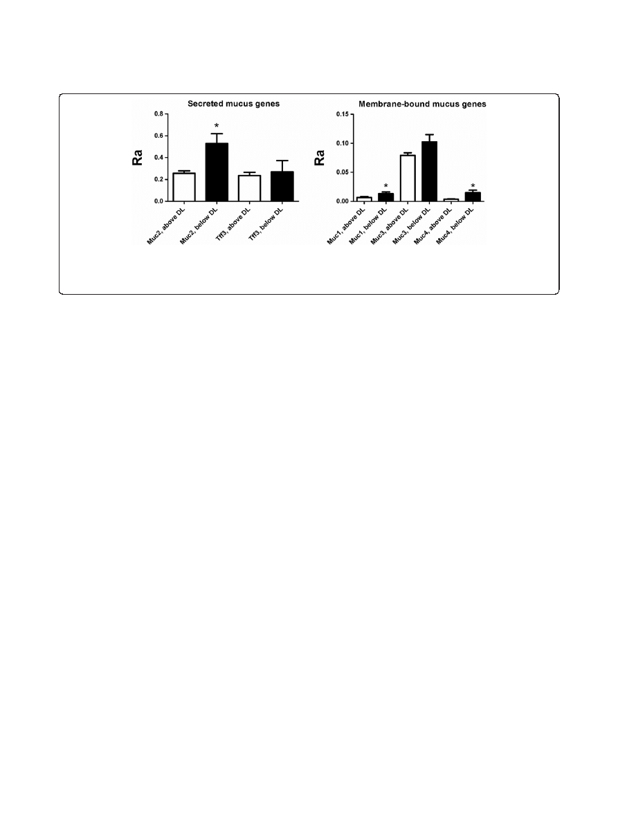
There was a significantly lower level of transcripts
(p < 0.05) of Muc1, Muc2 and Muc4 in the pups with de-
tectable amounts of lactobacilli on PND6 in the Lb
group than in pups with colonization below the detec-
tion limit (Figure 2). In other words, colonization with
relatively high levels of Lactobacilli in the pups had a
negative effect on mucin gene transcription on PND6.
For Muc2, pups colonized with Lactobacillus below the
detection limit in the Lb group were indeed comparable
to GF pups.
It has been established by others that degradation of
mucin in adult rats [38] as well as gene expression of
Muc1-4 and Tff3 in six week old mice [6], is different
Table 1 16S rRNA measured presence vs. absence of all
4 animal groups on each of days PND1 and PND6
PND1
PND6
Lactobacillus
16S
E. coli
16S
Lactobacillus
16S
E. coli
16S
GF
0/8
0/8
0/8
0/8
SPF
0/8
0/8
5/8
0/8
Ec
0/8
0/8
0/8
8/8
Lb
0/8
0/8
5/8
0/8
The fraction denotes number of samples significantly above detection limit
(DL) of the total number (N = 8 in each group).
Figure 1 Comparison of mucin gene expression between the four animal groups on PND1 and PND6. Transcription of Muc1, Muc2, Muc3,
Muc4 and Tff3 on postnatal days (PND) 1 and 6 for pups in groups: GF, SPF, Lb and Ec. Each column represents the average relative abundance
(Ra) (See Methods) of 8 animals. Error bars show SEM values.
*
p < 0.05,
**
p < 0.01,
***
p < 0.001 relative to PND1 (for the same group)
+
p < 0.05,
++
p < 0.01,
+++
p < 0.001 relative to GF (for the same day).
Bergström
et al. BMC Research Notes 2012, 5:402
Page 3 of 7
http://www.biomedcentral.com/1756-0500/5/402
when comparing GF and conventional animals. Clearly,
gene regulation induced by the colonizing microbiota is
a complex and continuous process occurring throughout
the first weeks of life, and as a more stable and adult-like
microbiota is probably not achieved until the end of
weaning process at approximately 21 days after birth
[39], the expression of the mucus regulating genes may
change not only in newborn animals, but also later in life
in response to periodic changes in the microbiota
Conclusions
In this manuscript, we show distinct differences between
the expression patterns of secreted (Muc2/Tff3) and
membrane-bound (Muc1/Muc3/Muc4) mucus-regulatory
genes in the very first days after birth. Presence of a full
microbiota (SPF) increased the relative level of transcrip-
tion of Muc2 and Tff3, which implies the two corre-
sponding secreted gene products, Muc2 and Tff3, to play
protective roles in the postnatal intestinal layer develop-
ment. The immediate activation of Muc2/Tff3 transcrip-
tion may provide a coating of the new born ileal
epithelial layer, allowing only passage of certain sub-
stances or organisms.
Methods
Animal experiments
GF Swiss Webster mice and SPF mice, containing con-
ventional microbiota, were purchased from Taconic (Lille
Skensved, Denmark) and kept in GF isolators or under
specific pathogen-free conditions, respectively [22]. Fecal
samples from GF mice, taken at sampling i.e. once a
week, were cultivated on non-selective LB medium and
under aerobic and anaerobic conditions to confirm steril-
ity of isolators. For breeding, pairs of female mice were
housed with one male until plugs were observed. Mono-
colonization of pregnant mice with Ec and Lb was
performed 7 days after mating by applying 5x10
8
CFU ml
-1
in 0.5 ml PBS suspension orally using a pipette
and 0.5 ml to the abdominal skin. Lb was grown anaer-
obically in de Man, Rogosa, and Sharpe broth (MRS,
Merck, Darmstadt, Germany) and Ec aerobically in
Luria-Bertani broth (LB, Merck) overnight at 37°C. The
cultures
were
harvested,
washed
twice
in
sterile
phosphate-buffered saline (PBS) (Lonza, Basel, Switzer-
land), re-suspended in 1/50 of the original culture
volume and frozen at
−80 °C until use. Prior to adminis-
tering bacteria to the mice, Ec suspensions were diluted
tenfold in PBS immediately to obtain 5x10
8
CFU ml
-1
.
Lb suspensions were not diluted. Four litters spontan-
eously delivered from 4 different mothers in each group;
SPF, GF, Lb and Ec, were used for the experiment. At
post-natal days 1 and 6, the pups were put down and
the distal ileum (segment from cecum and 3 cm up) was
removed from the small intestine of two pups per litter and
frozen in RNAlater (Qiagen, Hilden, Germany). No sep-
aration of mucosal from luminal content was performed.
Ethics
The mouse experiment was performed under a license to
Department of Microbiology, National Food Institute,
from the Danish Council for Animal Experimentation.
RNA isolation
Tissues were removed from RNAlater and homogenized
by a rotor strator in RLT buffer (Qiagen). RNA from
tissue homogenate was extracted using RNeasy Mini Kit
from Qiagen following the supplier's protocol.
Primer design and validation
A list of all primers used in this study is presented in
Table 2. All primers found in references were initially
checked with the Net Primer Software (http://www.
Figure 2 Difference in mucin gene expression between pups with detectable and undetectable
Lactobacillus 16S in Lb. Comparison of
Muc1, Muc2, Muc3, Muc4 and Tff3 transcription on PND6 between pups in the Lb group, where Lactobacillus 16S could be detected and could not
be detected, respectively. Each column represents the average relative abundance (Ra) of 5/8 (above DL) or 3/8 (below DL) animals. Error bars
show SEM values. DL = Detection Limit.
*
p < 0.05, denotes significant difference between detectably colonized and not colonized.
Bergström
et al. BMC Research Notes 2012, 5:402
Page 4 of 7
http://www.biomedcentral.com/1756-0500/5/402

premierbiosoft.com/netprimer/index.html). Primers not
scoring a rating of at least 90 % were not accepted and
new primers were then designed with NCBI
’s primer
designing tool (http://www.ncbi.nlm.nih.gov/tools/primer-
blast/) and the quality was again verified until satisfaction
with the Net Primer Software. All newly designed primers
were designed to span exon junctions to avoid amplifica-
tion of genomic DNA. The specificity of all primers was
evaluated in silico using nucleotide BLAST, (http://blast.
ncbi.nlm.nih.gov/Blast.cgi).
Quantitative PCR (qPCR)
Isolated ileal RNA was reverse transcribed into cDNA
using SuperScript
W
VILO
™ cDNA Synthesis Kit from
Invitrogen, Denmark. After verifying the quality of the
cDNA by spectroscopy (A
260
/A
280
= 1.8 ± 10 %) measured
on a NanoDrop ND-1000 Spectrophotometer (Saveen
Werner, Limhamn, Sweden), it was used as template in
quantitative real-time PCR using the ABI prism 7900HT
from Applied Biosystems. All cDNA concentrations were
within the range of 90-100 ng
μl
-1
. The amplification reac-
tions were carried out in a total volume of 20
μl containing
10
μl master mix (2x PerfeCTa
TM
SYBR
W
Green Super-
Mix, ROX from Quanta Biosciences
TM
), 0.4
μl of each
primer (10
μM), 2 μl template cDNA, and 7.2 μl nuclease-
free water (Qiagen GmbH, Germany) purified for PCR.
The amplification program consisted of one cycle of 50 °C
for 2 min; one cycle of 95 °C for 10 min; 40 cycles of 95 °
C for 15 s and 60 °C for 1 min; and finally one cycle of
dissociation curve analysis for assessing the amplification
products (95 °C for 15 s, 60 °C for 20s and increasing
ramp rate by 2 % until 95 °C for 15 s). These conditions
were selected based on preliminary qPCR experiments on
target DNA with similar concentrations (100 ng
μl
-1
).
Samples of all amplification products were further
subjected to gel electrophoresis in 2 % agarose, followed by
ethidium bromide staining in order to verify amplicon
sizes.
qPCR setup
Three separate qPCR experiments on ileal cDNA were
performed; 1) and 2) were separate replications of relative
quantifications on mucus gene transcription (Muc1-4 and
Tff3) with selected reference genes (see next paragraph)
and 3) on presence or absence of specific bacterial 16S
rRNA analysis (Lactobacillus, E.coli).
qPCR data analysis
All qPCR analysis was performed with the freely avail-
able LinRegPCR tool developed by Ruijter et al. [24,25].
The raw fluorescence data were exported from the
ABI prism 7900HT SDS-software, and the LinRegPCR
program was used to estimate baselines and individual
PCR efficiencies in order to calculate output as target
starting concentration, expressed in arbitrary fluores-
cence units N
0
, for each PCR sample by the formula
N
0
= threshold / (Eff
mean
Ct
), where Eff
mean
denotes the
optimal PCR mean efficiency/amplicon, threshold the
optimal
“cutoff” in the exponential region and C
t
is
the number, where each PCR sample exceeds this thresh-
old. Samples with no amplification, baseline error, too
much noise or without plateau were automatically
excluded by the LinRegPCR software. Subsequently, for
each amplicon the average of all remaining, successful
samples within 5 % of the mean value of all successful
samples/amplicon were used in the calculation of Eff
mean
for each amplicon. All N
0
-values were calculated as
means of double qPCR determinations.
For relative quantification of mucus gene transcripts,
two different eukaryotic reference genes were used namely
beta-actin [40] and neuroplastin, the latter suggested by
the Genevestigator software (https://www.genevestigator.
com) [41,42] based on microarray data on similar or-
ganism (M. musculus) and tissue (ileum). We used the
geometric mean of the two reference genes as previously
suggested [43]. Normalization to relevant reference gene
expression was then calculated according to the formula:
Table 2 Primers used for qPCR
Primer name
Fwd (5´-3´)
Rev (5´-3´)
Amplicon
size
Reference
Muc1
TCGTCTATTTCCTTGCCCTG
ATTACCTGCCGAAACCTCCT
185
This study
Muc2
CCCAGAAGGGACTGTGTATG
TTGTGTTCGCTCTTGGTCAG
276
Modified from [
Muc3
TGGTCAACTGCGAGAATGGA
TACGCTCTCCACCAGTTCCT
98
Modified from [
]
Muc4
GTCTCCCATCACGGTTCAGT
TGTCATTCCACACTCCCAGA
280
This study
Tff3
CTCTGTCACATCGGAGCAGTGT
TGAAGCACCAGGGCACATT
77
Neuroplastin
CGCTGCTCAGAACGAACCAAGAA
CTTACGGGTGGCAGTGAGTT
160
Modified from [
Beta-actin
GTCCACCTTCCAGCAGATGT
GAAAGGGTGTAAAACGCAGC
117
This study
Lactobacillus 16S rRNA
AGCAGTAGGGAATCTTCCA
a
CACCGCTACACATGGAG
b
341
a
[
]
b
]
E. coli 16S rRNA
CATGCCGCGTGTATGAAGAA
CGGGTAACGTCAATGAGCAAA
96
Bergström
et al. BMC Research Notes 2012, 5:402
Page 5 of 7
http://www.biomedcentral.com/1756-0500/5/402

Ra =Ratio = N
0
Sample
/ N
0
Reference
and averaged over the two
qPCR experiments.
Unspecific amplification of 16S rRNA bacterial genes
from GF mice was used to specify detection limits for
specific amplifications (Lactobacillus, E .coli). Cutoffs
for presence of either bacterium were defined by at
least 5 C
t
-values difference from the GF samples. No
normalization to reference genes and thus relative
quantification was used for the 16S analysis, since the
purpose was only to determine presence vs. absence of
detectable bacteria.
Statistics
All statistics was performed with GraphPad Prism 5.
One-way ANOVA followed by Dunnett
’s post hoc test
with GF as control group and Student
’s t-test was used to
compare mucus gene expression between the four ani-
mal groups and development from PND1 to PND6, re-
spectively. P-values lower than p = 0.05 were considered
statistically significant. Welch
’s correction for unequal
variances was applied, when necessary.
Competing interests
The authors declare that they have no competing interests.
Authors
’ contributions
AB performed the qPCR experiments, including cDNA syntheses, data
interpretation and statistical analysis, and wrote the manuscript. MBK and
SBM performed the animal experiments, including isolation of ileal tissue and
RNA purification. TRL, HF and LNF conceived of the study setup and
participated in its design and coordination. TRL, MBK, HF and MIB
contributed to data analysis and interpretation as well as preparation of the
manuscript. All authors read and approved the final manuscript.
Acknowledgements
The work was supported by Globalization Funds obtained from the Technical
University of Denmark, through The National Food Institute, and a post doc
grant for Anders Bergström from The Danish Agency for Science, Technology
and Innovation.
Author details
1
Gut Ecology Group, Department of Food Microbiology, National Food
Institute, Technical University of Denmark, Mørkhøj Bygade 19, Søborg 2860,
Denmark.
2
Department of Basic Sciences and Environment, Faculty of Life
Sciences, University of Copenhagen, Copenhagen, Denmark.
3
Center for
Biological Sequence Analysis, Department of Systems Biology, Technical
University of Denmark, Lyngby, Denmark.
Received: 16 November 2011 Accepted: 12 July 2012
Published: 2 August 2012
References
1.
Hollingsworth MA, Swanson BJ: Mucins in cancer: protection and control
of the cell surface. Nat Rev Cancer 2004, 4:45
–60.
2.
Adlerberth I, Wold AE: Establishment of the gut microbiota in Western
infants. Acta Paediatr 2009, 98:229
–238.
3.
Fanaro S, Chierici R, Guerrini P, Vigi V: Intestinal microflora in early
infancy: composition and development. Acta Paediatr Suppl 2003,
91:48
–55.
4.
Hooper LV, Wong MH, Thelin A, Hansson L, Falk PG, Gordon JI: Molecular
analysis of commensal host-microbial relationships in the intestine.
Science 2001, 291:881
–884.
5.
Favier CF, Vaughan EE, de Vos WM, Akkermans AD: Molecular monitoring
of succession of bacterial communities in human neonates. Appl Environ
Microbiol 2002, 68:219
–226.
6.
Comelli EM, Simmering R, Faure M, Donnicola D, Mansourian R, Rochat F,
Corthesy-Theulaz I, Cherbut C: Multifaceted transcriptional regulation of
the murine intestinal mucus layer by endogenous microbiota. Genomics
2008, 91:70
–77.
7.
Corfield AP, Myerscough N, Longman R, Sylvester P, Arul S, Pignatelli M:
Mucins and mucosal protection in the gastrointestinal tract: new
prospects for mucins in the pathology of gastrointestinal disease. Gut
2000, 47:589
–594.
8.
McGuckin MA, Linden SK, Sutton P, Florin TH: Mucin dynamics and enteric
pathogens. Nat Rev Microbiol 2011, 9:265
–278.
9.
Xing PX, Lees C, Lodding J, Prenzoska J, Poulos G, Sandrin M, Gendler S,
McKenzie IF: Mouse mucin 1 (MUC1) defined by monoclonal antibodies.
Int J Cancer 1998, 76:875
–883.
10.
Shekels LL, Hunninghake DA, Tisdale AS, Gipson IK, Kieliszewski M, Kozak CA,
Ho SB: Cloning and characterization of mouse intestinal MUC3 mucin: 3'
sequence contains epidermal-growth-factor-like domains. Biochem J 1998,
330(Pt 3):1301
–1308.
11.
van Klinken BJ, Einerhand AW, Duits LA, Makkink MK, Tytgat KM, Renes IB,
Verburg M, Büller HA: Gastrointestinal expression and partial cDNA
cloning of murine Muc2. Am J Physiol 1999, 276:G115
–G124.
12.
Desseyn JL, Clavereau I, Laine A: Cloning, chromosomal localization and
characterization of the murine mucin gene orthologous to human
MUC4. Eur J Biochem 2002, 269:3150
–3159.
13.
Salminen S, Isolauri E: Intestinal colonization, microbiota, and probiotics.
J Pediatr 2006, 149:S115
–S120.
14.
Savage DC, Dubos R, Schaedler RW: The gastrointestinal epithelium and its
autochthonous bacterial flora. J Exp Med 1968, 127:67
–76.
15.
Sanders ME, Klaenhammer TR: Invited review: the scientific basis of
Lactobacillus acidophilus NCFM functionality as a probiotic. J Dairy Sci
2001, 84:319
–331.
16.
Mack DR, Ahrne S, Hyde L, Wei S, Hollingsworth MA: Extracellular MUC3
mucin secretion follows adherence of Lactobacillus strains to intestinal
epithelial cells
in vitro. Gut 2003, 52:827–833.
17.
Dykstra NS, Hyde L, Adawi D, Kulik D, Ahrne S, Molin G, Jeppsson B,
Mackenzie A, Mack DR: Pulse probiotic administration induces
repeated small intestinal Muc3 expression in rats. Pediatr Res 2011,
69:206
–211.
18.
Vieira MA, Gomes TA, Ferreira AJ, Knobl T, Servin AL, Lievin-Le M: V: Two
atypical enteropathogenic Escherichia coli strains induce the production
of secreted and membrane-bound mucins to benefit their own growth
at the apical surface of human mucin-secreting intestinal HT29-MTX
cells. Infect Immun 2010, 78:927
–938.
19.
Wright CT, Klaenhammer TR: Calcium-induced alteration of cellular
morphology affecting the resistance of lactobacillus acidophilus to
freezing. Appl Environ Microbiol 1981, 41:807
–815.
20.
Nissle A: Mutaflor and its medical significance. Z Klin Med 1951, 2:68.
21.
Jacobi CA, Malfertheiner P: Escherichia coli Nissle 1917 (Mutaflor):
new insights into an old probiotic bacterium. Dig Dis 2011,
29:600
–607.
22.
Zeuthen LH, Fink LN, Metzdorff SB, Kristensen MB, Licht TR, Nellemann C,
Frøkiær H: Lactobacillus acidophilus induces a slow but more sustained
chemokine and cytokine response in naive foetal enterocytes compared
to commensal Escherichia coli. BMC Immunol 2010, 11:2.
23.
Fink LN, Metzdorff SB, Zeuthen LH, Nellemann C, Kristensen MB, Licht TR,
Frøkiær H: Establishment of tolerance to commensal bacteria requires a
complex microbiota and is accompanied by decreased intestinal
chemokine expression. Am J Physiol Gastrointest Liver Physiol 2012,
302(1):G55
–G65.
24.
Ramakers C, Ruijter JM, Deprez RH, Moorman AF: Assumption-free analysis
of quantitative real-time polymerase chain reaction (PCR) data. Neurosci
Lett 2003, 339:62
–66.
25.
Ruijter JM, Ramakers C, Hoogaars WM, Karlen Y, Bakker O, van den Hoff MJ,
Moorman AF: Amplification efficiency: linking baseline and bias in the
analysis of quantitative PCR data. Nucleic Acids Res 2009, 37:e45.
26.
Bustin SA: Absolute quantification of mRNA using real-time reverse
transcription polymerase chain reaction assays. J Mol Endocrinol 2000,
25:169
–193.
Bergström
et al. BMC Research Notes 2012, 5:402
Page 6 of 7
http://www.biomedcentral.com/1756-0500/5/402

27.
Veazey KJ, Golding MC: Selection of stable reference genes for
quantitative rt-PCR comparisons of mouse embryonic and extra-
embryonic stem cells. PLoS One 2011, 6:e27592.
28.
Bergstrom KS, Guttman JA, Rumi M, Ma C, Bouzari S, Khan MA, Gibson DL,
Vogl AW, Vallance BA: Modulation of intestinal goblet cell function during
infection by an attaching and effacing bacterial pathogen. Infect Immun
2008, 76:796
–811.
29.
Dignass AU, Sturm A: Peptide growth factors in the intestine. Eur J
Gastroenterol Hepatol 2001, 13:763
–770.
30.
Heazlewood CK, Cook MC, Eri R, Price GR, Tauro SB, Taupin D, Thornton DJ,
Png CW, Crockford TL, Cornall RJ, Adams R, Kato M, Nelms KA, Hong NA,
Florin TH, Goodnow CC, McGuckin MA: Aberrant mucin assembly in mice
causes endoplasmic reticulum stress and spontaneous inflammation
resembling ulcerative colitis. PLoS Med 2008, 5:e54.
31.
Boshuizen JA, Reimerink JH, Korteland-Van Male AM, Van HV, Bouma J,
Gerwig GJ, Koopmans MP, Büller HA, Dekker J, Einerhand AW: Homeostasis
and function of goblet cells during rotavirus infection in mice. Virology
2005, 337:210
–221.
32.
Taupin D, Podolsky DK: Trefoil factors: initiators of mucosal healing. Nat
Rev Mol Cell Biol 2003, 4:721
–732.
33.
Tran CP, Cook GA, Yeomans ND, Thim L, Giraud AS: Trefoil peptide TFF2
(spasmolytic polypeptide) potently accelerates healing and reduces
inflammation in a rat model of colitis. Gut 1999, 44:636
–642.
34.
Thim L, Madsen F, Poulsen SS: Effect of trefoil factors on the viscoelastic
properties of mucus gels. Eur J Clin Invest 2002, 32:519
–527.
35.
Scholven J, Taras D, Sharbati S, Schon J, Gabler C, Huber O, Meyer Zum
Büschenfelde D, Blin N, Einspainer R: Intestinal expression of TFF and
related genes during postnatal development in a piglet probiotic trial.
Cell Physiol Biochem 2009, 23:143
–156.
36.
Fanca-Berthon P, Michel C, Pagniez A, Rival M, Van SI, Darmaun D, Hoebler
C: Intrauterine growth restriction alters postnatal colonic barrier
maturation in rats. Pediatr Res 2009, 66:47
–52.
37.
Schaedler RW, Dubos R, Costello R: The development of the bacterial
flora in the gastrointestinal tract of mice. J Exp Med 1965, 122:59
–66.
38.
Midtvedt T, Carlstedt-Duke B, Hoverstad T, Midtvedt AC, Norin KE,
Saxerholt H: Establishment of a biochemically active intestinal ecosystem
in ex-germfree rats. Appl Environ Microbiol 1987, 53:2866
–2871.
39.
Davis CP, McAllister JS, Savage DC: Microbial colonization of the intestinal
epithelium in suckling mice. Infect Immun 1973, 7:666
–672.
40.
Huggett J, Dheda K, Bustin S, Zumla A: Real-time RT-PCR normalisation;
strategies and considerations. Genes Immun 2005, 6:279
–284.
41.
Laule O, Hirsch-Hoffmann M, Hruz T, Gruissem W, Zimmermann P: Web-
based analysis of the mouse transcriptome using Genevestigator. BMC
Bioinformatics 2006, 7:311.
42.
Zimmermann P, Hirsch-Hoffmann M, Hennig L, Gruissem W:
Genevestigator. Arabidopsis microarray database and analysis toolbox.
Plant Physiol 2004, 136:2621
–2632.
43.
Vandesompele J, De PK, Pattyn F, Poppe B, Van RN, De Paepe A, Speleman
F: Accurate normalization of real-time quantitative RT-PCR data by
geometric averaging of multiple internal control genes. Genome Biol
2002, 3(7):research0034.1-0034.11.
44.
Tai EK, Wu WK, Wong HP, Lam EK, Yu L, Cho CH: A new role for
cathelicidin in ulcerative colitis in mice. Exp Biol Med (Maywood) 2007,
232:799
–808.
45.
Liu J, Yu L, Tokar EJ, Bortner C, Sifre MI, Sun Y, Waalkes MP: Arsenic-induced
aberrant gene expression in fetal mouse primary liver-cell cultures. Ann
N Y Acad Sci 2008, 1140:368
–375.
46.
Kreutz MR, Langnaese K, Dieterich DC, Seidenbecher CI, Zuschratter W,
Beesley PW, Gundelfinger ED: Distribution of transcript and protein
isoforms of the synaptic glycoprotein neuroplastin in rat retina. Invest
Ophthalmol Vis Sci 2001, 42:1907
–1914.
47.
Walter J, Hertel C, Tannock GW, Lis CM, Munro K, Hammes WP: Detection of
Lactobacillus, Pediococcus, Leuconostoc, and Weissella species in human
feces by using group-specific PCR primers and denaturing gradient gel
electrophoresis. Appl Environ Microbiol 2001, 67:2578
–2585.
48.
Heilig HG, Zoetendal EG, Vaughan EE, Marteau P, Akkermans AD, De Vos
WM: Molecular diversity of Lactobacillus spp. and other lactic acid
bacteria in the human intestine as determined by specific amplification
of 16S ribosomal DNA. Appl Environ Microbiol 2002, 68:114
–123.
49.
Huijsdens XW, Linskens RK, Mak M, Meuwissen SG, Vandenbroucke-Grauls
CM, Savelkoul PH: Quantification of bacteria adherent to gastrointestinal
mucosa by real-time PCR. J Clin Microbiol 2002, 40:4423
–4427.
doi:10.1186/1756-0500-5-402
Cite this article as: Bergström et al.: Nature of bacterial colonization
influences transcription of mucin genes in mice during the first week of
life. BMC Research Notes 2012 5:402.
Submit your next manuscript to BioMed Central
and take full advantage of:
•
Convenient online submission
•
Thorough peer review
•
No space constraints or color figure charges
•
Immediate publication on acceptance
•
Inclusion in PubMed, CAS, Scopus and Google Scholar
•
Research which is freely available for redistribution
Submit your manuscript at
www.biomedcentral.com/submit
Bergström
et al. BMC Research Notes 2012, 5:402
Page 7 of 7
http://www.biomedcentral.com/1756-0500/5/402
Document Outline
- Abstract
- Background
- Results and discussion
- link_Tab1
- link_Fig1
- Conclusions
- Methods
- link_Fig2
- link_Tab2
- Competing interests
- Authors´ contributions
- Acknowledgements
- Author details
- References
- link_CR1
- link_CR2
- link_CR3
- link_CR4
- link_CR5
- link_CR6
- link_CR7
- link_CR8
- link_CR9
- link_CR10
- link_CR11
- link_CR12
- link_CR13
- link_CR14
- link_CR15
- link_CR16
- link_CR17
- link_CR18
- link_CR19
- link_CR20
- link_CR21
- link_CR22
- link_CR23
- link_CR24
- link_CR25
- link_CR26
- link_CR27
- link_CR28
- link_CR29
- link_CR30
- link_CR31
- link_CR32
- link_CR33
- link_CR34
- link_CR35
- link_CR36
- link_CR37
- link_CR38
- link_CR39
- link_CR40
- link_CR41
- link_CR42
- link_CR43
- link_CR44
- link_CR45
- link_CR46
- link_CR47
- link_CR48
- link_CR49
Wyszukiwarka
Podobne podstrony:
Shakespeare on the nature of life
national antisemitism in russia during the years of crisis (1914 1922)
The Fate of Psychiatrie Patients in Belarus During the German Occupation
nature of human language
The Nature of Mind Longchenpa
Nature of Survey Fragment, 1955
2009 Smith (01 Nature of Biotec Nieznany
Freud View On The Nature Of Man
To what extent does the nature of language illuminate the dif
The Nature Of Cancer, !!♥ TUTAJ DODAJ PLIK ⇪⇪⇪⇪⇪⇪⇪⇪⇪⇪⇪⇪⇪⇪
Lord of the Flies Analysis of Primitive Nature of Humanity
Frédéric Mégret The Nature of International Human Rights Obligations
The Nature of Experiment in Archaeology
Towards an understanding of the distinctive nature of translation studies
The Nature of Mind Longchenpa
Nature of order str 151
Sarbin Narrative psychology, the storied nature of human conduct str vii, ix, 3 63, 117 125
więcej podobnych podstron