
The pathogenesis of Shigella exneri infection:
lessons from in vitro and in vivo studies
Dana J. Philpott, Jonathan D. Edgeworth and Philippe J. Sansonetti
*
Unite¨ de Pathoge¨nie Microbienne Mole¨culaire, Institut Pasteur, 28 rue du Docteur Roux, 75724 Paris, France
Shigella £exneri is a Gram-negative facultatively intracellular pathogen responsible for bacillary dysentery
in humans. More than one million deaths occur yearly due to infections with Shigella spp. and the victims
are mostly children of the developing world. The pathogenesis of Shigella centres on the ability of this
organism to invade the colonic epithelium where it induces severe mucosal in£ammation. Much
information that we have gained concerning the pathogenesis of Shigella has been derived from the study
of in vitro models of infection. Using these techniques, a number of the molecular mechanisms by which
Shigella invades epithelial cells and macrophages have been identi¢ed. In vivo models of shigellosis have
been hampered since humans are the only natural hosts of Shigella. However, experimental infection of
macaques as well as the murine lung and rabbit ligated ileal loop models have been important in de¢ning
some of the immune and in£ammatory components of the disease. In particular, the murine lung model
has shed light on the development of systemic and local immune protection against Shigella infection. It
would be naive to believe that any one model of Shigella infection could adequately represent the
complexity of the disease in humans, and more sophisticated in vivo models are now necessary. These
models require the use of human cells and tissue, but at present such models remain in the developmental
stage. Ultimately, however, it is with such studies that novel treatments and vaccine candidates for the
treatment and prevention of shigellosis will be designed.
Keywords: Shigella; pathogenicity; mucosal immunity; immune response; animal models; in vitro models
1. INTRODUCTION
Infection with Shigella spp. is a serious cause of morbidity
and mortality especially in children of the developing
world. Recently, the World Health Organization esti-
mated that 1.1 million deaths per year are attributed to
shigellosis (Kotlo¡ et al. 1999). There are four species of
Shigella that cause these infections, with S. £exneri and, to a
lesser extent, S. sonnei, accounting for most of the endemic
disease. Epidemic disease is usually due to S. dysenteriae,
which displays the same invasive capacity as the other
species but in addition, secretes a potent cytotoxin, Shiga
toxin, that can cause haemolytic uraemic syndrome.
Existing antimicrobial treatments are becoming increas-
ingly compromised because of the growing occurrence of
antibiotic resistance among Shigella spp. In addition, the
cost of treating shigellosis with antibiotics, particularly in
the developing world, is impractical and stresses the need
for an e¤cient vaccine against this disease. Currently,
however, there is no vaccine available that can provide
adequate protection against the many di¡erent serotypes
of Shigella. Therefore, both the development of new treat-
ments and the design of innovative vaccines for the
prevention of shigellosis rely on an improved under-
standing of the pathogenesis of the disease. Our knowl-
edge of the pathogenesis of Shigella infection thus far and
what we hope to learn in the future has and continues to
depend on our ability to model the infection in vitro and to
validate these models with in vivo studies. This review
outlines our current understanding of the pathogenesis of
Shigella infection. Speci¢cally, ¢ndings from in vitro
systems will be compared to those gained from animal
models of shigellosis, while keeping in mind essential
features of the disease in humans. This is followed by a
discussion of the possibilities for future research and
where we believe further studies are required.
2.
SHIGELLA INFECTIONÐOVERVIEW
Shigella £exneri is a Gram-negative facultatively intra-
cellular pathogen that invades the colonic and rectal
mucosae of humans, causing bacillary dysentery. Shigel-
losis is highly infectious, with ingestion of as few as 100
organisms resulting in disease (Dupont et al. 1989), and is
transmitted by person-to-person contact or indirectly
through contaminated food or water. Shigellosis produces
a spectrum of clinical outcomes ranging from watery
diarrhoea to classic dysentery characterized by fever,
violent intestinal cramps and discharge of mucopurulent
and bloody stools. In£ammation of the infected tissue is a
key feature of shigellosis. Histopathological studies of
colonic biopsies from infected patients reveal in£amma-
tory cell in¢ltration into the epithelial layer, tissue
oedema and eroded regions of the colonic epithelium
(Mathan & Mathan 1991).
Since this organism is unable to invade epithelial cells
through the apical route, Shigella exploits M cells, the
specialized epithelial cells in the follicular associated
Phil. Trans. R. Soc. Lond. B (2000) 355, 575^586
575
© 2000 The Royal Society
*
Author for correspondence (psanson@pasteur.fr).
epithelium (FAE) that overlie lymphoid tissue, to gain
entry into the colonic epithelium (Wassef et al. 1989).
M cells allow intact Shigella to traverse into the under-
lying subepithelial pocket where macrophages reside.
Macrophages engulf Shigella, but instead of successfully
destroying the bacteria in the phagosome, the macro-
phage succumbs to apoptotic death (Zychlinsky et al.
1992). Prior to cell death, infected macrophages release
IL-1b through the direct activation of caspase-1 by
Shigella (Zychlinsky et al. 1994). The pro-in£ammatory
nature of this cytokine results in the recruitment of poly-
morphonuclear cells (PMNs) that in¢ltrate the infected
site and destabilize the epithelium (Perdomo et al.
1994a,b). Loss of integrity of the epithelial barrier allows
more bacteria to traverse into subepithelial space and
gives these organisms access to the basolateral pole of the
epithelial cells (Mounier et al. 1992). Shigella can then
invade the epithelial cells lining the colon, spread from
cell to cell and disseminate throughout the tissue. Cyto-
kines released by infected epithelial cells attract increased
numbers of immune cells to the infected site, thus
compounding and exacerbating the in£ammation.
3. INVASION OF EPITHELIAL CELLS
The link between epithelial cell invasion and expres-
sion of the virulent phenotype of Shigella was ¢rst made in
1964 (LaBrec et al. 1964). The Sere¨ny test, which is the
oldest animal model of shigellosis, was used as a model to
test Shigella invasiveness (Sere¨ny 1955). This assay consists
of inoculating a suspension of bacteria into the kerato-
conjunctival sac of guinea-pigs or mice. Pathogenic
Shigella invade the conjunctival epithelium causing
conjunctivitis and keratitis. This model proved useful for
identifying avirulent mutants of Shigella that are incapable
of expressing the invasive phenotype. However, the lack
of speci¢city of the response makes it impossible to discri-
minate among the various phenotypes of Shigella
including invasion of epithelial cells, cell-to-cell spread
and the initiation of an in£ammatory response.
Cultured epithelial cell lines have greatly aided the study
of the host-cell events involved in cell invasion by Shigella.
Examination of Shigella-infected cells by microscopic
methods has de¢ned the entry event as a macropinocytic-
like process that results in massive induction of host cell
membrane ru¥ingöchanges which are reminiscent of
those elicited by growth factors. In the case of invading
Shigella, however, the membrane ru¥es are con¢ned to the
site of bacterium^cell interaction. Cytoskeleton-mediated
membrane extensions are observed to rise up from the
surface of the cell and these projections eventually fuse to
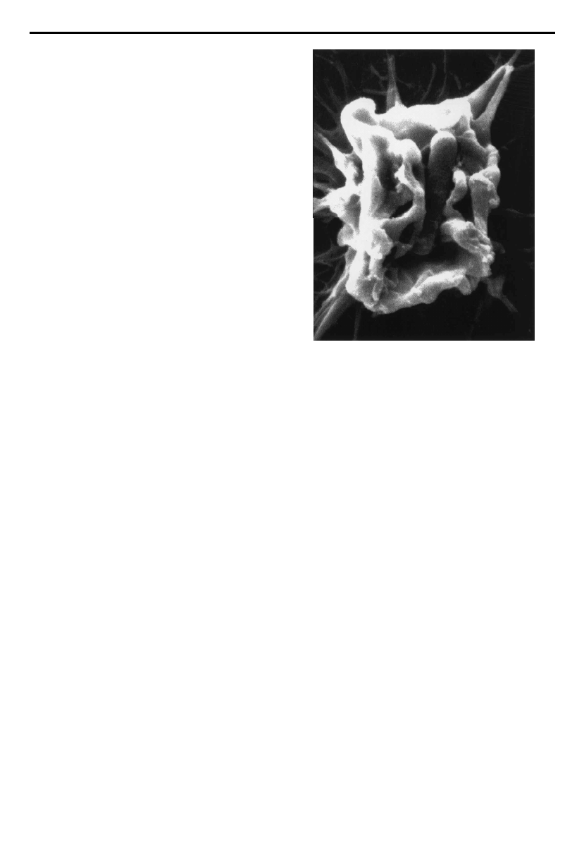
engulf the bacterial body (¢gure1).
Studies of epithelial-cell^Shigella interactions often use
poorly di¡erentiated and non-polarized epithelial cell
lines, such as HeLa or HEp-2 cells, grown in tissue culture
£asks. However, more sophisticated systems using human
intestinal cell lines grown on permeable ¢lter supports
with distinct upper (lumenal) and lower (basal) chambers
have been employed. Growing intestinal Caco-2 or T84
cell lines in this way allows the cells to grow as columnar
epithelial cells with a more or less organized brush border
(depending on the cell line) and to polarize with distinct
apical and basolateral membranes separated by inter-
cellular tight junctions. Bacterial infection of the apical
surface of cultured intestinal cells grown in this way more
closely mimics infection of the human intestinal epithelium.
Using this system, a surprising observation was noted as
apically infecting Shigella cannot invade polarized cells.
Only when intercellular junctions are disrupted by treat-
ment of the cells with ethylene glycol-bis (beta-aminoethyl
ether)-N,N,N’,N’-tetraacetic acid (EGTA) are Shigella able
to invade the ¢lter-grown Caco-2 cells. These studies indi-
cated that Shigella enter polarized Caco-2 cells almost
exclusively from the basolateral pole (Mounier et al.1992).
A methodological step forward was made in the study
of molecular mechanisms of bacterial invasion when it
was realized that the aminoglycoside antibiotic genta-
micin is membrane impermeable and thus bacteria that
are able to enter host cells survive antibiotic treatment of
an infected monolayer. This lack of accessibility of
gentamicin to intracellular bacteria forms the basis of the
`gentamicin protection assay’ whereby the capacity of an
organism to invade eukaryotic cells can be assessed repro-
ducibly and quantitatively. Using this assay, a number of
genes necessary for Shigella entry have been identi¢ed by
analysing mutants defective in surviving gentamicin treat-
ment. Genes encoding bacterial factors required for
Shigella entry reside on a 200 kb virulence plasmid of
wild-type S. £exneri. Strains lacking the plasmid are non-
invasive in vitro and also avirulent in animal models of
shigellosis. The e¡ectors of Shigella entry are the so-called
`invasion plasmid antigens’ or Ipa proteins which are
encoded in a 30 kb `entry region’. This region is composed
of two adjacent loci transcribed in opposite directions.
One locus is essentially composed of the ipa operon,
576 D. J. Philpott and others The pathogenesis of Shigella £exneri infection
Phil. Trans. R. Soc. Lond. B (2000)
Figure 1. Scanning electron micrograph of Shigella £exneri
inducing membrane ru¥es on the surface of an epithelial cell
prior to its uptake. Photograph is courtesy of Dr Ariel Blocker
(Institut Pasteur, France) and Dr Roger Webf (European
Molecular Biology Laboratory, Heidelberg, Germany).

which encodes four secreted proteins, IpaB, IpaC, IpaD
and IpaA, which are the e¡ectors of bacterial entry in
vitro. Mutations in the genes encoding IpaB, IpaC and
IpaD proteins render the bacteria non-invasive in cell
culture systems and are also avirulent in animal models;
these Shigella mutants are unable to provoke keratocon-
junctivitis in guinea pigs (Me¨nard et al. 1993). An IpaA
mutant of Shigella maintains a 10% invasion e¤ciency as
assessed in vitro; however, it is unable to induce £uid accu-
mulation in rabbit ligated loops, suggesting that the full
complement of Ipa proteins are necessary for e¤cient
translocation of Shigella across the epithelial barrier and
the initiation of an in£ammatory response. The other
locus in the entry region, the mxi/spa locus, comprises
genes that encode for a type-III secretion apparatus, an
evolutionary conserved bacterial system that is respon-
sible for the host-cell contact-dependent secretion of the
Ipa proteins, presumably into the host cell’s cytoplasm
(for a review, see Hueck 1998). Mutations in the genes
encoding the type-III secretion system are also avirulent
based on the Sere¨ny test, due to their inability to invade
(Sasakawa et al. 1988).
The detailed mechanisms by which the Ipa e¡ector
proteins bring about Shigella invasion have not yet been
fully de¢ned. The Ipa proteins are synthesized and stored
within the bacterial body and are secreted through the
type-III secretion system upon contact with the host cell
(Me¨nard et al. 1994). A complex formed by the associa-
tion of IpaB and IpaD is thought to regulate the £ux of
Ipa proteins through the secretion system (Me¨nard et al.
1996). Once secreted, IpaB and IpaC form a complex
interacting with the epithelial cell membrane. This
complex forms a pore through which it is presumed the
other Ipa proteins are translocated into the host cyto-
plasm (Blocker et al. 1999). IpaC and IpaA appear to
orchestrate the cytoskeletal rearrangements necessary to
direct uptake of the organism into the normally non-
phagocytic epithelial cell (Tran Van Nhieu et al. 1997,
1999; Bourdet-Sicard et al. 1999). Once the Shigella-
containing vacuole is formed within the infected cell,
IpaB mediates lysis of the vacuole and the bacterium is
then free in the cytosol (High et al. 1992).
4. CELL ADHESION RECEPTORS AND
SHIGELLA
ENTRY
A number of cell adhesion receptors have been impli-
cated in Shigella entry into epithelial cells. A secreted
complex of IpaB^C^D has been shown to bind a5b1 integ-
rins in vitro and this interaction appears to play a role in
Shigella entry since overexpression of a5b1 in Chinese
hamster ovary cells leads to e¤cient invasion compared to
non-transfected cells (Watari et al. 1996). a5b1 integrins
are present on the basolateral surface of epithelial cells
where they mediate interaction with the extracellular
matrix. Thus, the location of integrins is in agreement
with studies indicating that the basolateral membrane is
the point of entry of Shigella into epithelial cells. Since b1
integrins interact with the actin cytoskeleton through the
carboxy-terminal moiety of the b1 subunit, it was
suggested that the binding of Shigella to integrins induces
cytoskeletal rearrangements leading to the formation of
focal adhesion-like structures. Consistent with this idea,
the small GTPase Rho, which is important in stress ¢bre
and focal adhesion formation, was shown to be necessary
for invasion of epithelial cells by Shigella (Adam et al.
1996; Watari et al. 1997). Additionally, a number of
proteins normally associated with focal adhesions are
recruited to the site of Shigella entry. The focal adhesion
components vinculin and ezrin have been shown to be
associated with the Shigella-induced entry structure (Tran
Van Nhieu et al. 1997; Skoudy et al. 1999b).
More recently, another cell adhesion receptor was
shown to play a role in Shigella entry into epithelial cells.
The IpaB^C complex binds to CD44 during Shigella entry
of HeLa epithelial cells and this interaction also appears to
be important for invasion since blocking antibodies to
CD44 signi¢cantly reduce the uptake of Shigella into cells
(Skoudy et al. 1999a). CD44 is the receptor for hyaluronan,
a component of the extracellular matrix. Thus, CD44, like
b1 integrins, is likely to be expressed on the basolateral
membrane of epithelial cells, putting it in an optimal posi-
tion for the putative interaction with translocated Shigella.
Through its cytoplasmic domain, CD44 interacts with
ezrin, a protein belonging to the ezrin^radixin^moesin
(ERM) family of proteins that act to crosslink the plasma
membrane and the actin cytoskeleton. ERM proteins are
thought to be important in the dynamic regulation of cell
shape as they accumulate underneath the plasma
membrane in subcellular structures such as microvilli,
cell^cell contact sites as well as membrane ru¥es, ¢lo-
podia, microspikes and lamellipodia. Ezrin is also
enriched in the cellular protrusions that engulf invading
Shigella (Skoudy et al. 1999b). Moreover, it was shown that
the dynamic regulation of the cytoskeleton potentially
through ezrin is important for Shigella entry. Transfection
of cells with a dominant negative form of ezrin signi¢-
cantly reduced the ability of Shigella to invade. A role for
Rho GTPases is again indicated here since Rho can regu-
late the association of ERM proteins with the plasma
membrane (Takahashi et al. 1997).
Unfortunately, in vivo validation of the above-
mentioned in vitro experiments is lacking. Therefore, the
role played by either of a5b1 integrins or CD44 in Shigella
invasion in vivo is unknown and di¤cult to test directly.
However, it is likely that Shigella entry into host epithelial
cells is the result of a coordinate action of many di¡erent
signal transduction pathways and the use of any parti-
cular receptor may be redundant. In the case of integrins,
only the Ipa complex itself and not the bacterium bind to
integrins, questioning the role of this interaction in
Shigella entry. Additionally, cells that are de¢cient in either
integrins or CD44 are only partially defective in their
ability to be invaded by Shigella (Skoudy et al. 1999a). It
has been speculated that the IpaB^C complex transiently
associates with either integrins or CD44 and this
increases the e¤ciency by which these proteins are
inserted into the host membrane where they then act as a
pore through which the e¡ector Ipa proteins travel into
the host cytoplasm. Clearly, however, these proteins can
be inserted into host membranes and Shigella can invade
cells even in the absence of these cell-adhesion receptors.
This brings about the question of whether or not adhesion
is a necessary prerequisite to epithelial cell invasion by
Shigella. So far, adherence of Shigella to epithelial cells has
not been fully described and the recent sequencing of the
The pathogenesis of Shigella £exneri infection D. J. Philpott and others 577
Phil. Trans. R. Soc. Lond. B (2000)
virulence plasmid has not identi¢ed any putative adhesins
in Shigella (C. Parsot, personal communication). More-
over, the ability of Shigella to enter cells of many di¡erent
species argues against any particular species-speci¢c
receptor necessary for invasion. Secretion and insertion of
the IpaB^C pore into host membranes may be the rate-
limiting step in Shigella invasion and a receptor as such is
potentially unnecessary. This, however, is speculation
since an exhaustive search for a putative Shigella adhesin
awaits further research.
5. INTRA- AND INTERCELLULAR DISSEMINATION
Once inside the host cell cytoplasm, Shigella lyse the
membrane-bound vacuole and escape into the cytoplasm.
A direct consequence of this contact with the intracellular
milieu is intracellular motility. The outer membrane
protein, IcsA, is necessary and su¤cient to direct actin-
based motility of Shigella within the host cytoplasm
(Bernardini et al. 1989). The functional role of IcsA in
actin-based motility and the cellular partners involved
have been recently reviewed (Sansonetti et al. 1999a).
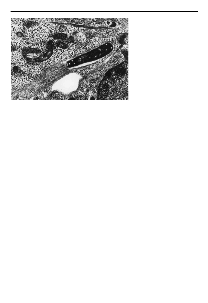
Intracellular Shigella use cytoskeletal components to
propel themselves inside the infected cell and when
contact occurs between the moving organism and the host
cell membrane, cellular protrusions are formed. These
protrusions are then engulfed by the neighbouring cell
thus permitting cell-to-cell spread of Shigella without the
bacterium ever leaving the con¢nes of the host epithelial
layer (¢gure 2).
Assays to study cell-to-cell spread have centred on two
techniques, the plaque assay and the infectious foci assay.
In the plaque assay, epithelial cells are infected with wild-
type Shigella and following a period of incubation,
medium containing gentamicin and agarose is added to
the infected cell monolayer in order to restrict reinfection
of cells from bacteria in the culture media. In this way,
bacteria must spread through the epithelial layer by
passing from one cell to the next. Two to three days later,
the agarose plug is removed and plaques can be observed
in the epithelial monolayer. These plaques correspond to
points of initial cellular infection and the resulting
destruction and clearing of infected cells (Oaks et al.
1985). Using this assay, a number of mutants have been
identi¢ed that are de¢cient in their ability to spread from
cell to cell and of these, IcsA has been best characterized.
The ability of IcsA to induce actin polymerization is
equally required for intracellular and intercellular spread.
Moreover, the IcsA phenotype is extremely relevant
during infection in vivo. Monkeys infected with an IcsA
mutant of Shigella develop only mild dysenteric symptoms
and show limited histopathological lesions of the colonic
and rectal mucosae (Sansonetti et al. 1991). These ¢ndings
stress the requirement for intercellular spread for full viru-
lence of Shigella during infection in vivo and perhaps point
to the role of the epithelial cell in the development of wide-
spread in£ammation (discussed in ½ 9).
E-cadherin, a key protein involved in intercellular
adhesion, has been shown to be an important cellular
component involved in the intercellular spread of Shigella.
Cell^cell contacts were thought to be necessary for inter-
cellular spread because of the observation that Shigella
passed from one cell to the next essentially at sites of the
intermediate junctions in Caco-2 cells (Vasselon et al.
1992). In addition, transmission electron microscopic
observations of various epithelial cell lines showed that
passage of Shigella protrusions from one cell to the next
occurred at sites where the two cells were closely
apposed, suggesting that cell^cell contacts were involved.
To test this directly, the infectious foci assay was devel-
oped. In this assay, cells are infected with Shigella for a
period of time and subsequently trypsinized and seeded
at a very low density with a population of uninfected cells
that are either cadherin-negative or stably expressing
cadherin. In the cells expressing cadherins, Shigella is e¤-
ciently transmitted from the originally infected cells to
the neighbouring cells such that large areas of the mono-
layer are observed to be infected. In contrast, cells that
are de¢cient in cadherins do not transmit Shigella and the
infection remains limited to the index cells only. Therefore
cell^cell contacts, dependent on the expression of
cadherins, are necessary for intercellular spread of
Shigella (Sansonetti et al. 1994). Further research is
required to identify whether or not Shigella interacts
578 D. J. Philpott and others The pathogenesis of Shigella £exneri infection
Phil. Trans. R. Soc. Lond. B (2000)
Figure 2. Transmission electron
micrograph of a Shigella £exneri-mediated
protrusion being taken up by a
neighbouring cell. Note the dense
accumulation of actin behind the moving
bacterium. Photograph is courtesy of
Dr Michelle Rathman (Institut Pasteur,
France).

directly with proteins at this junction to bring about
protrusion formation. Additionally, the role played by
intermediate junctions in the pathogenesis of Shigella
during infection in vivo needs to be addressed. Again,
because of the complexity of the system, such in vivo
evidence is di¤cult to obtain and will rely on the develop-
ment of novel model systems. In the meantime, however,
complementary techniques, such as the expression of
dominant negative proteins, the use of speci¢c inhibitors
as well as cell lines de¢cient in certain proteins, certainly
lends credence to these in vitro ¢ndings.
6. M CELLS: PORTS OF ENTRY INTO THE HOST
EPITHELIUM
One of the key events in the pathogenesis of enteroinva-
sive bacterial infections is the penetration of the intestinal
epithelium. Since Shigella cannot enter epithelial cells via
the apical pole, it uses M cells to gain entry into the host
epithelium. In fact, many Gram-negative bacteria that
cause enteric disease, including Salmonella and Yersinia,
have been shown to preferentially cross the epithelium via
specialized antigen sampling cells called M cells (for a
review, see Sansonetti & Phalipon 1999). M cells, which
stands for membranous or microfold cells, are modi¢ed
epithelial cells found within the FAE overlying lymphoid
follicles. These follicular lymphoid structures are scattered
throughout the small intestine in aggregates known as
Peyer’s patches and in the colon and rectum as isolated
solitary nodules. M cells are relatively rare, constituting
less than 0.1% of epithelial cells present in the lining of
the intestine and can be identi¢ed morphologically due to
the fact that they display (i) a poorly di¡erentiated brush
border compared with neighbouring absorptive epithelial
cells, and (ii) an irregular basolateral membrane border
containing invaginated lymphocytes. M cells have a
high endocytic activity which serves to transport soluble
and particulate lumenal antigens across the cytoplasm
and deliver them intact to the antigen-processing and
-presenting cells in the underlying follicle (Neutra et al.).
It is perhaps surprising that a cell so rare in the intestinal
epithelium can be the target of entry for many di¡erent
pathogens. How then do these pathogens seek out M cells
and use these cells to enter the host epithelium? It has
been suggested that the lack of both mucus and a well-
developed glycocalyx over the FAE facilitate non-speci¢c
interactions of pathogenic organisms with M cells.
Increased hydrophobic interactions may be favoured and
this could be the primary step that precedes what is likely
to be a non-speci¢c transport mechanism. In fact, it has
been shown that lectins, positively charged particles and
hydrophobic beads, all of which bind to the membrane
surface of M cells, are transported with increased e¤-
ciency (reviewed in Jepson et al. 1996). M cells may also
express characteristic surface molecules that could serve
as speci¢c receptors for pathogens. For example, M cells
express characteristic glycoconjugates, which vary
depending on the species and the location in the intestine
(Giannasca et al. 1994; Lelouard et al. 1999). Although no
speci¢c receptor engaged by a bacterial adhesin or
invasin has been identi¢ed, such a receptor may account
for tissue tropism of a particular pathogen as well as its
e¤cient uptake into the FAE.
7. M CELLS AND ENTRY OF
S. FLEXNERI INTO THE
HOST:
IN VIVO EVIDENCE
The ¢rst indication that S. £exneri exploits M cells to enter
the host epithelial layer came from studies using a rabbit
ligated ileal loop model. In this model, animals are anaes-
thetized and the intestine is externalized at laparotomy.
Sections of ileum are carefully ligated to preserve the
existing vasculature and, subsequently, a large inoculum of
bacteria, usually 10
9
colony forming units (CFU) ml
¡1
, is
injected into the intestinal loop. Invasive and in£ammatory
properties of the organisms can be observed following
sacri¢ce of the animals at a given time post-infection
(usually 2^8 h). Histological studies and measurements of
£uid accumulation within the infected loop can be
conducted. By isolating ileal loops with grossly identi¢able
Peyer’s patches, the role of the FAE in the initial steps of
epithelial translocation by Shigella has been assessed. Wild-
type S. £exneri was readily detected in the dome epithelium
of the FAE, whereas very few organisms were observed
within the villus epithelium. In addition, when the infected
loops were incubated for longer time periods, ulcerations
were observed preferentially over the dome regions of
Peyer’s patches suggesting that the FAE was the primary
site of entry (Wassef et al. 1989). These ¢ndings were recon-
¢rmed using the rabbit ileal loop model in a study indi-
cating that threefold more bacteria were present within the
infected tissue if Peyer’s patches were present within the
loop compared with those loops that lacked lymphoid folli-
cles (Perdomo et al. 1994b). Figure 3 shows wild-type Shigella
crossing the epithelial barrier by an M cell.
These ¢ndings were also con¢rmed in the macaque
monkey model of shigellosis. Macaques in particular are
one of the few animals that develop a dysentery-like
disease following oral or gastric inoculation of Shigella,
although a dose of 10
10
organisms is typically required for
the development of disease. Using this model, it was
observed that when monkeys were infected with an icsA
mutant of S. £exneri, which does not spread intra- or inter-
cellularly, animals do not develop clinical symptoms but
small ulcers corresponding to the presence of lymphoid
follicles are observed on the colonic lining (Sansonetti et
al. 1991). These ¢ndings suggest that the icsA mutant is
The pathogenesis of Shigella £exneri infection D. J. Philpott and others 579
Phil. Trans. R. Soc. Lond. B (2000)
enterocyte
M cell
Figure 3. Transmission electron micrograph of Shigella £exneri
crossing the intestinal epithelium by an M cell. Photograph
adapted from Sansonetti & Phalipon (1999).

capable of entry into the FAE but owing to its inability to
spread from the initial entry site, only a local ulceration
at the point of the FAE is observed. These ¢ndings
con¢rm that the FAE serves as the site of bacterial entry
into the epithelium and also reiterates the role of intra-
and intercellular spread in the development of widespread
in£ammation during wild-type Shigella infection.
Another important observation made using the ileal
loop model was that a signi¢cantly greater number of
wild-type Shigella were found in the dome epithelium
compared with non-pathogenic strains or heat-killed
organisms, suggesting that the presence of virulence
factors play a role in the increased uptake of the wild-
type strain into the FAE (Wassef et al. 1989). This
observation was further characterized using strains of
S. £exneri expressing either an invasive or an adhesive but
non-invasive phenotype in the rabbit ileal loop model.
The adhesiveness of the latter strain is mediated by the
expression of an Escherichia coli adhesin that mediates
attachment of the organism to rabbit M cells (Inman &
Cantey 1984). By immunostaining for lipopolysaccharide
(LPS), it was shown that the amount of bacterial material
associated with the FAE and the dome of the lymphoid
follicle was essentially equivalent in loops infected with
either the adhesive^non-invasive or the invasive strain,
whereas very few control organisms, i.e. non-adhesive
and non-invasive, could be isolated from similarly
infected loops (Sansonetti et al. 1996). These data suggest
that either speci¢c adhesion to M cells or an invasive
capacity to enter M cells is required for an organism to
be transported through the epithelium and into the
subepithelial space. What was also clear from this study
was that once the bacteria gain access to the subepithelial
space they have very di¡erent fates depending on their
virulence capacities. Whereas infection with wild-type,
fully invasive Shigella results in rapid in£ammation and
subsequent destruction of the FAE, the adhesive yet non-
invasive strain is sequestered and destroyed within
lysosomes of macrophages present within the dome.
Although the animal models discussed above have been
useful for studying some aspects of Shigella^M-cell inter-
actions, there are signi¢cant drawbacks and their direct
relevance to human infection can be questioned. Most
obvious is the fact that oral or gastric inoculation of
rabbits does not lead to dysenteric symptoms. Also, shigel-
losis in humans is a disease of the distal colon and
rectum, whereas in the rabbit model it is the ileum that is
studied. Therefore, this model does not take into account
the tissue speci¢city that is seen in human infection thus
ignoring the potential of a speci¢c interaction between
Shigella and M cells of the human colon. In addition to
having ethical and ¢nancial drawbacks, the macaque
model is also not ideal since the infectious dose required
for the animals to develop dysentery is ten million to 100
million times higher that the infectious dose in humans.
The questionable relevance of such a model is particularly
apparent in the context of testing the tolerance of attenu-
ated vaccine candidates or doing challenge experiments
in vaccinated animals.
Despite these drawbacks, however, many of the obser-
vations made in animal studies do correlate with what we
know from human infections with Shigella. In fact, clinical
observations of patients su¡ering from shigellosis support
the idea that the FAE is the primary route of entry of
Shigella into the host tissues. In patients examined endo-
scopically within two days of the start of infection, early
in£ammatory lesions resembling aphtoid ulcers are
present in the rectum and distal colon, and on histopatho-
logical observation these lesions are found to correspond
to lymphoid follicles (Mathan & Mathan 1991). Addition-
ally, in£ammation is observed to be con¢ned to follicular
regions of the rectum and distal colon early in the course
of infection, but is later detected in the surrounding villi
and can be seen to extend proximally (Islam et al. 1994).
There are a number of aspects of M cell^Shigella inter-
actions, however, that cannot be addressed adequately
using these in vivo systems and therefore require in vitro
modelling. Recently, such a model was developed in
which Caco-2 cells, a human intestinal cell line, were
induced to switch to an M-cell phenotype when co-
cultured with lymphocytes isolated from the Peyer’s patch
(Kerne¨is et al. 1997). Using this model, it was shown that
Vibrio cholerae O:1, a non-invasive pathogen transported
exclusively by M cells (Owen et al. 1986), could also be
transported across the model epithelium by the in vitro-
induced M cells. This model will assist studies into
Shigella^M-cell interactions and may help to identify
speci¢c receptors or adhesive factors on M cells that facil-
itate the uptake of Shigella by these cells. Additionally, this
model will allow the determination of the virulence
factors of Shigella that are necessary for entry and trans-
port across M cells.
At later time-points of infection, in£ammation disrupts
the integrity of epithelium and this may be a secondary
means by which pathogenic Shigella translocate across the
epithelial barrier in order to reach the basolateral pole of
epithelial cells, which they can then e¤ciently invade.
The possibility of this mode of Shigella translocation was
modelled in vitro (Perdomo et al. 1994a). This system was
again based on the culture of monolayers of human intest-
inal cells on ¢lter supports; however, another layer of
complexity to this system was added so that the early
immune responses to invasive bacteria could be investi-
gated. In this system, isolated human PMNs are added to
the basolateral side of polarized T84 cells, which are then
infected apically with pathogenic Shigella. A rapid para-
cellular transmigration of the PMNs which disrupts the
barrier function of the epithelium is observed, as
measured by a drop in transepithelial electrical resistance.
Subsequent to these events, Shigella is able to pass through
the disrupted tight junctions and thus gains access to the
basolateral pole of the cells (Perdomo et al. 1994a). These
studies suggest that lumenal Shigella can induce epithelial
cells to produce potent chemotactic signals that elicit
transepithelial transmigration of PMNs. In fact, the
epithelial cell has been shown to play a signi¢cant role in
innate immunity against enteroinvasive bacterial infec-
tions (Jung et al. 1995) and, in the case of Shigella infec-
tion, is important in intiating the in£ammatory response
(Sansonetti et al. 1999b; see ½ 9).
8. MACROPHAGE APOPTOSIS IN RESPONSE TO
SHIGELLA INVASION
Bacteria that have crossed the epithelial layer via M cells
are likely to be phagocytosed by resident macrophages
580 D. J. Philpott and others The pathogenesis of Shigella £exneri infection
Phil. Trans. R. Soc. Lond. B (2000)
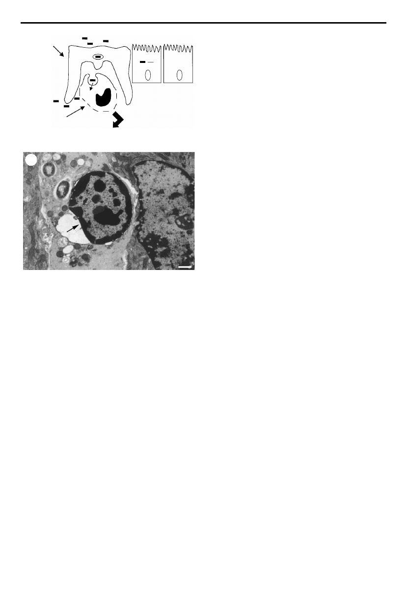
within the subepithelial dome overlying the lymphoid folli-
cles (see ¢gure 4a).The uptake of Shigella by macrophages in
vitro does not require the virulence plasmid and presumably
occurs by normal phagocytic mechanisms. The fate of
S. £exneri following phagocytosis was ¢rst studied in the late
1980s using the murine macrophageJ774 cell line (Clerc et
al. 1987). It was noted that uptake of Shigella resulted in lysis
of the phagocytic vacuole and rapid killing of the infected
cell. It was not until ¢ve years later, however, that macro-
phage cell death was shown to occur by apoptosis
(Zychlinsky et al. 1992; ¢gure 4b). Apoptosis was not seen
with plasmid-cured strains, which led to the identi¢cation
of the plasmid-encoded protein, IpaB, as the mediator of
cell death (Zychlinsky et al. 1994). IpaB gains access to the
cytosol, where it binds and activates caspase-1, also known
as interleukin(IL)-1-converting enzyme (Chen et al. 1996).
Activation of caspase-1 is absolutely required for Shigella-
induced apoptosis, since cell death is not seen in caspase-1
knockout mice (Hilbi et al. 1998). The downstream events
promoting apoptosis following caspase-1activationby IpaB
are unknown.
Apoptosis is generally considered to be an immunologi-
cally silent cell death process unaccompanied by in£am-
mation, however, this is not the case with caspase-1-
dependent apoptosis. Caspase-1 cleaves and activates the
pro-in£ammatory cytokines IL-1b and IL-18 (Ghayur
et al. 1997). Murine macrophages have been shown to
release large amounts of mature IL-1b during the Shigella-
induced apoptotic process (Zychlinsky et al. 1994). Given
that mucosal in£ammation is the hallmark of shigellosis,
these observations made largely in murine macrophage
cell lines prompted the search for evidence that apoptosis
and the consequent cytokine production play a role in
Shigella infection in vivo.
Apoptotic cells have been identi¢ed in the subepithelial
dome and lymphoid follicles in a rabbit ligated ileal loop
model of Shigella infection (Zychlinsky et al. 1996). Apop-
totic cells were not seen in the mucosa when challenged
with plasmid-cured Shigella or plasmid-cured strains trans-
fected with an E. coli adhesin, which allowed the bacteria to
penetrate into the subepithelial space in comparable
numbers to the wild-type Shigella. Apoptotic cells have also
been seen in rectal mucosal biopsies from patients acutely
infected with Shigella (Islam et al. 1997). Together these
observations provide evidence for apoptosis in vivo during
Shigella infection, and suggest that this phenomenon is due
to the presence of the virulence plasmid.
There have recently been some reports that Shigella can
kill macrophages by an alternative mechanism termed
oncosis, and that this process does not involve caspase-1
(Fernandez-Prada et al. 1997; Nonaka et al. 1999). In the
latter report a di¡erentiated human monocyte-like cell
line, U937, was used to show that Shigella infection could
result in apoptosis or oncosis depending on the di¡eren-
tiation stimulus used. Evidence of oncosis in vivo in Shigella
infection and whether it contributes towards the disease
manifestations has yet to be investigated.
9. ENCOUNTERS WITH THE INNATE IMMUNE
RESPONSE
The innate immune response provides an early defence
against bacterial infection, which serves to limit bacterial
proliferation, localize the infection and also both activate
and regulate the subsequent adaptive immune response.
Many cell types and soluble proteins, including phago-
cytic cells (neutrophils, macrophages and dendritic cells),
lymphocytes (natural killer (NK) cells and gd T cells),
cytokines (most notably, IL-1, Il-6, IL-12, tumour
necrosis factor a (TNFa) and IFNg) and liver-derived
serum proteins such as complement factors contribute
towards innate immunity. In addition to these classical
immune components, non-immune cells such as epithelial
cells recognize and respond to bacterial invasion by
producing chemokines that can attract and activate
immune cells (Jung et al. 1995). The net result of the
complex interaction between these many factors is usually
manifested as acute in£ammation.
The study of the innate immune response in shigellosis
has largely focused on the mechanisms involved in regu-
lating the in£ux of neutrophil into the infected site. The
neutrophil response can be separated into two stages: an
initial in£ux focused in the region of the lymphoid follicles;
and a later phase of massive neutrophil in£ux into intestinal
The pathogenesis of Shigella £exneri infection D. J. Philpott and others 581
Phil. Trans. R. Soc. Lond. B (2000)
M cell
macrophage
IpaB
(a)
IL-1
(b)
Figure 4. (a) Model of Shigella £exneri penetration of the
intestinal epithelium by M cells and subsequent contact with
the underlying macrophages at the site of the follicular
lymphoid tissue. Shigella is phagocytosed by resident
macrophages; however, the organism escapes from the phago-
cytic vacuole and induces macrophage apoptosis via
interaction of bacterial IpaB with host cell caspase-1.
Activated caspase-1 cleaves and activates pro-IL-1 that is
released in large quantities from the dying macrophage.
(b) Apoptosis of a Shigella-infected macrophage in vivo. Arrow
points to the condensation of chromatin at the periphery of
the nucleus which is a characteristic of apoptotic cell death.
Photograph adapted from Sansonetti & Phalipon (1999).
villi and crypts, which produces large areas of epithelial
destruction and mucosal ulceration that extend far beyond
the initial site of bacterial entry (Mathan & Mathan 1991).
It is likely that these two stages re£ect di¡erent processes.
IL-1 released from Shigella-infected apoptotic macrophages
may be responsible for the ¢rst stage; in the rabbit ileal loop
infection model, treatment with an IL-1 receptor antago-
nist prior to infection with virulent S. £exneri signi¢cantly
decreased the in£ammation and tissue destruction within
the lymphoid follicles (Sansonetti et al. 1995).
Observations that the second stage of in£ammation
occurs at some distance from the follicles and that infected
epithelial cells secrete pro-in£ammatory cytokines
prompted an investigation into the role of IL-8 as a
mediator of in£ammation in this second phase (Sansonetti
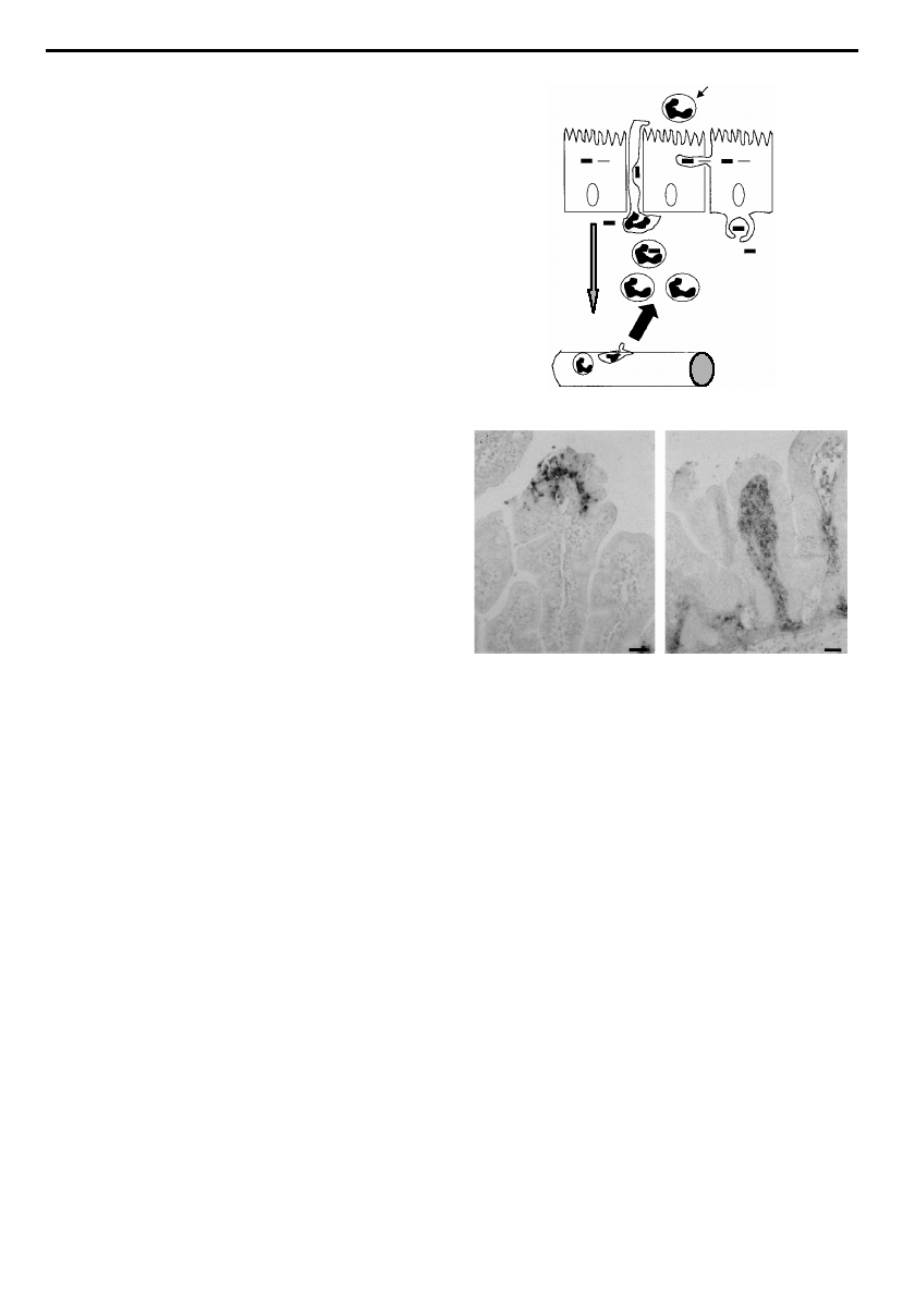
et al. 1999b; see ¢gure 5a for model). Again using the in vivo
rabbit ileal loop model, a neutralizing anti-IL-8 mono-
clonal antibody was found to considerably reduce the
neutrophil in£ux entering via the lamina propria into the
intestinal villi and to attenuate the consequent epithelial
destruction. In vitro studies have shown that IL-8 produc-
tion by epithelial cells induces neutrophil migration across
polarized epithelial monolayers and this can occur with or
without invasion of the epithelial cells by Shigella (Beatty
& Sansonetti 1997; McCormick et al. 1998). Thus, bacterial
interaction with epithelial cells appears to be a require-
ment for this second phase. The rapid extension of in£am-
mation to sites distant from the follicles stresses the
importance of cell-to-cell spread by Shigella, and is further
supported by experimental Shigella infection of macaque
monkeys with the icsA mutant, capable of epithelial cell
invasion but unable to spread from one cell to another
(Sansonetti et al. 1991). The inability of the icsA mutant to
spread through the epithelial layer restricts the contribu-
tion of epithelial chemokine release and consequently
limits the in£ammation seen in infected animals.
It has been noted that blocking the neutrophil in£ux
using anti-b1-integrin antibodies, IL-1 receptor antago-
nists or anti-IL-8 antibodies, all result in decreased epi-
thelial destruction implicating the neutrophil rather than
Shigella as the direct cause of mucosal damage (Perdomo et
al. 1994b; Sansonetti et al. 1995, 1999b). Neutrophils can
kill opsonized Shigella in vitro (Mandic-Muleg et al. 1997),
and the neutrophil in£ammatory response localizes the
bacteria to the epithelium. When neutrophil in£ux is
blocked, bacteria migrate deep into the lamina propria
and mesenteric blood vessels, con¢rming the importance
of neutrophils in localizing bacterial infection (Sansonetti
et al. 1999b). Thus, neutrophil in£ux appears to be respon-
sible for the majority of tissue destruction associated with
shigellosis, and yet is vital for preventing the systemic
spread of bacteria (¢gure 5b).
The murine lung model of shigellosis, although not rele-
vant with regard to the organ speci¢city of the disease, has
been useful for exploring details of the immune and
in£ammatory components, as well as some aspects of the
systemic and local immune response against Shigella infec-
tion (Mallett et al. 1993; Verg et al. 1995). In this model, an
inoculum of wild-type Shigella is administered intranasally
resulting in invasion of the tracheo-bronchial tract
resulting in an in£ammatory broncho-tracheo-alveolitis
(Voino-Yasenetsky & Voino-Yasenetskaya 1961). Using this
model, the role of cytokines in the innate immune response
has been investigated. TNFa and IFNg are both produced
locally during the ¢rst 24 hours of infection. Sublethal
inoculation into IFNg knockout mice results in over-
whelming local proliferation of bacteria and death,
compared with a steady decline in bacterial numbers in
wild-type controls (Way et al. 1998). Histology of lungs
from knockout mice showed an obliterative neutrophilic
bronchiolitis suggesting that neutrophils alone, in the
absence of IFNg are unable to clear the infection. The
course of infection in ab and gdT-cell knockout mice was
582 D. J. Philpott and others The pathogenesis of Shigella £exneri infection
Phil. Trans. R. Soc. Lond. B (2000)
(a)
PMN
IL-8
(b)
Figure 5. (a) Shigella £exneri-infected epithelial cells are a
source of interleukin-8 (IL-8), a potent chemotactic
chemokine that is responsible for the recruitment of PMNs
into the infected site. PMNs migrate between adjacent
epithelial cells, break intercellular junctions and thus
compromise the integrity of the epithelial barrier. This causes
destruction of the mucosal surface by allowing invasion of
further organisms from the colonic lumen. Conversely, the
neutrophil in£ux is necessary in order to control the
proliferation of organisms locally and prevent systemic
bacterial dissemination. (b) Photographs of intestinal sections
stained for LPS from Shigella £exneri-infected rabbit ileal loops.
The left panel shows a tissue section from an ileal loop infected
with S. £exneri in a rabbit pre-treated with a control antibody
showing abscess formation and local tissue destruction. The
right panel shows a tissue section from a rabbit in which IL-8
was neutralized using speci¢c antibodies prior to infection.
Although the epithelium is spared, bacterial di¡usion into the
lamina propria is observed. This stresses the important role
for IL-8-mediated neutrophil in£ux in preventing bacterial
translocation. Scale bars, 10 mm. Photographs adapted from
Sansonetti et al. (1999b).

the same as wild-type controls suggesting neither cell type
acted as a source for this cytokine. Conversely, NK-cell-
de¢cient mice had an increased susceptibility implicating
the NK cell as the source of IFNg. There are probably
multiple roles for IFNg, including inhibition of bacterial
proliferation within epithelial cells (Way et al. 1998),
enhanced macrophage killing of bacteria and perhaps
inhibition of macrophage apoptosis induced by Shigella
(Hilbi et al. 1997).
10. THE ADAPTIVE IMMUNE RESPONSE
During the course of infection, Shigella exists in both an
extracellular and intracellular location. This implies a
requirement for both humoral and cellular immune
responses for e¡ective sterilizing immunity. The fact that
mice do not display intestinal infection following challenge
with Shigella has hampered study of the adaptive immune
response. Some information, however, has been obtained
from study of the murine pulmonary inoculation model
and serological studies of infected humans. In the
pulmonary model, isotype-speci¢c secretory IgA-anti LPS
antibodies targeted to the mucosa from subcutaneous hybri-
domas, provides protection against challenge with a lethal
dose of organisms (Phalipon et al. 1995). This underscores
the importance of local IgA in providing protection, and
supports observations in humans suggesting that protective
immunity is isotype speci¢c and therefore directed pre-
dominantly against LPS (DuPont et al. 1972). Using the
pulmonary challenge model, immunized mice were used to
de¢ne the characteristics of a protective humoral response.
Sublethal infection induces local IgG and IgA responses
directed against LPS, and some Ipa proteins, but responses
are slow to develop (Verg et al. 1995). Short-lived, protective,
serotype-speci¢c humoral responses have been generated,
although this predominantly consists of an IgM response
and is T-cell independent (Way et al. 1999a,b). Again, it is
not clear whether these results accurately re£ect the situa-
tion in the intestinal mucosa.
Our understanding of how the mucosal immune system
manages any Gram-negative bacteria, including Shigella, to
obtain LPS in a form that can be presented to B, and
perhapsT, cells for induction of a high-a¤nity IgA response
is minimal. Recently, it was shown that Shigella LPS can be
tra¤cked through polarized intestinal cells and thus poten-
tially processed and presented in an immunologically
active form (Beatty et al. 1999). Lipoglycans such as LPS are
clearly dealt with di¡erently from proteins, but apart from
observations that CD1-restricted CD4^CD8 double nega-
tive T cells can be generated, reacting with mycobacterial
lipoglycans, there is little information (Porcelli & Modlin
1999). There is an urgent need for studies into the immune
responses against bacterial LPS. The situation with cellular
immunity in shigellosis is equally uncertain. T-cell clones
have been produced against Shigella (Zwillich et al. 1989)
and activated T cells have been isolated from the blood of
patients with shigellosis, but their function is unknown
(Islam et al. 1995,1996).
11. CONCLUSION AND PERSPECTIVES
Our understanding of the pathophysiology of shigel-
losis is largely based on studying the invasion of epithelial
cell monolayers and macrophages in vitro, and the experi-
mental infection of exteriorized rabbit ileal loops. Infor-
mation has also been obtained from the murine
pulmonary model and from rectal biopsies of macaque
monkeys and humans after experimental and natural
infection, respectively. The fact that Shigella does not
cause intestinal infection in mice, which denies the use of
the many murine-speci¢c reagents and genetic manipula-
tions, has probably inhibited a more detailed investigation
of the cytokine and cellular mechanisms involved. None-
theless, the application of knockout mice in the murine
lung model of shigellosis has added, and will continue to
add, to our knowledge of the innate and adaptive
immune responses to Shigella infection. Future studies
using transgenic animals expressing human-speci¢c
factors will also open up new possibilities for investigating
the immune response in shigellosis.
In vitro and in vivo studies have allowed the formulation
of a detailed model of the disease process, however, many
questions still remain to be addressed. An analysis into
the timing of the in£ammatory response in terms of the
cell types and mediators that are recruited and secreted
at the site of infection needs to be conducted. For
example, it will be important to determine the relative
contribution of resident macrophages versus newly
recruited monocytes/macrophages to the disease process
and which in£ammatory mediators are responsible for
this induction during infection in vivo. Improved techni-
ques that combine multiple immune staining and confocal
microscopy of infected tissue sections will help to identify
the early players in the development of in£ammation
following infection with Shigella. These techniques could
also be used to observe the fate of bacterial virulence
factors, such as LPS, in the infected tissue during the
course of infection. Techniques to measure cytokines in
situ with placement of microdialysis probes in the infected
site (Bruce et al. 1999) have the potential to identify new
in£ammatory mediators and perhaps point to a novel
means of treatment by targeting these molecules and
modulating their function during in vivo infection.
Another possible research avenue that remains to be
explored is the potential for di¡erential gene expression,
in both the host and the bacterium, during Shigella infec-
tion. One example of a host gene speci¢cally upregu-
lated during infection with Shigella is IL-8 and its
regulation by the eukaryotic transcription factor, NFkB,
has recently been demonstrated (Philpott et al. 1999).
However, a more comprehensive approach to identify
the expression of Shigella-induced host genes would be
the
application of
DNA
microarray
technology
(reviewed in Khan et al. 1999). This approach will lead
to the identi¢cation of gene products up- or down-
regulated during Shigella infection. Additionally, this
approach could be used to attribute a particular pheno-
type to Shigella mutants that remain uncharacterized. By
comparing the pattern of gene expression from wild-
type versus mutant infected cells, a particular function
could be ascribed for the gene product missing in these
mutants. Conversely, genes expressed in the bacterium
during infection of the host could also be examined. The
potential to apply techniques such as signature-tagged
mutagenesis (Hensel et al. 1995) to Shigella infection also
remains unexplored.
The pathogenesis of Shigella £exneri infection D. J. Philpott and others 583
Phil. Trans. R. Soc. Lond. B (2000)

It is unwise to assume that any particular, or indeed a
combination of, animal models will reveal all the compo-
nents involved in producing the human disease. Infections
usually exhibit a restricted host range, and in the case of
many important human infections such as shigellosis, the
disease is essentially con¢ned to humans. Therefore,
human-speci¢c factors that allow expression of the disease
phenotype probably exist. Furthermore, in the past,
bacterial infections have infected the majority of the
population and caused signi¢cant mortality in children,
providing the potential for skewing the surviving popula-
tion towards genetic expression of factors that probably
in£uence the host response to disease. Such factors are
likely to act at the level of the innate immune response
and may be represented only in humans. Identi¢cation of
such factors would shed light on natural resistance and,
for example, help to explain why in human Shigella the
challenge studies, a maximum of only 70% of volunteers
get the clinical disease (DuPont et al. 1969).
For these reasons, in shigellosis, as much as in any other
bacterial infection, there is a need to develop experimental
models that can more closely mimic human disease, using
human cells and tissues. At present, such models remain in
the developmental stage. One possibility is the further
development of techniques for maintaining the viability of
human tissue samples such as intestinal biopsies, which
could be used to study the response of resident cells to inva-
sion of Shigella. A second possibility is the re¢nement of
techniques for grafting human mucosal tissues into SCID
mice (Yan et al. 1993) and then repopulating the bone
marrow with autologous bone marrow cells. Infection of
such xenografts could then be assessed in the context of
both the human intestine and immune system yet would
be amenable to the manipulations achievable in the mouse.
Ultimately, such studies as those described here will help
form a basis of knowledge by which improved treatments
and novel vaccine candidates for the prevention of shigel-
losis will be designed.
We thank members of the Sansonetti laboratory past and pre-
sent whose work was discussed in this manuscript. We are
grateful to Dr Claude Parsot for helpful discussions, Dr Michelle
Rathman and Dr Maria Mavris for careful review of the manu-
script. D.J.P. is supported by a Marie Curie Fellowship from the
European Community; J.D.E. is supported by the Wellcome
Trust. Research from the Sansonetti Laboratory is supported by
grants from the Ministe©re de l’Education Nationale de la
Recherche et de la Technologie (Programme BIOTECH and
Programme de Recherche Fondamentale en Microbiologie).
REFERENCES
Adam, T., Giry, M., Boquet, P. & Sansonetti, P. J. 1996 Rho-
dependent membrane folding causes Shigella entry into epithe-
lial cells
. EMBO J.
Beatty, W. L. & Sansonetti, P. J. 1997 Role of lipopolysaccharide
in signaling to subepithelial polymorphonuclear leukocytes.
Beatty, W. L., Me¨resse, S., Gounon, P., Davoust, J., Mounier, J.,
Sansonetti, P. J. & Gorvel, J.-P. 1999 Tra¤cking of Shigella
lipopolysaccharide in polarized intestinal epithelial cells.
Bernardini, M. L., Mounier, J., d’Hauteville, H., Coquis-
Rondon, M. & Sansonetti, P. J. 1989 Identi¢cation of icsA, a
plasmid locus of Shigella £exneri that governs bacterial intra-
and intercellular spread through interaction with F-actin.
Proc. Natl Acad. Sci. USA 86, 3867^3871.
Blocker, A., Gounon, P., Larquet, E., Niebuhr, K., Cabiaux, V.,
Parsot, C. & Sansonetti, P. J. 1999 The tripartite type III
secreton of Shigella £exneri inserts IpaB and IpaC into host
membranes
Bourdet-Sicard, R., Rudiger, M., Jockusch, B. M., Gounon, P.,
Sansonetti, P. J. & Tran Van Nhieu, G. 1999 Binding of the
Shigella protein IpaA to vinculin induces F-actin depolymer-
ization
Bruce, S. R., Tack, C. J. J. & Goldstein, D. S. 1999
Microdialysis for measurement of extracellular £uid concen-
trations of catechols in human skeletal muscle and adipose
tissue. In Monitoring molecules in neuroscience (ed. H. Rollema, E.
Abercrombie, D. Sulzer & J. Zachheim), pp. 104^109.
Newark, NJ: Rutgers.
Chen, Y., Smith, M. R., Thirumalai, K. & Zychlinsky, A. 1996
A bacterial invasin induces macrophage apoptosis by directly
binding ICE.
Clerc, P. L., Ryter, A., Mounier, J. & Sansonetti, P. J. 1987
Plasmid-mediated early killing of eukaryotic cells by Shigella
£exneri as studied by infection of J774 macrophages.
DuPont, H. L., Hornick, R. B., Dawkins, A. T., Snyder, M. J. &
Formal, S. B. 1969 The response of man to virulent Shigella
£exneri 2a
DuPont, H. L., Hornick, R. B., Snyder, M. J., Libonati, J. P.,
Formal, S. B. & Gangarosa, E. J. 1972 Immunity in shigel-
losis. II. Protection induced by oral live vaccine or primary
infection
DuPont, H. L., Levine, M. M., Hornick, R. B. & Formal, S. B.
1989 Inoculum size in shigellosis and implications for
expected mode of transmission.
J. Infect. Dis. 159, 1126^1128.
Fernandez-Prada, C. M., Hoover, D. L., Tall, B. D. &
Venkatesan, M. M. 1997 Human monocyte-derived macro-
phages infected with virulent Shigella £exneri in vitro undergo
a rapid cytolytic event similar to oncosis but not apoptosis.
Ghayur, T. (and 13 others) 1997 Caspase-1 processes IFN-
gamma-inducing factor and regulates LPS-induced IFN-
gamma production.
Giannasca, P. J., Giannasca, K. T., Falk, P., Gordon, J. I. &
Neutra, M. R. 1994 Regional di¡erences in glycoconjugates of
intestinal M cells in mice: potential targets for mucosal
vaccines. Am. J. Physiol.
267, G1108^G1121.
Hensel, M., Shea, J. E., Gleeson, C., Jones, M. D., Dalton, E. &
Holden, D. W. 1995 Simultaneous identi¢cation of bacterial
virulence genes by negative selection.
High, N., Mounier, J., Pre¨vost, M. C. & Sansonetti, P. J.
1992 IpaB of Shigella £exneri causes entry into epithelial
cells and escape from the phagocytic vacuole. EMBO J.
11,
1991^1999.
Hilbi, H., Chen, Y., Thirumalai, K. & Zychlinsky, A. 1997 The
interleukin-1b-converting enzyme, caspase-1, is activated
during Shigella £exneri-induced apoptosis in human monocyte-
derived macrophages.
Hilbi, H., Moss, J. E., Hersh, D., Chen,Y., Arondel, J., Banerjee,
S., Flavell, R. A., Yuan, J., Sansonetti, P. J. & Zychlinsky, A.
1998 Shigella-induced apoptosis is dependent on caspase-1which
binds to IpaB.
J. Biol. Chem. 273, 32 895^32900.
Hueck, C. J. 1998 Type III secretion systems in bacterial
pathogens of animals and plants. Microbiol. Mol. Biol. Rev.
62,
379^433.
Inman, L. & Cantey, J. R. 1984 Peyer’s patch lymphoid follicle
epithelial adherence of a rabbit enteropathogenic Esherichia
coli (strain RDEC-1). Role of plasmid-mediated pili in initial
adherence
584 D. J. Philpott and others The pathogenesis of Shigella £exneri infection
Phil. Trans. R. Soc. Lond. B (2000)

Islam, M. M., Azad, A. K., Bardhan, P. K., Raqib, R. & Islam, D.
1994 Pathology of shigellosis and its complications.
Islam, D., Bardham, P. K., Lindberg, A. A. & Christensson, B.
1995 Shigella infection induces cellular activation of T and B
cells, and distinct species-related changes in peripheral blood
lymphocyte subsets during the course of the disease.
Islam, D., Wretlind, B., Lindberg, A. A. & Christensson, B. 1996
Changes in the peripheral blood Tcell receptorVb repertoire in
vivo and in vitro during shigellosis.
Islam, D., Veress, B., Bardhan, P. K., Lindberg, A. A. &
Christensson, B. 1997 In situ characterisation of in£ammatory
responses in the rectal mucosae of patients with shigellosis.
Jepson, M. A., Clark, M. A., Foster, N., Mason, C. M., Bennett,
M. K., Simmons, N. L. & Hirst, B. H. 1996 Targeting to
intestinal M cells.
Jung, H. C., Eckmann, L., Yang, S. K., Panja, A., Fierer, J.,
Morzycka-Wroblewska, E. & Kagno¡, M. F. 1995 A distinct
array of proin£ammatory cytokines is expressed in human
epithelial cells in response to bacterial invasion.
Khan, J., Bittner, M. L., Chen, Y., Meltzer, P. S. & Trent, J. M.
1999 DNA microarray technology: the anticipated impact on
the study of human disease. Biochim. Biophys. Acta
1423,
M17^M28.
Kerne¨is, S., Bogdanova, A., Kraehenbuhl, J.-P. & Pringault, E.
1997 Conversion by Peyer’s patch lymphocytes of human
enterocytes into M cells that transport bacteria.
Kotlo¡, K. L., Winicko¡, J. P., Ivano¡, B., Clemens, J. D.,
Swerdlow, D. L., Sansonetti, P. J., Adak, G. K. & Levine, M. M.
1999 Global burden of Shigella infections: implications for
vaccine development and implementation of control strate-
gies.WHO Bull.
77, 651^666.
LaBrec, E. H., Schneider, H., Magnani, T. J. & Formal, S. B.
1964 Epithelial cell penetration as an essential step in the
pathogenesis of bacillary dysentery. J. Bacteriol.
88, 1503^1518.
Lelouard, H., Reggio, H., Mangeat, P., Neutra, M. &
Montcourrier, P. 1999 Mucin-related epitopes distinguish M
cells and enterocytes in rabbit appendix and Peyer’s patches.
McCormick, B. A., Siber, A. M. & Maurelli, A. T. 1998
Requirement of the Shigella £exneri virulence plasmid in the
ability to induce tra¤cking of neutrophils across polarised
monolayers of the intestinal epithelium.
Mallett, C., Verg, L. v. d., Collins, H. & Hale, T. 1993
Evaluation of Shigella vaccine safety and e¤cacy in an intra-
nasal challenged mouse model.
Mandic-Muleg, I., Weiss, J. & Zychlinsky, A. 1997 Shigella £exneri
is trapped in polymorphonuclear leukocyte vacuoles and e¤-
ciently killed.
Mathan, M. M. & Mathan, V. I. 1991 Morphology of rectal
mucosa of patients with shigellosis. Rev. Infect. Dis.
13
(Suppl. 4), S314^S318.
Me¨nard, R., Sansonetti, P. J. & Parsot, C. 1993 Nonpolar muta-
genesis of the ipa genes de¢nes IpaB, IpaC and IpaD as
e¡ectors of Shigella £exneri entry into epithelial cells.
Me¨nard, R., Sansonetti, P. J., Parsot, C. & Vasselon, T. 1994
Extracellular assoication and cytoplasmic partitioning of
the IpaB and IpaC invasins of Shigella £exneri
Me¨nard, R., Sansonetti, P. J. & Parsot, C. 1996 The secretion of
the Shigella £exneri Ipa invasins is induced by the epithelial
cell an controlled by IpaD
. EMBO J.
Mounier, J., Vasselon, T., Hellio, R., Lesourd, M. & Sansonetti,
P. J. 1992 Shigella £exneri enters human colonic Caco-2 epithelial
cells throughthe basolateralpole.
Neutra, M. R., Pringault, E. & Kraehenbuhl, J.-P. 1996
Antigen sampling across epithelial barriers and induction of
mucosal immune responses.
Nonaka, T., Kuwae, A., Sasakawa, C. & Imajoh-Ohmi, S. 1999
Shigella £exneri YSH6000 induces two di¡erent types of cell
death, apoptosis and oncosis, in the di¡erentiated human
monoblastic cell line U937
Oaks, E., Wing¢eld, M. & Formal, S. 1985 Plaque formation by
virulent Shigella £exneri
Owen, R. L., Pierce, N. F., Apple, R. T. & Cray Jr, W. C. 1986
M cell transport of Vibrio cholerae from the intestinal lumen into
Peyer’s patches: a mechanism for antigen sampling and for
microbial transepithelial migration.
Perdomo, J. J., Gounon, P. & Sansonetti, P. J. 1994a
Polymorphonuclear leukocyte transmigration promotes inva-
sion of colonic epithelial monolayer by Shigella £exneri
. J. Clin.
Perdomo, O. J., Cavaillon, J. M., Huerre, M., Ohayon, H.,
Gounon, P. & Sansonetti, P. J. 1994b Acute in£ammation
causes epithelial invasion and mucosal destruction in experi-
mental shigellosis.
Phalipon, A., Kaufmann, M., Michetti, P., Cavaillon, J. M.,
Huerre, M. & Sansonetti, P. J. 1995 Monoclonal immunoglo-
bulin A antibody directed against serotype-speci¢c epitope of
Shigella £exneri lipopolysaccharide protects against murine
experimental shigellosis.
Philpott, D. J., Yamaoka, S., Isral, A. & Sansonetti, P. J. 1999
Invasive Shigella £exneri activates NFkB through an innate
intracellular response and leads to IL-8 expression in epithe-
lial cells. J. Immunol. (Submitted.)
Porcelli, S. A. & Modlin, R. L. 1999 The CD1 system: antigen
presenting molecules for Tcell recognition of lipids and glyco-
lipids.
Sansonetti, P. J. & Phalipon, A. 1999 M cells as ports of entry for
enteroinvasive pathogens: mechanisms of interaction, conse-
quences for the disease process.
Sansonetti, P. J., Arondel, J., Fontaine, A., d’Hauteveille, H. &
Bernadini, M. L. 1991 OmpB (osmo-regulation) and icsA
(cell-to-cell spread) mutants of Shigella £exneri: vaccine candi-
dates and probes to study the pathogenesis of shigellosis.
Sansonetti, P. J., Mournier, J., Pre¨vost, M. C. & Mere©ge, R. M.
1994 Cadherin expression is required for the spread of Shigella
£exneri between epithelial cells.
Sansonetti, P. J., Arondel, A., Cavaillon, J. M. & Huerre, M.
1995 Role of IL-1 in the pathogenesis of experimental shigel-
losis.
Sansonetti, P. J., Arondel, J., Cantey, R. J., Pre¨vost, M. C. &
Huerre, M. 1996 Infection of rabbit Peyer’s patches by Shigella
£exneri: e¡ect of adhesive or invasive bacterial phenotypes on
follicular-associated epithelium.
Sansonetti, P. J., Tran Van Nhieu, G. & Egile, E. 1999a Rupture
of the intestinal epithelial barrier and mucosal invasion by
Shigella £exneri
Sansonetti, P. J., Arondel, J., Huerre, M., Harada, A. &
Matsushima, K. 1999b Interleukin-8 controls bacterial trans-
epithelial translocation at the cost of epithelial destruction in
experimental shigellosis.
Sasakawa, C., Adler, B., Tobe, T., Okada, N., Nagai, S.,
Komatsu, K. & Yoshikawa, M. 1988 Virulence-associated
genetic regions comprising 31 kilobases of the 230-kilobase
plasmid in Shigella £exneri 2a
Sere¨ny, B. 1955 Experimental Shigella conjunctivitis. Acta
Microbiol. Acad. Sci. Hungary 2, 293^295.
The pathogenesis of Shigella £exneri infection D. J. Philpott and others 585
Phil. Trans. R. Soc. Lond. B (2000)

Skoudy, A., Aru¡o, A., Gounon, P., Sansonetti, P. J. & TranVan
Nhieu, G. 1999a CD44 binds to the Shigella IpaB protein and
participates in bacterial invasion of epithelial cells. Cell.
Microbiol. (In the press.)
Skoudy, A., Tran Van Nhieu, G., Mantis, N., Arpin, M.,
Mounier, J., Gounon, P. & Sansonetti, P. 1999b A functional
role for ezrin during Shigella £exneri entry into epithelial cells.
Takahashi, K., Sasaki, T., Mammoto, A., Takaishi, K.,
Kameyama,T., Tsukita, S., Tsukita, S. & Takai,Y. 1997 Direct
interaction of the RhoGDP dissociation inhibitor with ezrin/
radixin/moesin initiates the activation of the Rho small G
protein
371^23375.
Tran Van Nhieu, G., Ben-Ze’ev, A. & Sansonetti, P. J. 1997
Modulation of bacterial entry into epithelial cells by associa-
tion between vinculin and the Shigella IpaA invasin. EMBO J.
16, 2717^2729.
Tran Van Nhieu, G., Caron, E., Hall, A. & Sansonetti, P. J.
1999 IpaC induces actin polymerization and ¢lopodia forma-
tion during Shigella entry into epithelial cells.
Vasselon, T., Mounier, J., Hellio, R. & Sansonetti, P. J. 1992
Movement along actin ¢laments of the perijunctional area
and de novo polymerization of cellular actin are required for
Shigella £exneri colonizaion of epithelial Caco-2 cell mono-
Verg, L. L. v. d., Mallett, C. P., Collins, H. H., Larsen, T.,
Hammack, C. & Hale, T. L. 1995 Antibody and cytokine
responses in a mouse pulmonary model of Shigella £exneri sero-
type 2a infection.
Voino-Yasenetsky, M. V. & Voino-Yasenetskaya, M. K. 1961
Experimental pneumonia caused by bacteria of the Shigella
group. Acta Morphol.
XI, 440^454.
Wassef, J., Keren, D. F. & Mailloux, J. L. 1989 Role of M cells
in initial bacterial uptake and in ulcer formation in the rabbit
intestinal loop model in shigellosis.
Watari, M., Funato, S. & Sasakawa, C. 1996 Interaction of Ipa
proteins of Shigella £exneri with a5b1 integrin promotes entry of
the bacteria into mammaliancells.
Watari, M., Kamata, Y., Kozaki, S. & Sasakawa, C. 1997 Rho,
a small GTP-binding protein, is essential for Shigella invasion
of epithelial cells.
Way, S. S., Borczuk, A. C., Dominitz, R. & Goldberg, M. C.
1998 An essential role for gamma interferon in innate resis-
tance to Shigella £exneri infection
Way, S. S., Borczuk, A. C. & Goldberg, M. B. 1999a Adaptive
immune response to Shigella £exneri 2a cydC in immunocompe-
tent mice and mice lacking immunoglobin A.
Way, S. S., Borczuk, A. C. & Goldberg, M. B. 1999b Thymic
independence of adaptive immunity to the intracellular
pathogen Shigella £exneri serotype 2a.
Yan, H.-C., Juhasz, J., Pilewski, J., Murphy, G. F., Herlyn, M.
& Albeelda, S. M. 1993 Human/severe combined immuno-
de¢cient mouse chimeras.
Zwillich, S. H., Duby, A. D. & Lipsky, P. E. 1989 T lymphocyte
clones responsive to Shigella £exneri
Zychlinsky, A., Pre¨vost, M. C. & Sansonetti, P. J. 1992 Shigella
£exneri induces apoptosis in infected macrophages.
Zychlinsky, A., Fitting, C., Cavaillon, J. M. & Sansonetti, P. J.
1994a Interleukin-1 is released by murine macrophages
during apoptosis induced by Shigella £exneri
Zychlinsky, A., Kenny, B., Me¨nard, R., Pre¨vost, M. C.,
Holland, I. B. & Sansonetti, P. J. 1994b IpaB mediates macro-
phage apoptosis induced by Shigella £exneri
Zychlinsky, A., Thirumalai, K., Arondel, J., Cantey, J. R.,
Aliprantis, A. O. & Sansonetti, P. J. 1996 In vivo apoptosis in
Shigella £exneri infections.
Discussion
C. W. Keevil (Centre for Applied Microbiology and Research,
Porton Down, Wiltshire, UK). With respect to determining
the infectious dose or LD
50
of a gastro-intestinal
pathogen, it may be worth considering the recent publica-
tion of James & Keevil (1999). This paper showed that
verocytotoxigenic Escherichia coli 0157 attaches more
avidly to enterocytes, with actin ¢lament rearrangement,
when grown microaerophilically or anaerobically rather
than aerobically. Regrettably, many laboratories do not
consider growing facultatively anaerobic pathogens under
physiologically relevant conditions, especially low redox
potential, prior to their in vitro or in vivo challenge studies.
Could you comment on what anaerobic inoculum experi-
ments have been performed by laboratories when exam-
ining the pathogenesis of Shigella spp.? One possible
interpretation of some of your present data is that Shigella
spp. express an anaerobic phenotype capable of enhanced
attachment to epithelial cells; once they become intracel-
lular, their phenotype will change in response to local
nutrients, particularly oxygen concentration, making
them ¢t for subsequent tissue invasion and dissemination
to macrophages.
P. J. Sansonetti. I agree with Dr Keevil that not much
attention has so far been paid to the e¡ect of anaerobic
growth conditions on the invasive capacity of Shigella.
With regard to in vitro assays of cell invasion, growth
conditions have been selected with the aim of optimizing
bacterial entry into cells. It turns out that optimal condi-
tions are the middle exponential phase of growth with
aeration achieved by shacking. We are far away from
anaerobiosis under such circumstances! Still, in those
conditions, bacteria need to be centrifuged over the cell
surface in order to achieve an intimate interaction, as no
signi¢cant adherence system has ever been identi¢ed.
Our preliminary evidence however, based on the
sequence and annotation of the S. £exneri virulence
plasmid, does not show any gene with a homology indi-
cating a candidate for encoding and adhesin. This does
not preclude the possibility that a pathogenicity island on
the Shigella chromosome may encode an adherence
system. In any event, the anaerobic growth conditions
de¢nitely need to be tested.
The situation seems di¡erent with regard to animal
models: none of them, except infection in the macaque
monkey, really re£ects the situation of colonic infection
that prevails in humans. In consequence, when macaque
monkeys are infected intraperitoneally, the growth condi-
tions do not really matter as the bacteria ¢rst need to
survive gastric acidity, then transit through the small
intestine and ¢nally establish infection in the colon.
Under such neutral conditions, bacteria have time to
adapt and express any putative speci¢c adhesin. We
believe that the best way to identify this putative adhesin
will be a combination of genomics and signature-tagged
mutagenesis.
586 D. J. Philpott and others The pathogenesis of Shigella £exneri infection
Phil. Trans. R. Soc. Lond. B (2000)
Wyszukiwarka
Podobne podstrony:
Interruption of the blood supply of femoral head an experimental study on the pathogenesis of Legg C
Interruption of the blood supply of femoral head an experimental study on the pathogenesis of Legg C
The impact of Microsoft Windows infection vectors on IP network traffic patterns
A proteogenomic analysis of Sh flexneri
Modification of Intestinal Microbiota and Its Consequences for Innate Immune Response in the Pathoge
Frino, Mcinish And Toner The Liquidity Of Automated Exchanges New Evidence From German Bund Futures
Exposure of Sh flexneri to acid stress and heat shock enhances acid tolerance
The dynamics of computer virus infection
On the frequency of protein glycolysation as deduced from analysis ofthe SWISS PROT database
The use of Merit Pay Scales as Incentives in Health?re
22 The climate of Polish Lands as viewed by chroniclers, writers and scientists
the role of interpersonal trust for enterpreneurial exchange in a trnsition economy
The Effect of DNS Delays on Worm Propagation in an IPv6 Internet
Nadelhoffer; On The Importance of Saying Only What You Believe in the Socratic Dialogues
The Relationship of ACE to Adult Medical Disease, Psychiatric Disorders, and Sexual Behavior Implic
social networks and planned organizational change the impact of strong network ties on effective cha
The Contribution of Early Traumatic Events to Schizophrenia in Some Patients
The Rights of Persons Belonging to National or Ethnic, Religious and Linguistic Minorities, Annual R
więcej podobnych podstron