
* Corresponding author. Tel.: #81-43-270-3778; fax: #81-73-270-
3780.
E-mail address: yosinari@tdc.ac.jp (M. Yoshinari).
Biomaterials 22 (2001) 2043}2048
In#uence of surface modi"cations to titanium on antibacterial
activity in vitro
M. Yoshinari
*, Y. Oda , T. Kato, K. Okuda
Department of Dental Materials Science and Oral Health Science Center, Tokyo Dental College, 1-2-2, Masago, Mihama-ku, Chiba 261-8502, Japan
Department of Microbiology and Oral Health Science Center, Tokyo Dental College, 1-2-2, Masago, Mihama-ku, Chiba 261-8502, Japan
Received 31 October 2000; accepted 3 November 2000
Abstract
The antibacterial e!ect of surface modi"cations to titanium on Porphyromonas gingivalis ATCC 33277 and Actinobacillus
actinomycetemcomitans ATCC 43718 was evaluated. Surface modi"cations were performed with dry processes including ion
implantation (Ca
>, N>, F>), oxidation (anode oxidation, titania spraying), ion plating (TiN, alumina), and ion beam mixing (Ag, Sn,
Zn, Pt) with Ar
> on polished pure titanium plates. F>-implanted specimens signi
"cantly inhibited the growth of both
P. gingivalis
and A. actinomycetemcomitans than the polished titanium. The other surface-modi"ed specimens did not exhibit e!ective antibacterial
activity against both bacteria. No release of the #uorine ion was detected from F-implanted specimens under dissolution testing. This
result and the characterization of the F
>-implanted surfaces suggested that the possible antibacterial mechanism of the F>-implanted
specimen was caused by the formation of a metal #uoride complex on the surfaces. In addition, F
>-implanted surfaces did not inhibit
the proliferation of "broblast L929-cells. These "ndings indicate that surface modi"cation by means of a dry process is useful in
providing antibacterial activity of oral bacteria to titanium implants exposed to the oral cavity.
2001 Elsevier Science Ltd.
All rights reserved.
Keywords: Surface modi"cation; Titanium; Implant; Oral bacteria; Antibacterial activity
1. Introduction
Microbial plaque accumulation surrounding dental
implants may develop into peri-implantitis or peri-im-
plantoclasia, which is de"ned as in#ammation or infec-
tion around an implant, with accompanying bone loss. It
is important to maintain plaque-free surfaces on both
supra- and sub-gingival portions of dental implants
to prevent peri-implantitis. There are at least two
methods of inhibiting the formation of microbial plaque.
The "rst method is to inhibit the initial adhesion of oral
bacteria. The second method is to inhibit the coloniz-
ation of oral bacteria, which involves surface antibac-
terial activity.
Microbial colonization and antibacterial activity on
metallic and ceramic implant materials have been re-
ported under in vitro and in vivo tests [1}6]. Titanium
itself has no antibacterial activity [7], but there is a prob-
able risk of plaque formation on titanium implants [8].
Nevertheless, few experiments have been conducted on
the surface modi"cation of titanium implants to inhibit
the colonization of oral bacteria [9].
The modi"ed surfaces must resist wear, because these
are the parts that are brushed as a means of plaque
control. Surface modi"cations using a dry process have
been utilized in the medical and dental "elds as suitable
methods for providing good resistance to wear as well as
creating thin and adhesive "ne ceramics [10}14].
In our previous study [8], the initial adherence of oral
bacteria on cp-titanium and surface-modi"ed titanium
with a dry process was investigated. The results showed
that comparatively large amounts of Porphyromonas gin-
givalis and Actinobacillus actinomycetemcomitans, which
are major periodontopathic bacteria, adhered to polished
cp-titanium. These "ndings indicate that there is a prob-
able risk of bacterial adhesion to titanium surfaces at
the supra- and sub-gingival portions of implants, and
surface modi"cation to inhibit the adherence of oral
bacteria is required. The data showed that some surface
0142-9612/01/$ - see front matter
2001 Elsevier Science Ltd. All rights reserved.
PII: S 0 1 4 2 - 9 6 1 2 ( 0 0 ) 0 0 3 9 2 - 6
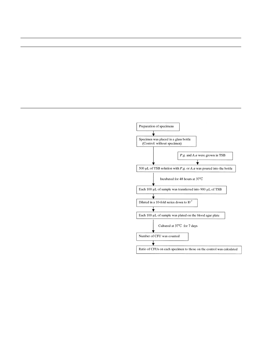
Table 1
Cp-titanium and surface-modi"ed specimens with dry process
Classi"cation
Condition
Code
Titanium
Cp-Ti plate (10
;10;1 mm)
Polished with 0.3
m alumina
Ti-polished
Ion implantation
Ca
>, Acc voltage: 41 kV
Ca-implanted
Dose: 5
;10 ions/cm
N
>, Acc voltage: 15 kV
N-implanted
Depth: 30 nm
F
>, Acc voltage: 19 kV
F-implanted
Oxidation
Anode oxidation
Anode-oxidized
Titania spraying
Titania-sprayed
Ion plating
TiN-ion plating
TiN-coated
Alumina-ion plating
Alumina-coated
Ion beam mixing
Ag, Acc voltage: 40 kV
Ag-IBM
Ar
>
Sn, Acc voltage: 25 kV
Sn-IBM
Dose: 5
;10 ions/cm
Zn, Acc voltage: 34 kV
Zn-IBM
Depth: 20 nm
Pt, Acc voltage: 50 kV
Pt-IBM
Fig. 1. A #ow chart of antibacterial activity test.
modi"cation with a dry process is useful in controlling
the initial adhesion of oral bacteria.
It is also required to provide antibacterial activity for
maintaining plaque-free surfaces on titanium implants
exposed to the oral cavity. The present study was there-
fore designed to investigate the in#uence of surface modi-
"cation to titanium on the colonization of oral bacteria
as an index of antibacterial activity in vitro. In addition,
this study evaluated the release of #uorine ions and
cytotoxicity of the L929 cells on F
>-implanted titanium
surfaces that exhibited remarkable antibacterial activity.
2. Materials and methods
2.1. Preparation of specimens
Commercially pure wrought titanium (cp-Ti) plates
(99.9 mass% Ti, 10
;10;1 mm) were used as the substra-
te material for modi"cation. They were ground down to
1200 grit, then polished using 0.3
m alumina, and
"nally,
ultrasonically cleaned with acetone and distilled water as
the control material. The polished titanium surfaces were
modi"ed with ion implantation (Ca
>, N>, F>), oxidation
(anode oxidation, titania spraying), ion plating (TiN,
alumina), and ion beam mixing (Ag, Sn, Zn, Pt) with Ar
>,
as shown in Table 1.
2.2. Antibacterial activity test
The antibacterial activity of the surface-modi"ed speci-
mens was demonstrated against P. gingivalis ATCC
33277 (P.g.) and A. actinomycetemcomitans ATCC 43718
(A.a.) as summarized in Fig. 1. These strains were main-
tained anaerobically on blood agar plates containing
trypticase soy agar (Becton Dickinson Microbiology Sys-
tem, Cockeysville, MD) supplemented with 10% de"b-
rinated horse blood, hemin (5
g/ml; Sigma Chemical
Co., St. Louis, MO) and menadione (0.5
g/ml; Wako
Pure Chemical Industries, Osaka).
Polished titanium and surface-modi"ed specimens
were placed in a 15-mm-diameter glass bottle with a #at-
bottom surface with the modi"ed surface of the specimen
placed facing upward. They were then incubated anaer-
obically in trypticase soy broth (TSB) of 0.5 ml with both
P.g. and A.a. of 1
;10 cells/ml for 48 h at 37
3C
as
shown in Fig. 1. After incubation, a 100-
l sample was
2044
M. Yoshinari et al. / Biomaterials 22 (2001) 2043}2048
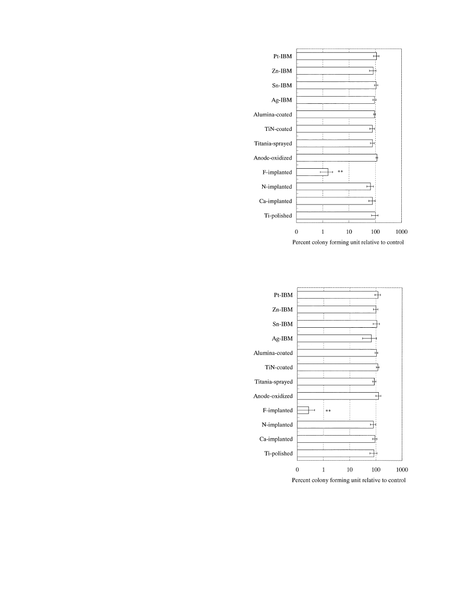
Fig. 2. The percent colony-forming units relative to control against P.
gingivalis ATCC 33277 on 1-cm
plates for 48 h (**p(0.01 to Ti-
polished).
Fig. 3. The percent colony forming units relative to control against A.
actinomycetemcomitans ATCC 43718 on 1-cm
plates for 48 h
(**p(0.01 to Ti-polished).
immediately transferred into 900
l of TSB and diluted in
a 10-fold series to l0
\. The TSB used as the dilution
solution was prepared with the addition of 0.8-mm glass
beads. Each 100
l of sample was plated on a blood agar
plate, spread evenly with a Conrage stick, and cultured at
373C for 7 days. At the end of the culture period, the
number of colonies (colony-forming unit: CFU) was
counted.
Antibacterial activity was expressed as the ratio of
CFUs on each specimen to those on the control that was
incubated without specimens. Each colonization test was
run in triplicate and repeated at "ve separate times. The
ratio of CFUs was analyzed via one-way ANOVA. The
CFU ratios were then compared with those of the control
by the Fisher PLSD (protected least signi"cant di!er-
ence) test.
2.3. Fluorine ion release test
The F
>-implanted specimens (10;10;1 mm) were
immersed in a 0.9% NaCl solution of 2 ml at 373C for
1 week. The F-ion concentration in the solution was then
measured using an ion electrode-type concentration
measuring instrument (IA-100, TOA Electric, Tokyo,
Japan). Five specimens were prepared.
2.4. Proliferation test of L929 cells
Mouse "broblast cell line L929 was used for the
cytotoxicity test. L929 cells were cultured with Eagle's
minimal essential medium (MEM) containing 10 vol%
fetal bovine serum (FBS) and 2 vol% antibiotic (penicillin
and streptomycin). F
>-implanted and Ti-polished speci-
mens (10
;10;1 mm) were placed in 24-well polystyrene
tissue-culture plates (well diameter of 16 mm), then 1 ml
of cell suspension containing 1
;10 cells was seeded into
each well. The plates were then placed inside the incuba-
tor at 373C in a humidi"ed atmosphere of 5% CO in air,
and incubated for 3}10 days. The culture medium was
changed every 3 days. At the end of the incubation peri-
od, the specimens were removed from the wells and the
cells were suspended using 0.05% trypsin}0.02% EDTA
solution of 1 ml. After centrifugation, the supernatant
#uid was discarded, and the remaining cells were sus-
pended with PBS(!) of 1 ml. The number of cells was
counted using a Coulter counter (Z-series, COULTER,
USA). Each cell proliferation test was run in triplicate
and repeated at three separate times.
3. Results
The percentage of CFUs relative to the control against
P.g. and A.a. are shown in Figs. 2 and 3, respectively.
Analysis of the data via one-way ANOVA revealed sig-
ni"cant di!erences (p(0.01). F
>-implanted specimens
signi"cantly inhibited the growth of both P.g. and A.a.
(p(0.01) than Ti-polished specimen. Other surface-
modi"ed specimens did not show any inhibition of the
growth of either bacteria.
M. Yoshinari et al. / Biomaterials 22 (2001) 2043}2048
2045
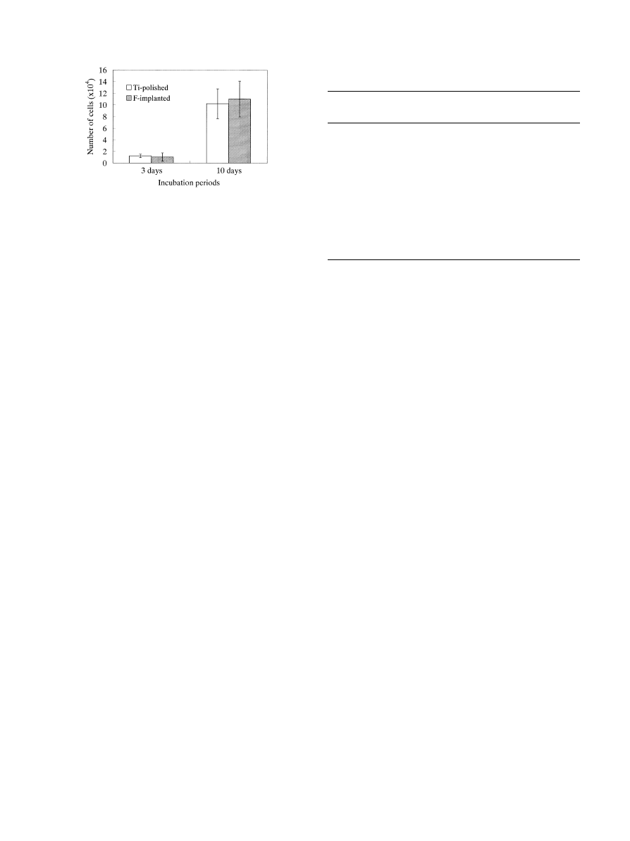
Fig. 4. The number of L929 cells proliferated on 1-cm
plates.
Table 2
Characterization of the modi"ed surfaces
Code
Identi"ed compounds
of modi"ed layer
Maximum thickness
of modi"ed layer
Ti-polished
TiO
30 nm
Ca-implanted
CaTiO, TiO, TiO
150 nm
N-implanted
TiN, TiN, TiO
300 nm
F-implanted
TiF, TiOF, TiO,TiO
150 nm
Anode-oxidized
TiO (brookite), TiO
300 nm
Titania-sprayed
TiO (rutile, anatase)
'
3
m
TiN-coated
TiN
3
m
Alumina-coated
AlO (corundum)
3
m
Ag-IBM
Ag, TiO TiOV
100 nm
Sn-IBM
Sn, TiO TiOV
150 nm
Zn-IBM
Zn, TiO TiOV
100 nm
Pt-IBM
Pt, TiO
150 nm
Chemical compounds except Ti identi"ed by X-ray photoelectron
spectroscopy (XPS) and thin-"lm X-ray di!ractometry (XRD).
Equivalent thickness to sputtering rate of titanium by Ar-ion etching
on XPS depth analysis.
The concentration of #uorine ions released from F
>-
implanted specimens in 0.9% NaCl solution was less
than 0.10 mg/l of the detection limit.
The number of L929 cells proliferated on 10
;10 mm
plates is shown in Fig. 4. No signi"cant di!erences were
observed between Ti-polished and F-implanted speci-
mens at either the 3- or 10-day intervals.
4. Discussion
It is generally believed that rapid osseointegration with
titanium compared with that of other metallic implants is
due to the ease with which calcium phosphates and
serum proteins are adsorbed on titanium surfaces. This
implies, however, that the calcium and pellicle in saliva
are adsorbed and form on titanium surfaces, and then,
that oral bacteria adhere and colonize on titanium surfa-
ces. This situation leads to the probable risk of plaque
formation to titanium implants exposed to the oral cav-
ity. Therefore, it is important to provide a source of
antibacterial activity as well as to inhibit the initial ad-
hesion of oral bacteria to titanium surfaces.
The surface modi"cations used in this study employed
the same methods as in our previous studies for ensuring
good wear resistance [8,14]. The surfaces were character-
ized using X-ray photoelectron spectroscopy and thin-
"lm X-ray di!ractometry as shown in Table 2. Brie#y,
ion-implanted surfaces (Ca
>-, N>-, and F>-implanted)
consisted of Ti compounds with the implanted elements,
titanates of the implanted elements, as well as titanium
oxides. Ti}F compounds were present on the F-im-
planted surfaces. The titanium oxides, mainly rutile, were
created on oxide "lms through titania spraying and
anodic oxidation. Anatase was also included in the oxide
"lms of the titania spraying. Ion-plated surfaces, includ-
ing TiN- and alumina-ion plating, had similar composi-
tions to those of the raw materials used for coating. The
surfaces treated with ion beam mixing (Ag-, Sn-, Zn-, and
Pt-IBM) consisted of raw metal elements and titanium
oxides.
Fluoride is widely used as a highly e!ective anticaries
agent in dental "elds, and #uorine ions released from
#uoride can a!ect bacterial metabolism as an enzyme
inhibitor. Metal}#uoride complexes are also responsible
for #uoride inhibition of proton-translocating F-AT-
Pases and are thought to act by mimicking phosphate to
form complexes with ADP at the reaction centers of the
enzymes [15}17].
The photocatalytic reaction of titania, especially
anatase, was found by Fujishima and Honda [18]. The
antibacterial activity of titania was also reported in the
biomedical "elds [19}21]. Initially, TiO photocatalysis
promoted peroxidation of the polyunsaturated phos-
pholipid component of the lipid membrane and induced
major disorder in the bacteria. This photocatalytic activ-
ity becomes e!ective when irradiated with near-UV light
and coupled with a conductor such as platinum [19].
Ag
> inhibits the DNA synthesis with direct binding on
the bacterial DNA. Ag
> also adsorbs the protein on the
surface of the bacterial membrane, in#uencing membrane
synthesis with S}Ag bonds [22]. Silver is used in a mix-
ture with silica glass [23], silver coating, or to adsorb
zeolite [24]. However, halogenated silver such as AgCl or
an oxide such as AgO reduces antibacterial activity. In
general, Sn and Zn potentiate the antiseptic agents [25],
Stannous #uoride, stannous chloride, and zinc chloride
inhibited all strains tested [26}28]. Zn is also used in
zeolite with Ag [29], and as an amalgam component
[30]. Platinum complexes have been shown to be e!ec-
tive inhibitors of bacterial DNA, RNA, and protein syn-
thesis [31]. Metal chelates of the platinum group exhibit
signi"cant activity against a wide spectrum of microor-
ganisms at di!erent concentrations [32].
From characterizations of the modi"ed surfaces
studied previously and the above-mentioned reports, the
F
>-implanted, titania-sprayed, and Ag-, Sn-, Zn-,
and Pt-IBM specimens were expected to be e!ective in
2046
M. Yoshinari et al. / Biomaterials 22 (2001) 2043}2048

promoting surface antibacterial activity. The results of
the experiments, however, showed that only the F
>-im-
planted specimen e!ectively exhibited antibacterial activ-
ity against both P.g. and A.a.
There are two possible explanations for antibacterial
mechanism of the F
>-implanted specimen. One on hand,
the action of the #uorine ions could be responsible for
this mechanism; on the other hand, the action of the
metal}#uoride complexes could be responsible, as men-
tioned above. The concentration of #uorine ions released
from F
>-implanted specimens in 0.9% NaCl solution
was less than 0.10 mg/l of the detection limit. In our
previous study [8,14], the #uoride complex was observed
on the F
>-implanted specimen with thin
"lm X-ray dif-
fractometry and X-ray photoelectron spectroscopy. It
has been reported that several #uoride salts with poly-
valent cations such as Cu
>, Sn>, and Al> exhibit
a direct antibacterial e!ect, and titanium tetra#uoride
seeded with bacteria had similar growth inhibition zones
to those of these salts [33]. Accordingly, the current
results indicate that the latter mechanism, caused by the
metal}#uoride complexes, is primarily responsible for the
antibacterial activity on the F
>-implanted specimen. In
the previous study [8], the initial adherence of bacteria
was not reduced on the F
>-implanted surface compared
to that of cp-Ti. In conclusion, the mode of action inhibi-
ting bacterial growth on F
>-implanted specimens is most
likely as follows: at "rst, the oral bacteria adhered to the
F
>-implanted surfaces. Then, the bacteria were injured
by the pharmacological e!ect of the metal}#uoride
complexes with their inhibition of enzymatic activity.
Incidentally, it was con"rmed that F
>-implanted surfa-
ces did not in#uence the proliferation of mouse-"broblast
cells.
Titania-sprayed specimens generated no antimicrobial
activity despite the anatase that formed on the surfaces.
This may be because no UV light was used, and no
coupling metals were used for stimulating photocatalytic
reactions. The Ag-, Sn-, Zn-, and Pt-IBM specimens also
did not exhibit any antibacterial activity. Ag, Sn, and Zn
work e!ectively on the surface of the bacterial membrane
as metal ions. The formation of halogenations, oxides or
sul"des of these elements markedly reduces antibacterial
activity. We believe that these compounds such as AgCl,
AgO or AgS were formed during the bacterial coloniz-
ation test. Platinum chelates, which were reported to
exhibit signi"cant activity against bacteria, seemed not to
be formed on the Pt-IBM specimens. Further investiga-
tions are necessary to clarify these phenomena. In this
study, we evaluated antibacterial activity on surfaces
treated with a dry process as surface modi"cations
with wear resistance. Other antibacterial treatments
are also considered e!ective such as a sulfated poly-
saccharide extracted from seaweed funoran, or 3-
(trimethoxysilyl)-propyldimethyloctadecyl
ammonium
chloride [34,35].
Acknowledgements
This study was supported in part by a Grant-in-Aid for
Scienti"c Research No. 10085839 from The Ministry of
Education, Science, Sports and Culture in Japan.
References
[1] Nakazato G, Tsuchiya H, Sato M, Yamauchi M. In vivo plaque
formation on implant materials. Int J Oral Maxillofac Implants
1989;4:321}6.
[2] Wolinsky LE, de Camargo PM, Erard JC, Newman MG. A study
on in vitro attachement of Streococcus sangius and Actinomyces
viscosus to saliva-treated titanium. Int J Oral Maxillofac Imp
1989;4:27}31.
[3] Drake DR, Paul J, Keller JC. Primary bacterial colonization of
implant surfaces. Int J Oral Maxillofac Imp 1999;12:226}32.
[4] Leonhardt A, Olsson J, Dahlen G. Bacterial colonization on
titanium, hydroxyapatite, and amalgam surfaces in vivo. J Dent
Res 1995;74:1607}12.
[5] Berry CW, Moore TJ, Safsr JA, Henry CA, Wagner MJ. Antibac-
terial activity of dental implant metals. Implant Dentistry
1992;1:59}65.
[6] Gatewood RR, Cobb CM, Killoy WJ. Microbial colonization on
natural tooth structure compared with smooth and plasma-
sprayed dental implant surfaces. Clin Oral Impl Res 1993;4:53}64.
[7] Leonhardt A, Dahlen G. E!ect of titanium on selected oral
bacterial species in vitro. Eur J Oral Sci 1995;103:382}7.
[8] Yoshinari M, Oda Y, Kato T, Okuda K, Hirayama A. In#uence of
surface modi"cations to titanium on oral bacterial adhesion
in vitro. J Biomed Mater Res 2000;52:388}94.
[9] Bellanda M, Cassinelli C, Morra M. Reduced plaque accumula-
tion on hydrocarbon thin "lm deposited on restorative acrylic
polymers. J Biomed Mater Res 1997;36:216}22.
[10] Buchanan RA, Rigney ED, Williams JM. Ion implantation of
surgical Ti-6Al-4V for improved resistance to wear accelerated
corrosion. J Biomed Mater Res 1987;21:355}66.
[11] Yoshinari M, Ozeki K, Sumii T. Properties of hydroxyapatite-
coated Ti-6Al-4V alloy produced by the ion-plating method. Bull
Tokyo Dent Coll 1991;32:147}56.
[12] Yoshinari M, Watanabe Y, Ohtsuka Y, DeHrand T. Solubility
control of thin calcium}phosphate coating with rapid heating.
J Dent Res 1997;76:1486}95.
[13] Yoshinari M, Hayakawa T, Wolke JCG, Nemoto K, Jansen JA.
In#uence of rapid heating with infrared radiation on RF magnet-
ron sputtered calcium phosphate coatings. J Biomed Mater Res
1997;37:60}7.
[14] Miyayama N, Yoshinari M, Oda Y. Surface modi"cation of
titanium implants with dry process * surface characterization.
Jpn J Dent Mater 1999;18:109}21 (in Japanese).
[15] Marquis RE. Antimicrobial actions of #uoride for oral bacteria.
Can J Microbiol 1995;41:955}64.
[16] Guha-Chowdhury N, Clark AG, Sissons CH. Inhibition of puri"-
ed enolases from oral bacteria by #uoride. Oral Microbiol Immu-
nol 1997;12:91}7.
[17] Forss H, Jokinen J, Spets-Happonen S, Seppa L, Luoma H.
Fluoride and mutans streptococci in plaque grown on glass
ionomer and composite. Caries Res 1991;25:454}8.
[18] Fujishima A, Honda K. Electrochemical photolysis of water at
a semiconductor electrode. Nature 1972;238:37}8.
[19] Maness PC, Smolinski S, Blake DM, Huang Z, Wolfrum EJ,
Jacoby WA. Bactericidal activity of photocatalytic TiO(2) reac-
tion: toward an understanding of its killing mechanism. Appl
Environ Microbiol 1999;65:4094}8.
M. Yoshinari et al. / Biomaterials 22 (2001) 2043}2048
2047

[20] Ireland JC, Klostermann P, Rice EW, Clark RM. Inactivation of
Escherichia coli by titanium dioxide photocatalytic oxidation.
Appl Environ Microbiol 1993;59:1668}70.
[21] Saito T, Iwase T, Horie J, Morioka T. Mode of photocatalytic
bactericidal action of powdered semiconductor TiO on mutans
streptococci. J Photochem Photobiol 1992;14:369}79.
[22] Modak SM, Fox Jr CL. Binding of silver sulfadiazine to the
cellular components of Pseudomonas aeruginosa. Biochem Phar-
macol 1973;22:2391}404.
[23] Kawashita M, Tsuneyama S, Miyaji F, Kokubo T, Kozuka H,
Yamamoto K. Antibacterial silver-containing silica glass pre-
pared by sol}gel method. Biomaterials 2000;21:393}8.
[24] Cook G, Costerton JW, Darouiche RO. Direct confocal micros-
copy studies of the bacterial colonization in vitro of a silver-
coated heart valve sewing cu!. Int J Antimicrob Agents
2000;13:169}73.
[25] Zeelie JJ, McCarthy TJ. E!ects of copper and zinc ions on the
germicidal properties of two popular pharmaceutical antiseptic
agents cetylpyridinium chloride and povidone-iodine. Analyst
1998;123:503}7.
[26] Eisenberg AD, Young DA, Fan-Hsu J, Spitz LM. Interactions of
sanguinarine and zinc on oral streptococci and Actinomyces spe-
cies. Caries Res 1991;25:185}90.
[27] Mayhew RR, Brown LR. Comparative e!ect of SnF, NaF, and
SnCl on the growth of Streptococcus mutans. J Dent Res
1981;60:1809}14.
[28] Attramadal A, Svatun B. In vivo antibacterial e!ect of tin on the
oral micro#ora. Scand J Dent Res 1984;92:161}4.
[29] Hotta M, Nakajima H, Yamamoto K, Aono M. Antibacterial
temporary "lling materials: the e!ect of adding various ratios of
Ag}Zn}zeolite. J Oral Rehabil 1998;25:485}9.
[30] Morrier JJ, Suchett-Kaye G, Nguyen D, Rocca JP, Blanc-Benon
J, Barsotti O. Antimicrobial activity of amalgams, alloys and their
elements and phases. Dent Mater 1998;14:150}7.
[31] Kohl HH, Haghighi S, McAuli!e CA. Inhibitory studies of
DNA, RNA and protein synthesis in Escherichia coli by platinum
containing
complexes.
Chem
Biol
Interact
1980;29:
327}33.
[32] O$ong OE, Etok C, Martelli S. Synthesis and biological activity
of platinum group metal complexes of o-vanillin thiosemicar-
bazones. Farmaco 1996;51:801}8.
[33] Skartveit L, Selvig KA, Myklebust S, Tveit AB. E!ect of TiF
solutions on bacterial growth in vitro and on tooth surfaces. Acta
Odontol Scand 1990;48:169}74.
[34] Saeki Y, Kato T, Naito Y, Takazoe I, Okuda K. Inhibitory e!ects
of funoran on the adherence and colonization of Mutans Strepto-
cocci. Caries Res 1996;30:119}25.
[35] Saito T, Takatsuka T, Kato T, Ishihara K, Okuda K. Adherence
of oral Streptococci to an immobilized antimicrobial agent. Archs
Oral Biol 1997;42:539}45.
2048
M. Yoshinari et al. / Biomaterials 22 (2001) 2043}2048
Wyszukiwarka
Podobne podstrony:
6 63 76 Influence of Surface Heat Treatment on Thermal Fatique Behaviour
70 1003 1019 Influence of Surface Engineering on the Performance of Tool Steels for Die Casting
Influence of titanium surfaces on attachment of osteoblast
Laser surface modification of hydroxyapatite and glass
07 Kolar K i inni Influence of separation agents on quality of concrete surface
The Influence of` Minutes
Capability of high pressure cooling in the turning of surface hardened piston rods
20 255 268 Influence of Nitrogen Alloying on Galling Properties of PM Tool Steels
In the Flesh The Cultural Politics of Body Modification
3 The influence of intelligence on students' success
72 1031 1039 Influence of Thin Coatings Deposited by PECVD on Wear and Corrosion Resistance
Influence Of Magnetic Field On Two Phase Flow Convective Boiling Of Some Refrigerant Mixtures
Zen in the Influence of the Sword
więcej podobnych podstron