ABC of diseases of liver, pancreas, and biliary system
Liver tumours
I J Beckingham, J E J Krige
Tumours of the liver may be cystic or solid, benign or
malignant. Most are asymptomatic, with patients having normal
liver function, and they are increasingly discovered incidentally
during ultrasonography or computed tomography. Although
most tumours are benign and require no treatment, it is
important for non-specialists to be able to identify lesions that
require further investigation and thus avoid unnecessary biopsy.
Modern imaging combined with recent technical advances in
liver surgery can now offer many patients safe and potentially
curative resections for malignant, as well as benign, conditions
affecting the liver.
Cystic liver lesions
Cystic lesions of the liver are easily identified by
ultrasonography. Over 95% are simple cysts. Asymptomatic
cysts are regarded as congenital malformations and require no
further investigation or treatment as complications are rare.
Aspiration and injection of sclerosants should be avoided as it
may cause bleeding and infection and does not resolve the cyst.
Rarely, simple cysts can grow very large and produce
compressive symptoms. These are managed by limited surgical
excision of the cyst wall (cyst fenestration), which can usually be
done laparoscopically.
About half of patients with simple cysts have two or more
cysts. True polycystic liver disease is seen as part of adult
polycystic kidney disease, an uncommon autosomal dominant
disease that progresses to renal failure. Patients nearly always
have multiple renal cysts, which usually precede development of
liver cysts. Liver function is normal, and most patients have no
symptoms. Occasionally the cysts cause pain because of
distension of the liver capsule, and such patients may require
cyst fenestration or partial liver resection.
Thick walled cysts and those containing septa, nodules, or
echogenic fluid may be cystic tumours (cystadenoma,
cystadenocarcinoma) or infective cysts (hydatid cysts and
abscesses; see later article in this series), and patients should be
referred for specialist surgical opinion. Cystic dilatations of the
bile ducts (Caroli’s disease) are important as they may produce
cholangitis and are premalignant with the potential to develop
into cholangiocarcinoma.
Benign tumours
Benign liver tumours are common and are usually
asymptomatic. Although most need no treatment, it is
important to be able to differentiate them from malignant
lesions.
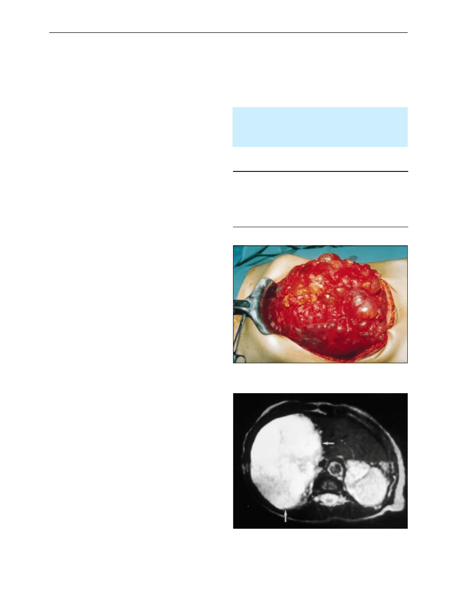
Haemangiomas
Haemangiomas are the commonest benign solid tumours of
the liver, with an incidence in the general population of around
3%. Those over 10 cm in diameter occasionally produce
non-specific symptoms of abdominal discomfort and fullness
and, rarely, fever, thrombocytopenia, and hypofibrinogenaemia
due to thrombosis in the cavernous cavities. Malignant
transformation and spontaneous rupture are rare. Contrast
enhanced computed tomography is usually sufficient to
Liver biopsy of a tumour mass should be reserved for
patients with suspected malignancy who are not suitable
for surgery and in whom the diagnosis may have clinical
impact—for example, ovarian or neuroendocrine tumours,
carcinoid, or lymphoma
Characteristics of simple cysts
x Thin walled
x Contain clear fluid
x Contain no septa or debris
x Surrounded by normal liver tissue
x Usually asymptomatic
x Present in 1% of population
Polycystic liver disease
T2 weighted magnetic resonance image of large benign haemangioma
showing light bulb sign
Clinical review
477
BMJ VOLUME 322 24 FEBRUARY 2001 bmj.com
diagnose most haemangiomas, and in equivocal cases magnetic
resonance imaging or technetium-99 labelled red blood cell
scintigraphy will confirm the diagnosis. Angiography and
biopsy are seldom required. Resection is indicated only for large
symptomatic tumours.
Liver cell adenoma and focal nodular hyperplasia
These uncommon tumours occur predominantly in women of
childbearing age. Liver cell adenoma became more prevalent
with the widespread use of oral contraceptives in the 1960s, but
the reduced oestrogen content of modern contraceptives has
made it less common. Most patients present with pain due to
rapid tumour growth, intratumour haemorrhage, or the
sensation of a mass. The risk of rupture is 10%, and malignant
transformation is found in 10% of resected specimens. Patients
should have liver resection to prevent these events.
Focal nodular hyperplasia is not related to use of oral
contraceptives, is usually asymptomatic, and is not
premalignant. Mass lesions usually contain a central stellate scar
on computed tomography and magnetic resonance imaging. It
does not require treatment unless symptomatic.
In a small proportion of patients a firm radiological
diagnosis cannot be reached and the distinction from a
malignant liver tumour is uncertain. Histological distinction
between focal nodular hyperplasia and cirrhosis and between
liver cell adenoma and well differentiated hepatocellular
carcinoma can be difficult with tru-cut biopsy or fine needle
aspiration samples, and biopsy has the added risk of bleeding
and tumour seeding. The histology should therefore be
determined by surgical resection, which in specialist centres has
a mortality of < 1%.
Malignant tumours
Hepatocellular carcinoma
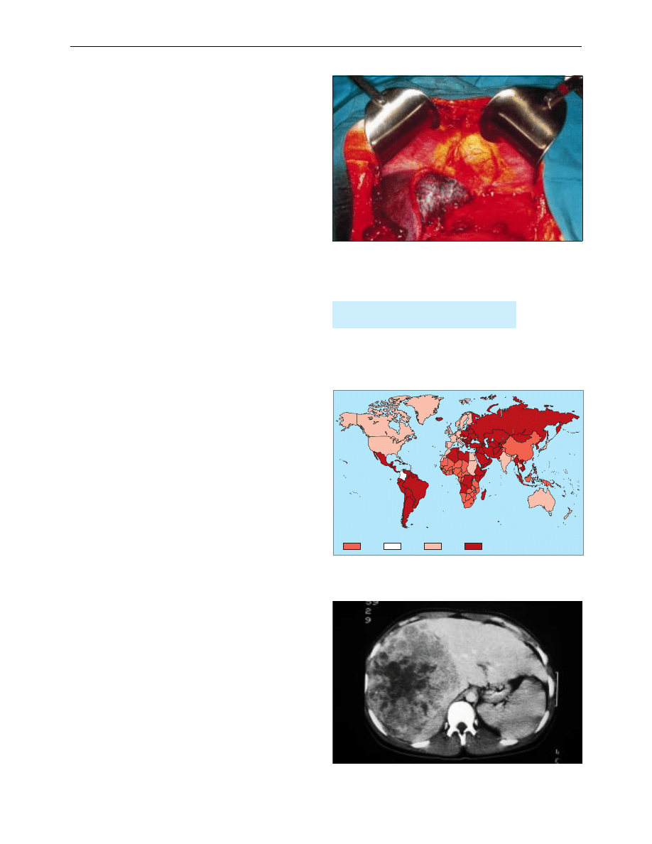
Hepatocellular carcinoma is uncommon in the United
Kingdom and accounts for only 2% of all cancers. Worldwide
there are over one million new cases a year, with an annual
incidence of 100 per 100 000 men in parts of South Africa and
South East Asia. The incidence of hepatocellular carcinoma is
increased in areas with high carrier rates of hepatitis B and C
and in patients with haemochromatosis. More than 80% of
hepatocellular carcinomas occur in patients with cirrhotic livers.
Once viral infection is established it takes about 10 years for
patients to develop chronic hepatitis, 20 years to develop
cirrhosis, and 30 years to develop carcinoma. In African and
Asian countries aflatoxin, produced as a result of contamination
of imperfectly stored staple crops by Aspergillus flavus, seems to
be an independent risk factor for the development of
hepatocellular carcinoma, probably through mutation of the
p53 suppressor gene. Seasonal variation in incidence is seen in
these countries.
In patients with cirrhosis, the diagnosis should be suspected
when there is deterioration in liver function, an acute
complication (ascites, encephalopathy, variceal bleed, jaundice),
or development of upper abdominal pain and fever.
Ultrasonography will identify most tumours, and the presence
of a discrete mass within a cirrhotic liver, together with an
á fetoprotein concentration above 500 ng/ml is diagnostic.
Biopsy is unnecessary and should be avoided to reduce the risk
of tumour seeding. Surgical resection is the only treatment that
can offer cure. However, owing to local spread of tumour and
severity of pre-existing cirrhosis, such treatment is feasible in
less than 20% of patients. Average operative mortality is 12% in
cirrhotic patients, and five year survival is around 15%.
Hepatocellular carcinoma is the
commonest malignant tumour worldwide
Intraoperative view after left hepatectomy—raw surfaces of liver are coated
with fibrin glue after resection to aid haemostasis and prevent small bile
leaks
10-15
Annual incidence (cases per 100 000)
3-10
1-3
Undefined
Distribution of hepatocellular carcinoma
Computed tomogram of large hepatocellular carcinoma
Clinical review
478
BMJ VOLUME 322 24 FEBRUARY 2001 bmj.com
Patients with cirrhosis and small ( < 5 cm) tumours should
have liver transplantation. Injection of alcohol or
radiofrequency ablation can improve survival in patients with
small tumours who are unsuitable for transplantation. For
larger tumours, transarterial embolisation with lipiodol and
cytotoxic drugs (cisplatin or doxorubicin) may induce tumour
necrosis in some patients.
In patients without cirrhosis, hepatocellular carcinomas
usually present late with an abdominal mass and abnormal liver
function. Computed tomography has a greater sensitivity and
specificity than ultrasonography, particularly for tumours
smaller than 1 cm.
á Fetoprotein concentrations are raised in
80% of patients but may also be raised in patients with testicular
or germ cell tumours.
Fibrolamellar carcinoma is an important subtype of
hepatocellular carcinoma. It occurs in patients without cirrhosis
or previous hepatitis infection. It accounts for 15% of
hepatocellular carcinoma in the Western hemisphere. The
prognosis is better than for other hepatocellular carcinomas,
with a five year survival of 40-50% after resection.
Metastatic tumours
Liver metastases are common and are found in 40% of all
patients dying from cancer. They are most frequently associated
with carcinomas of the gastrointestinal tract (colorectal, pancreas,
and stomach) but are nearly as common in carcinomas of the
bronchus, breast, ovary, and lymphoma. With the exception of
liver metastases of colorectal cancer, tumour deposits are almost
always multiple and seldom amenable to resection.
Colorectal liver metastases
Around 8-10 % of patients undergoing curative resection of
colorectal tumours have isolated liver metastases suitable for
liver resection, equivalent to around 1000 patients in the United
Kingdom a year. Half will have metastases at the time of
diagnosis of the primary tumour (synchronous metastases) and
most of the rest will develop metastases within the next three
years (metachronous metastases).
Without surgical resection the five year survival rate for all
patients with liver metastases is zero, compared with an overall
five year survival after resection of 30%. Patients most suited for
resection are those with fewer than three or four metastases in
one lobe of the liver, but tumours need not be confined to one
lobe. The principle of complete tumour removal, however,
remains a prerequisite, and one limitation is the need to leave
enough liver to function. This depends both on the extent and
distribution of the tumour burden and the general fitness of the
patient and his or her liver. The liver has an enormous capacity
for regeneration. A fit patient with a healthy liver will regenerate
a 75% resection within three months. Age is only a relative
contraindication, and several series have reported low mortality
in septuagenarians.
Liver resection
Liver resection has advanced rapidly over the past two decades
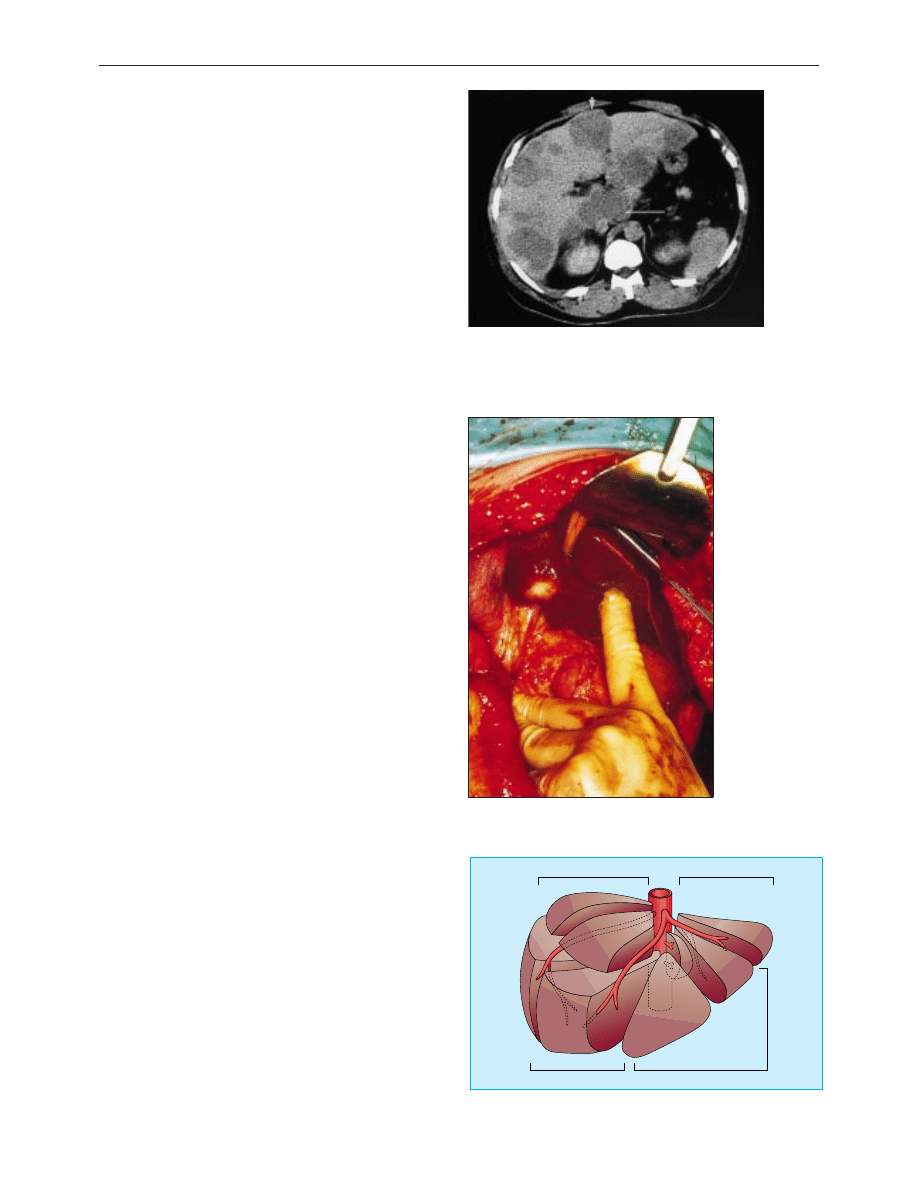
because of several important developments. The segmental
anatomy of the liver, with each of the eight segments supplied
by its own branch of the hepatic artery, portal vein, and bile
duct, was first described by Couinaud in 1957. It is now possible
to remove each of these segments individually when required,
reducing the amount of normal liver unnecessarily removed.
Subsequently surgical techniques have been developed to
divide the liver parenchyma, either by crushing with a clamp or
by ultrasonic dissection, allowing the vascular and biliary radicals
Inoperable extensive liver metastases
Solitary metastasis in segment IV of liver
Right lateral (posterior) sector
Right medial (anterior) sector
Left medial
(anterior) sector
Left lateral (posterior) sector
VII
VIII
VI
V
IV
III
II
I
Couinaud’s segmental anatomy of liver
Clinical review
479
BMJ VOLUME 322 24 FEBRUARY 2001 bmj.com

to be individually ligated. Blood loss has been reduced by
occlusion of the vascular inflow (Pringle manoeuvre) and where
possible the appropriate hepatic vein, together with lowering of
the central venous pressure during resection, and blood
transfusion is now unnecessary in 60% of major liver resections.
Improvements have also occurred in anaesthetic and
postoperative care, including epidural anaesthesia to reduce
postoperative pain and chest complications and the ability to
manage postoperative fluid or bile collections by radiological or
endoscopic drainage. These developments mean that the
median hospital stay for patients having liver resection is now
7-10 days and mortality is around 5%. Liver resection has
evolved from a hazardous bloody procedure into a routine
operation.
J E J Krige is associate professor of surgery, Groote Schuur Hospital
and University of Cape Town, South Africa
The ABC of diseases of liver, pancreas, and biliary system is edited by
I J Beckingham, consultant hepatobiliary and laparoscopic surgeon,
department of surgery, Queen’s Medical Centre, Nottingham
(Ian.Beckingham@nottingham.ac.uk). The series will be published as
a book later this year.
BMJ 2001;322:477-80
Lesson of the week
Splenic trauma complicating cardiopulmonary resuscitation
A Fitchet, R Neal, P Bannister
Cardiopulmonary resuscitation can result in trauma
to abdominal organs. We report two cases of splenic
rupture causing life threatening haemorrhage.
Case reports
Case 1—A 64 year old woman who had undergone cor-
onary artery bypass grafting 10 years previously had a
cardiorespiratory arrest at a railway station late one
night. Cardiopulmonary resuscitation was started
immediately by bystanders and continued for 20 min-
utes until paramedics arrived. Ventricular fibrillation
was confirmed, and she was externally defibrillated. On
arrival at hospital she was alert and breathing sponta-
neously but hypotensive with a blood pressure of
80/40 mm Hg and a sinus tachycardia of 100
beats/min. Clinical examination suggested hypovolae-
mia with lowered central venous pulse pressure,
normal heart sounds, and clear breath sounds. Electro-
cardiography confirmed an acute inferior myocardial
infarction. Thrombolysis was not given because of pro-
longed resuscitation. She clinically improved on
challenge with intravenous fluid. The central venous
pulse became visible and her blood pressure rose to
120/70 mm Hg. Over the next hour progressive hypo-
tension recurred, once again with clinical evidence of
hypovolaemia. Blood pressure was restored with
further
intravenous
fluid.
An
echocardiogram
excluded major pericardial effusion, showing a
non-dilated left ventricle with inferior wall akinesia and
overall moderate function. At this stage the patient
complained of left sided abdominal pain, with tender-
ness elicited over the left hypochondrium. Chest x ray
films taken in the erect position showed no evidence of
rib fractures or subdiaphragmatic gas. Ultrasonogra-
phy showed free fluid in the abdominal cavity, and
aspiration of this fluid confirmed blood. Computed
tomography of the abdomen showed a tear in the
upper pole of the spleen (figure). At emergency
laparotomy 2 litres of free blood were found, and the
ruptured but histologically normal spleen was
removed. She made a full recovery after a prolonged
postoperative course, and she was discharged from
hospital six weeks later.
Case 2—A 50 year old man attended the casualty
department
with
general
malaise. Examination
revealed lower limb cellulitis. He had a fever at 37.9°C,
and his blood pressure was 114/70 mm Hg and heart
rate 120 beats/min. Initial investigations showed a
haemoglobin concentration of 36 g/l (normal range
130-180) (mean corpuscular volume 96 fl (80-97)),
white cell count of 2
×
10
9
/l (4-11), and platelet count
of l34
×
10
9
/l (150-400); a bone marrow aspirate later
confirmed megaloblastic anaemia. Intravenous pipera-
cillin and gentamicin were started. Soon after this he
developed bradycardia followed by a cardiorespiratory
arrest requiring two brief episodes of cardiopulmonary
resuscitation and insertion of a temporary pacing wire.
He was transferred to the intensive care unit and was
transfused 12 units of blood over the next 48 hours. He
was given folate and vitamin B-12 supplementation.
Blood cultures taken before insertion of the pacing
wire confirmed Staphylococcus aureus septicaemia.
Haemoglobin concentration increased to 90 g/l with
transfusion over 48 hours and then decreased to 66 g/l
over the next 24 hours. Ultrasonography and
Summary points
x Simple liver cysts are common, benign, and require no treatment
x Patients with solitary liver masses should be referred to a
hepatobiliary surgeon and liver biopsy avoided
x Liver resection is a safe procedure in non-cirrhotic patients, with a
mortality around 5%
x 10% of patients with colorectal cancer develop potentially curable
liver metastases and should have six monthly liver ultrasonography
or computed tomography
x Five year survival after resection of colorectal metastases is > 30%
Further reading
x Blumgart LH, Jarnogin W, Fang Y. Liver resection. In: Blumgart LH,
ed. Surgery of the liver and biliary tract. London: WB Saunders,
2000:1639-1714
x Launois B, Jamieson GG. Modern operative techniques in liver surgery.
Edinburgh: Churchill Livingstone, 1993
x Neeleman N, Andersson R. Repeated liver resection for recurrent
liver cancer. Br J Surg 1996;83:885-92
Unexplained
hypotension
after
cardiopulmonary
resuscitation
might be due to
intra-abdominal
trauma and
concealed
haemorrhage
Manchester Heart
Centre, Manchester
Royal Infirmary,
Manchester
M13 9WL
A Fitchet
specialist registrar in
cardiology
Blackburn Royal
Infirmary, Bolton
Road, Blackburn
BB2 3LR
R Neal
specialist registrar in
elderly medicine
Department of
Medicine,
Manchester Royal
Infirmary
P Bannister
consultant physician
Correspondence to:
A Fitchet
Alan.Fitchet@mhc.
cmht.nwest.nhs.uk
BMJ 2001;322:480–1
Clinical review
480
BMJ VOLUME 322 24 FEBRUARY 2001 bmj.com
Wyszukiwarka
Podobne podstrony:
ABC Liver and pancreatic trauma
ABC Pancreatic tumours
ABC Liver abscesses and hydaid disease
ABC Of Liver,Pancreas and Gall Bladder
ABC Other causes of parenchymal liver disease
ABC Investigation of liver correct
ABC Transplantation of the liver and pancreas
ABC Investigation of liver and biliary disease
2014 ABC DYDAKTYKIid 28414 ppt
Amortyzacja pozycki ABC
ABC mądrego rodzica droga do sukcesu
ABC praw konsumenta demo
abc 56 58 Frezarki
ABC Madrego Rodzica Inteligencja Twojego Dziecka
ABC Neostrada
ABC trzylatka przewodnik
więcej podobnych podstron