
Open Life Sci. 2015; 10: 119–129
DOI 10.1515/biol-2015-0012
Received January 13, 2014; accepted August 20, 2014
1 Introduction
Cyanobacteria that are able to produce hepatotoxins
known as microcystins are the key indicators of increasing
eutrophication caused by the excessive inflow of nutrients
into freshwater aquatic environments [1]. Thus, a limitation
in nutrient inflow from the catchment must be the first
step in reducing cyanobacterial blooms [2-5]. However,
the investigation and selection of methods for removing
nutrients requires time and specific physicochemical and
biological data for a particular body of water. Therefore,
it is important to develop methods to treat areas where
toxic cyanobacteria already exist and affect the quality of
drinking and recreational water resources. For this task,
implementation of biological methods with the use of
controlling agents such as bacteria capable of microcystins
removal seems to be promising.
In the study of Ho et al. [6] the rapid biological
sand filtration with natural indigenous bacteria (with
domination of Sphingopyxis sp. LH21) aggregated in the
biofilm was reported as an effective treatment process for
the complete removal of microcystins. Also, Bourne et al.
[7] reported the usefulness of applying selected cultured
bacteria Sphingomonas sp. MJ-PV strain for removing of
microcystin-LR (MC-LR) in sand filtration columns.
An example of possible microcystins removal from
surface water was described in the pilot study of Ji et al.
[8]. In a meso-scale experiment performed in Lake Taihu
(China), artificial media were submerged in the flowing
water from the lake. The biofilm containing indigenous
bacteria (with domination of Pseudomonas spp. and
Bacillus spp.), which was created on artificial media, was
able to degrade microcystins.
As indicated by cited studies, the removal of
microcystins by a diverse community of bacteria is
considered to be the dominant proces responsible for the
disappearance of cyanobacterial-derived hepatotoxins
Abstract: Water blooms dominated by cyanobacteria
are capable of producing hepatotoxins known as
microcystins. These toxins are dangerous to people and
to the environment. Therefore, for a better understanding
of the biological termination of this increasingly
common phenomenon, bacteria with the potential to
degrade cyanobacteria-derived hepatotoxins and the
degradative activity of culturable bacteria were studied.
Based on the presence of the mlrA gene, bacteria with a
homology to the Sphingopyxis and Stenotrophomonas
genera were identified as those presenting potential for
microcystins degradation directly in the water samples
from the Sulejów Reservoir (SU, Central Poland). However,
this biodegrading potential has not been confirmed in in
vitro experiments. The degrading activity of the culturable
isolates from the water studied was determined in more
than 30 bacterial mixes. An analysis of the biodegradation
of the microcystin-LR (MC-LR) together with an analysis of
the phylogenetic affiliation of bacteria demonstrated for
the first time that bacteria homologous to the Aeromonas
genus were able to degrade the mentioned hepatotoxin,
although the mlrA gene was not amplified. The maximal
removal efficiency of MC-LR was 48%. This study
demonstrates a new aspect of interactions between the
microcystin-containing cyanobacteria and bacteria from
the Aeromonas genus.
Keywords: microcystins, biodegradation, mlrA gene,
Aeromonas, Stenotrophomonas, Sphingopyxis
Research Article
Open Access
© 2015 J. Mankiewicz-Boczek et al., licensee De Gruyter Open.
This work is licensed under the Creative Commons Attribution-NonCommercial-NoDerivs 3.0 License.
Mankiewicz-Boczek J.*, Gągała I., Jurczak T., Jaskulska A., Pawełczyk J., Dziadek J.
Bacteria homologus to Aeromonas capable of
microcystin degradation
*Corresponding author: Joanna Mankiewicz-Boczek: European Re-
gional Centre for Ecohydrology of the Polish Academy of Sciences, 3
Tylna Str., 90-364 Łódź, Poland, E-mail: j.mankiewicz@erce.unesco.
lodz.pl
Gągała I., Jaskulska A.: European Regional Centre for Ecohydrology
of the Polish Academy of Sciences, Łódź, 90-364, Poland
Mankiewicz-Boczek J., Jurczak T., Jaskulska A.: Department of
Applied Ecology, Faculty of Biology and Environmental Protection,
University of Lodz, Łódź, 90-237, Poland
Pawełczyk J., Dziadek J.: Institute for Medical Biology of the Polish
Academy of Sciences, Łódź, 93-232, Poland
Brought to you by | Uniwersytet Lodzki
Authenticated
Download Date | 8/31/15 9:07 AM
120
J. Mankiewicz-Boczek et al.
2 Experimental Procedures
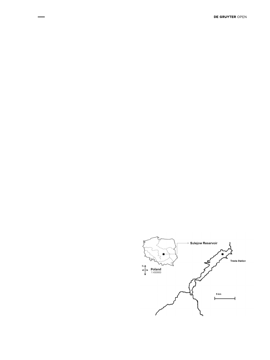
2.1 The study site
In the present study, water samples were collected from
the Sulejów Reservoir at Tresta Station located near the
dam in the lacustrine zone of the reservoir (+51°27′42.53″,
+19°58′40.88″). The reservoir located in Central Poland was
formed by damming at 138.9 km of the Pilica River (Fig. 1).
This reservoir is used for flood control, recreation, fishing
and power generation. The Sulejów Reservoir is also used
as an alternative source of drinking water for the city of
Lodz. It is an example of a dam reservoir with progressive
anthropogenic eutrophication, in which cyanobacterial
blooms dominated by toxic M. aeruginosa appear regularly
every year [17-23]. During the bloom accumulation, the total
microcystins concentration (intra- and extracellular) in the
water could increase to 30 µg L
-1
[19].
2.2 Preparation and molecular analysis of
environmental samples
Integrated water samples were collected every 2 weeks
during the summer season from May to October 2010.
To obtain material for DNA analysis, each water sample
(100 mL) was filtered using a sterile filter with a pore size
of 0.45 µm for the analysis of cyanobacteria or a pore size
of 0.22 µm for the analysis of other bacteria (Millipore,
USA). The filters were placed in 2 mL of lysis buffer (40 mM
EDTA, 400 mM NaCl, 0.75 M sucrose and 50 mM Tris-HCl,
pH 8.3) and stored at -20°C until DNA extraction. The DNA
was isolated by hot phenol extraction from the filters
based on the protocol by Giovannoni et al. [24] with the
modifications described in Mankiewicz-Boczek et al. [20].
in water. Therefore this biological termination of
microcystins by bacteria is currently being intensively
studied. Bacteria capable of microcystins degradation
belong to the genus: Pseudomonas (Australia, Japan,
China), Sphingomonas – including Sphingosinicella
(Japan, Argentina, New Zealand), Sphingopyxis (Australia,
China), Novosphingobium (China), Stenotrophomonas
(China),
Ochrobactrum (China), Methylobacillus
(China), Methylosinus (China), Ralstonia (China),
Bacillus (Saudi Arabia), Morganella (USA), Rhizobium
(USA), Microbacterium (USA), Burkholderia (Brazil),
Methylotenera (USA) and various Burkholderiales,
including Bordetella (USA, China) [9-12].
In Europe, there is limited data on the specific bacteria
capable of degrading cyanobacterial hepatotoxins in
fresh water. The first strain of bacteria was isolated from
sediment of Lake Vihnusjärvi in 2005 and classified
as a novel bacterium: Paucibacter toxinivorans [13]. In
Scotland, three new strains of bacteria were discovered:
Arthrobacter sp., Brevibacterium sp. and Rhodococcus
sp. These species were isolated from Lake Rescobie, Lake
Forfar, and the River Carron [14-15].
The process of microcystins degradation, as was
already mentioned, can be performed by different groups
of bacteria, but the only described and continuously
studied route of degradation of microcystin molecule was
presented by Bourne et al. [16]. This 3-step sequential
enzymatic process was based on proteolytic hydrolysis
of peptide bonds, in which a crucial role is played by
the mlr gene cluster, consisting of the genes: mlrA, mlrB,
mlrC and mlrD, coding intracellular enzymes. The first
step of this process (activation of mlrA gene) involves the
linearization of the microcystin molecule. The product of
the first enzymatic step was reported to be 160-fold less
reactive than the cyclic microcystin. Both the second and
third steps involved the gradual cutting of the linearized
microcystin chain, which resulted in degradation into its
individual components.
The objectives of the present study were: 1) to assess
the co-occurrence of bacteria with the potential for
microcystins degradation (based on mlrA genes presence)
and microcystin-producing cyanobacteria (based of
mcyE gene presence), together with determination of
the concentration of cyanobacteria-derived hepatotoxins
in Sulejów Reservoir (SU), the lowland dam reservoir in
Central Poland; and 2) to identify culturable bacteria
isolated from the reservoir actively degrading microcystin
molecules, and determine their respective removal
efficiencies. The phylogenetic affiliation of culturable
bacteria based on sequencing of the 16S rRNA gene
fragment was also performed.
Figure 1: Study site. Sampling point located in Tresta Station,
Sulejów Reservoir, between Tresta Gulf and Borki Gulf.
Brought to you by | Uniwersytet Lodzki
Authenticated
Download Date | 8/31/15 9:07 AM

Degradation of microcystin by Aeromonas
121
2.2.1 Amplification of mcyE gene
Molecular analysis using polymerase chain reaction (PCR)
was performed to determine the presence of potential
microcystin-producers via the amplification of the mcyE
gene with mcyE-R1/mcyE-S1 primers (Table 1). In the
present study, the primers were designed, using Vector
NTI Advance™ 9 software (Invitrogen), to hybridize to
the mcyE consensus sequence - a sequence of DNA having
similar structure and function in microcystin-producing
cyanobacteria: Microcystis aeruginosa, Planktothrix
agardhii and Anabaena sp. (currently Dolichospermum).
The cyanobacterial mcyE gene takes part in the synthesis
and integration of the Adda moiety (3-amino-9-methoxy-
2,6,8-trimethyl-10-phenyl-4(E),6(E)-decadienoic acid) into
the microcystin molecule. The Adda moiety is required
for microcystin toxicity and binding the hepatotoxin
to protein phosphatases [25]. The amplification of the
mcyE gene fragments was performed for 11 isolated DNA
samples.
The PCR was performed in a 20 µL volume reaction
containing 1x PCR buffer, 0.25 μM each primer, 3 mM MgCl
2
,
0.25 mM dNTP, 0.1 mg mL
-1
BSA and 1 U of Taq polymerase
(Qiagen). For one reaction, 1 µL of cyanobacteria DNA was
used (DNA concentration range from 25 – 1,116 ng µL
-1
).
The PCR consisted of an initial denaturation step at 95°C
for 5 min, followed by 30 cycles of DNA denaturation at
94°C for 30 s, primer annealing at 59°C for 30 s, and strand
extension at 72°C for 1 min, and a final extension step at
72°C for 10 min.
The PCR products were separated on a 1.5% agarose
gel by electrophoresis using a constant voltage (70 V),
and the DNA was visualized using ethidium bromide
(2 µg mL
-1
).
2.2.2 Amplification of mlrA gene
For amplification of the mlrA gene fragments specific to
the microcystin-degrading bacteria, primers designed
by Saito et al. [26] were used. The mlrA gene encoding
methylopeptidase (MlrA enzyme) catalyzes the first step
of bacterial degradation of cyanobacterial hepatotoxin
associated with hydrolysis and ring opening of microcystin
molecule at the Adda-Arg peptide-bond formation site
[16]. Both mlrA gene fragments were amplified in 5 of 11
isolated DNA samples. To amplify the longer fragment
of the mlrA gene (807 bp), the first set of primers MF/MR
were used (Table 1). The PCR reaction was performed
according to Saito et al. [26] with minor modifications.
The PCR reaction was performed in a final volume of
20 µL containing 1x PCR buffer, 5 μM each MF/MR primer,
2.5 mM MgCl
2
(Qiagen), 0.2 mM dNTP, 0.1 mg mL
-1
BSA
(Fermentas), and 0.5 U of Taq polymerase (Qiagen). For
each reaction, 1 µL of bacterial DNA was diluted 20 times
(DNA concentration range from 3 – 113 ng µL
-1
). The PCR
protocol consisted of an initial denaturation step at 94°C
for 1 min, followed by 35 cycles of DNA denaturation at
94°C for 20 s, primer annealing at 60°C for 10 s, and strand
extension at 72°C for 30 s, and a final extension step at
72°C for 10 min.
In the second stage, a nested PCR was performed with
the products of the mlrA gene amplification containing
fragments 807 bp in length (11 samples in total).
Amplification of the shorter fragment of the mlrA gene,
with a length of 453 bp, was performed using the primer
pairs MF2/MR (Table 1). The PCR reaction was performed
in a final volume of 20 µL containing 1x PCR buffer,
5 μM each primer MF2/MR, 2.5 mM MgCl
2
, 0.2 mM dNTP,
0.1 mg mL
-1
BSA (Fermentas), and 0.5 U of Taq polymerase
Table 1: Molecular markers and primer sequences used in the present study.
Genes & Primers
Sequence (5’ to 3’)
Size [bp]
Source
mcyE
405
Present study
mcyE-R1
ATAGGATGTTTAGAGAGAATTTTTTCCC
mcyE-S1
GGGACGAAAAGATAATCAAGTTAAGG
16S rRNA
1300-1400
[28]
B27F
AGAGTTTGATCCTGGCTCAG
U1492R
GGTTACCTTGTTACGACTT
mlrA
453 and 807
[26]
MF
GACCCGATGTTCAAGATACT
MF2
TCGCCATTTATGTGATGGCTG
MR
CTCCTCCCACAAATCAGGAC
Brought to you by | Uniwersytet Lodzki
Authenticated
Download Date | 8/31/15 9:07 AM

122
J. Mankiewicz-Boczek et al.
(Qiagen). Instead of the DNA, for each reaction, 1 µL of the
mlrA PCR product (807 bp) from the previous reaction was
used. The initial denaturation step was performed at 94°C
for 1 min followed by 35 cycles of DNA denaturation at
94°C for 20 s, primer annealing at 58°C for 10 s, and strand
extension at 72°C for 20 s, and a final extension step at
72°C for 5 min. Visualization of the results was performed
as described above.
For the sequence analysis of the mlrA gene, the shorter
PCR product (453 bp) obtained with specific MF2/MR
primers (Table 1) was used. The PCR product was initially
purified using a QIAEX® II Gel Extraction Kit (Qiagen) and
then cloned into a pJET1.2/blunt vector (MBI Fermentas),
followed by sequencing. Homology searches were
performed using the National Center for Biotechnology
Information microbial and nucleotide BLAST network
service (http://blast.ncbi.nlm.nih.gov/Blast.cgi) [27] and
Vector NTI Advance™ 9 software (Invitrogen).
2.3 In vitro experiments with environmental
culturable bacteria
2.3.1 Preparation of bacterial cultures
Immediately after water sample collection, 100 µL of the
unfiltered water taken on July 13
th
2010 from Sulejów
Reservoir, was placed on nutrient broth medium (8 g L
-1
NB medium, 10 g L
-1
glucose, 2 mL L
-1
Tween 80, 1.5% agar)
at dilutions made with distilled water: 0, 10
-1
, and 10
-2
.
One sample dilution was used for one plate. The plates
were incubated at 25°C in the dark. The initial plating
of the water samples resulted in bacterial colonies with
different morphologies. After 3 days of incubation, the
bacterial colonies were washed from the plate, suspended
in liquid NB medium, and mixed with sterile glycerol
(final concentration 25%). The bacterial stocks prepared
from the 0, 10
-1
, and 10
-2
dilutions containing the total
pool of culturable bacteria were stored at -70°C. In further
analysis with the total pool (experiment no. 1) or selected
bacteria (experiment no. 2), only bacterial stocks prepared
from the undiluted water sample was used. This plate
contained the highest variability of bacterial colonies
based on morphological characteristics.
2.3.2 Experiment with total pool of culturable
environmental bacteria – no. 1
Before starting the in vitro experiment with MC-LR standard
(Alexis®, USA), the previously prepared bacterial stocks
were thawed and plated on solid NB medium in a volume
of 50 µL. The plate was incubated at 25°C for 3 days. After
passaging the bacteria from the thawed glycerol stocks
(stored at -70°C), only morphologically homogenous
colonies were obtained.
In the first experiment, the distilled water aliquots
were spiked with MC-LR standard (Alexis®, USA) at a
final concentration of 10 µg mL
-1
. A high concentration
of MC-LR was used to determine hepatotoxin levels with
an analytical method (HPLC-DAD, High Performance
Liquid Chromatography with Diode Array Detection). The
bacteria isolated from the plate were added to the prepared
MC-LR water solutions. As an experimental control, sterile
distilled water without added bacteria was spiked with
MC-LR standard. The prepared samples and controls were
incubated with continuous shaking (50 rpm) in the dark
at 25°C for 2 weeks. To determine the remaining MC-LR
concentration, 400 µL subsamples were taken after 7 and
14 days.
2.3.3 Experiment with selected culturable environmental
bacteria – no. 2
Bacteria from the stocks were prepared with undiluted
water samples and plated on agar plates. The plates
were incubated in the dark at 25°C for 3 days. Serial
dilutions of the bacteria (dilutions in distilled water
from 0 to 10
-5
) were plated to obtain single bacterial
colonies. The material originating from 192 individually
grown bacterial colonies was randomly pooled into mix
containing 6 colonies (cultivated bacteria were scratched
from plate). Each bacterial mix was suspended in 100 µL
of distilled water, and the suspensions were used in
experiment no. 2. This process created 32 bacterial mixes.
The control without bacteria was spiked with MC-LR and
incubated according to the description in experiment
no. 1. Subsamples from each individual bacterial colony
from experiment no. 2 were stored in glycerol stocks
(final concentration 25%) for further cultivation. Other
subsamples from experiment no. 2 were taken for further
phylogenetic analysis using molecular methods (see
next subsection).
Similar in vitro experiments with individual bacterial
colonies were also performed. However, passaging the
bacteria from thawed glycerol stocks reduced the growth
of individual colonies. As a result, no MC-LR degradation
was observed in the experiments with individual bacterial
colonies. Therefore, this part of the study was not included
in the Results section.
Brought to you by | Uniwersytet Lodzki
Authenticated
Download Date | 8/31/15 9:07 AM

Degradation of microcystin by Aeromonas
123
2.4 Preparation and molecular analysis of
culturable bacteria
The bacterial colonies from mixes 2, 3, 8, 10 and 12 (chosen
due to their high degrading potential >40% in experiment
no. 2) were subjected to chromosomal DNA isolation and
further phylogenetic analysis to identify bacteria capable of
MC-LR degradation. Additionally, the bacteria from mixes 22
and 23 were selected as samples with low potential (<10%)
for MC-LR degradation. The bacteria were suspended in
200 µL of TE buffer (10 mM Tris-HCl, 1 mM EDTA, pH 8)
containing 0.1 mm diameter zirconia/silica beads (BioSpec
Products, Bartlesville, OK). The cells were lysed using a
Mini-BeadBeater-8 cell disruptor (BioSpec Products). An
equal volume of DNAzol ® reagent (Invitrogen) was added,
and the DNA was then extracted from the lysate using
chloroform:isoamyl alcohol (24:1). After centrifugation
(15 minutes at 4°C, 12,000×g), the upper aqueous phase was
collected and ethanol precipitated by adding 3 volumes
of 96% ethanol in the presence of 0.1 volumes of 5 M
CH
3
COOK. The DNA was incubated at -70°C for 30 minutes.
After drying, the precipitate was dissolved in 200 μL of
sterile deionized water.
2.4.1 Amplification of 16S rRNA gene specific for
bacteria
The amplification of the 16S rRNA gene fragment
(approximately 1300 to 1400 bp) was performed in
40 bacterial isolates using the specific primer pairs
B27F/U1492R, as described by Orphan et al. [28] (Table 1).
The PCR reaction was performed in a final volume of 25 μL
per reaction. The PCR mix contained 1x PCR buffer with
dNTP (Buffer A, no. 11), 7.5 µM each primer, and 0.5 U of
Accu Prime™ Taq Polymerase High Fidelity (Invitrogen).
Each reaction contained approximately 25 ng of DNA
isolated from bacterial samples selected based on in vitro
experiments with MC-LR. The initial denaturation step
was at 94°C for 1 min. This step was followed by 35 cycles
of DNA denaturation at 94°C for 30 s, primer annealing
at 58°C for 30 s and strand extension at 68°C for 1.5 min.
Visualization of the DNA was performed as previously
described.
The amplification products were purified using
Wizard ® SV Gel and PCR Clean-Up System (Promega)
according to the manufacturer’s instructions. The purified
products were subjected to sequencing, and the homology
searches were performed using BLAST and Vector NTI
Advance™ 9 software (Invitrogen), as described for mlrA
sequence analysis.
Rectangular phylogram representing the phylogenetic
distance between the 16S rDNA sequence of Aeromonas
and other microcystin-degrading bacteria was generated
using ClustalW2 with Neighbour-joining clustering
method and visualized by Dendroscope V3.2.9 software
[29].
2.5 Determination of microcystins
concentration
2.5.1 Environmental samples
One liter water samples from the Sulejów Reservoir
(11 samples in total) were filtered through GF/C filters
(Whatman) immediately after sampling. The microcystins
concentration in both forms (cell-bound and dissolved in
water) after extraction were identified using the HPLC-DAD
(model 1100, Hewlett Packard) according to Jurczak et al.
[18]. Microcystins in the suspended material were extracted
in 75% aqueous methanol [18]. To analyze the dissolved
microcystins, the filtered water samples were concentrated
using solid phase extraction (SPE) [18]. The identification
of microcystins were based on the comparison of retention
times of MC-LR, -RR and -YR standards and UV spectra. In
the present study focus was put on the above-mentioned
variants because, as described in previous studies
[18], they are main variants of microcystin found in the
Sulejow Reservoir. The microcystins concentrations were
calculated automatically by calibration curves prepared
for standards of MC-RR and MC-LR (Calbiochem). The limit
of detection (LOD) was 4 ng of microcystin per injection
(20 µL). The limit of quantification (LOQ) was 10 ng of
microcystin per injection (20 µL).
2.5.2 Samples from bacterial experiments
Subsamples (400 µL) were collected after the 1
st
and 2
nd
weeks of the bacterial experiments from the total pool
of bacteria (experiment no. 1) and selected culturable
environmental bacteria in 32 mixes (experiment no. 2).
The samples were stored at -20°C until further analysis.
Prior to analysis, the subsamples were prepared similar
to the environmental samples with some modifications.
The subsamples were evaporated to dryness at 40°C using
the vacuum centrifuge SC 110A SpeedVac Plus1 (Thermo-
Savant). The dried subsamples were reconstituted in the
same volume of 400 µL of 75% methanol and then filtrated
through a Gelman GHP Acrodisc 13 mm syringe filter (with
0.45 mm GHP membrane and minispike outlet; East Hills,
NY, USA). The samples were analyzed as described with
Brought to you by | Uniwersytet Lodzki
Authenticated
Download Date | 8/31/15 9:07 AM
124
J. Mankiewicz-Boczek et al.
the MC-LR standard. The LOD and LOQ were the same as
those for the environmental samples.
2.6 Nucleotide sequence accession numbers
In the present study, sequencing results showed high
homology with sequences deposited in GeneBank with
accession numbers: AB468058, AB468058 and JF490063.
3 Results and Discussion
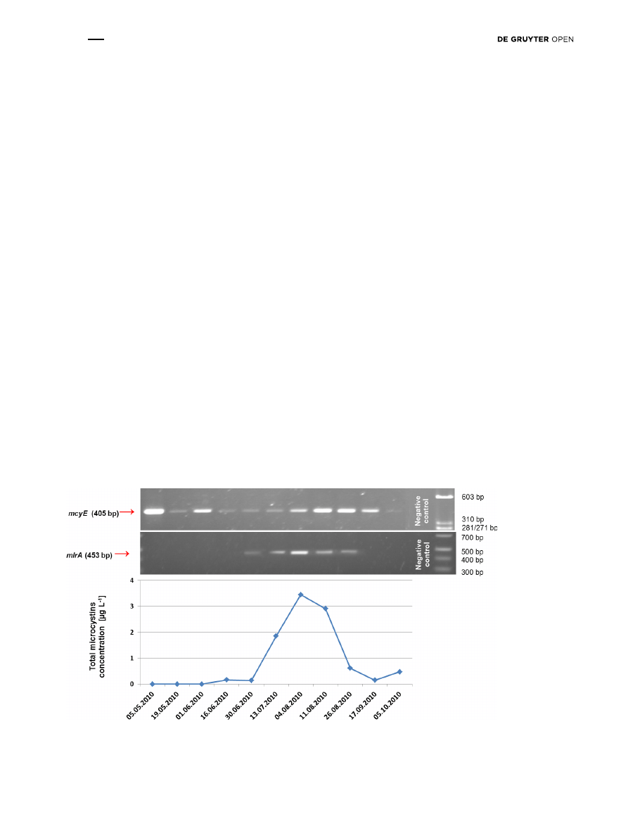
To assess the co-occurrence of bacteria with potential
for microcystins degradation and microcystin-producing
cyanobacteria, the identification of the mlrA and the mcyE
genes respectively was performed in summer season
of 2010. Bacteria with the potential to degradation of
microcystin molecule were identified directly in the water
collected from the lowland Sulejów Reservoir (Fig. 2). The
molecular analysis of mlrA in the water samples from the
reservoir confirmed the presence of bacteria from late June
to the end of August 2010 (Fig. 2). The mcyE gene, which
indicates the presence of microcystin-producers, was
amplified in all 11 samples in the summer season from May
until October 2010 (Fig. 2). In turn, the microcystins were
present from June until the end of the monitoring period on
October 2010, with maximum concentration of 3.45 µg L
-1
on August 4 (Fig. 2). It was observed that bacteria with
the potential to degrade microcystins were found in water
samples in which cyanobacteria-derived hepatotoxins were
also detected (Fig. 2), and physico-chemical conditions
favored the development of phytoplankton [30]. According
to Orr and Jones [31], products of microcystin molecule
degradation can be utilized as the source of carbon and
nitrogen. In consequence, this process provides energy
necessary for growth of planktonic bacteria associated
with cyanobacterial blooms.
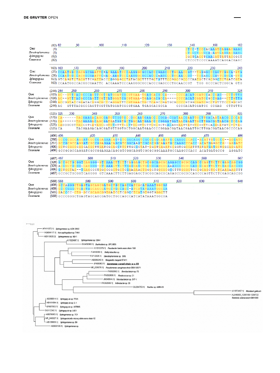
To determine the bacteria with the potential to
degrade microcystin molecule, an analysis of the mlrA
gene sequence was performed. The nucleotide sequence
of the PCR products was blasted with a DNA database. The
results showed 95% homology with the mlrA gene of the
Sphingopyxis strain C-1 (GeneBank AB468058.1) and the
Stenotrophomonas sp. strain EMS (GeneBank GU224277.1)
(Fig. 3). These bacteria genera had been previously
isolated from Chines lakes [32-33] (Fig. 4). Collectively, our
genetic study of water samples obtained directly from the
Sulejów Reservoir showed that bacteria comparable to the
Sphingopyxis sp. C-1 strain and/or Stenotrophomonas sp.
EMS may be responsible for microcystins degradation.
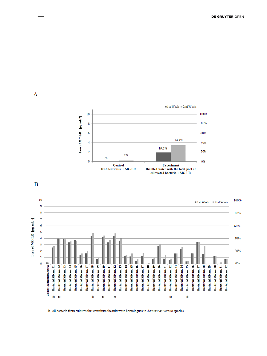
To assess the actual ability to degrade microcystins,
we analyzed the cultures of pelagic bacteria collected
from the Sulejów Reservoir in July 2010. First, the in vitro
experiment no. 1 was performed with the total pool of
bacteria and standard MC-LR. After one week, the MC-LR
level was reduced by 19% compared to the control sample.
After two weeks, the level of MC-LR degradation by the
total pool of culturable bacteria reached 34% (Fig. 5A).
Next, in experiment no. 2, the active degradation of MC-LR
Figure 2: The results of: 1) determination of microcystins concentration, 2) molecular monitoring of microcystin-producing cyanobacte-
ria – presence of mcyE gene, and 3) molecular monitoring of microcystin-degrading bacteria – presence of mlrA gene, in Tresta Station, in
Sulejów Reservoir.
Brought to you by | Uniwersytet Lodzki
Authenticated
Download Date | 8/31/15 9:07 AM
Degradation of microcystin by Aeromonas
125
Figure 3: Homology analysis of mlrA gene fragment (453 bp) amplified in sample from Tresta Station, Sulejów Reservoir. (Query – obtained
sequence; Sphingopyxis – strain C1 AB468058.1; Stenotrophomonas - strain EMS GU224277.1).
Figure 4: The approximate phylogenetic distance between the 16S rDNA sequence of Aeromonas sp. and other microcystin-degrading
bacteria.
Brought to you by | Uniwersytet Lodzki
Authenticated
Download Date | 8/31/15 9:07 AM
126
J. Mankiewicz-Boczek et al.
was determined in 32 bacterial mixes (6 colonies per mix).
The level of MC-LR degradation was dependent on the
bacterial mix used. After one week, the bacterial mixes
1-5, 8, 10-13, 20 and 24 reduced MC-LR levels by more than
20% (Fig. 5B). After two weeks, degradation was also
observed in mixes 27 and 28. The highest degradation
after two weeks was identified in mixes 8 and 12, in which
the loss of MC-LR reached 48% (Fig. 5B). In the control mix
without bacteria, there was a 2% degradation of MC-LR
after both the first and second week of the experiment
(Fig. 5B).
Taking into account the maximal 48% loss of MC-LR
(from 10 µg mL
-1
to 5.2 µg mL
-1
) in relation to the duration
of the experiment (14 days) it could be established
that the degradation rate reached up 0.4 µg mL
-1
per
day. Previous studies on the identification of bacteria
capable of degrading of mentioned cyanobacterial
hepatotoxin and assessment of its activity demonstrated
Figure 5: The results of the analysis of MC-LR degradation in in vitro experiments with: A) total pool of culturable bacteria – experiment no. 1,
and B) mixes of selected culturable environmental bacteria – experiment no. 2.
Brought to you by | Uniwersytet Lodzki
Authenticated
Download Date | 8/31/15 9:07 AM

Degradation of microcystin by Aeromonas
127
even 100 % degradation of MC-LR for bacteria mainly
of the family Sphingomonadaceae [6, 33-39]. The rate of
MC-LR degradation was determined from 0.0015 µg mL
-
1
up to 101.5 µg mL
- 1
per day (depending on the initial
amount of bacteria and the concentration of MC-LR) [6;
36-38]. The reason that the degradation of MC-LR did not
exceed 50 % could have been influenced by high initial
concentration of MC-LR (10 µg mL
-1
). The application of
high initial concentration was dictated by the sensitivity
of the available HPLC–DAD method to ensure accurate
and reliable measurement.
To determine the phylogenetic affiliation of culturable
bacteria from the mixes, the 16S rRNA gene fragment
was amplified and sequenced. The results indicated that
regardless of the ability to cause MC-LR degradation, the
40 bacterial isolates belonged to the Aeromonas genus
(100% homology) (Figs 4 and 5B). This phenomenon
could partly result from the activity of various pathogenic
factors associated with Aeromonas, such as exotoxins,
extracellular lytic enzymes, iron-binding and secretion
systems, or an ability to survive low temperatures
[40-42]. These factors might facilitate the total domination
of Aeromonas in laboratory cultures. An interesting
conclusion was formulated in the study of Gaoshan et
al. [43], which demonstrated that the crude microcystin
may be an important factor stimulating the transition of
Aeromonas sobria from the VBNC state (viable but non-
culturable) to the active growth stage. Therefore, it was
presumed that in the present experiments (no. 1 and 2),
entering the VBNC state could contribute to the great
variability in MC-LR degradation.
The analysis of the sequences showed that isolates
represented the strain of Aeromonas veronii w-s-03
(GenBank record number JF490063.1) (Fig. 4). According to
our knowledge, no one has yet demonstrated directly that
bacteria of the genus Aeromonas (family Aeromonadaceae)
are capable of MC-LR degradation.
Aeromonas belongs to the class of
Gammaproteobacteria, which contains three types
of bacteria capable of degrading microcystins:
Pseudomonas, Stenotrophomonas and Morganella (see
Introduction). Previous studies indicated that the bacteria
originating from the Aeromonas genus might coexist
with cyanobacterial blooms [44-45]. Østensvik et al. [46]
and Bomo et al. [47] reported antibacterial activity of
Microcystis aeruginosa extracts on Aeromonas hydrophila.
On the other hand, Liu et al. [48] observed a strong
algicidal effect of bacterium Aeromonas sp. strain FM
against cyanobacterium M. aeruginosa.
When it comes to research directly associated with the
relationship between cyanobacteria-derived hepatotoxins
and Aeromonas, Lee et al. [49] identified Aeromonas
among the pool of different bacteria potentially capable
of degrading microcystins. These bacteria were absorbed
on a GAC (granular active carbon) filter from a water
treatment facility, creating a biofilm. When the biofilm
was used as an inoculum in the experiment, bacteria
were found capable of microcystin molecule degradation.
However, Aeromonas itself was not isolated nor tested for
the potential to remove microcystins from water.
To verify whether Aeromonas, isolated in the present
study, contained the mlrA gene, a genetic analysis was
performed. The mlrA gene amplification product was not
detected in either of the cultivated bacteria belonging to
the Aeromonas genus. It is likely that these bacteria might
be able to degrade MC-LR differently than described by
Bourne et al. [7, 16]. In general, the fate of the degradation
products and enzymatic character of the decomposition
process in different types of microcystin-degrading
species are still relatively unknown [50].
The mlr genes were also found to be absent in other
microcystin-degrading bacteria, including Burkholderia
sp. [51], Paucibacter toxinivorans [13], Methylobacillus
sp. [52], Pseudomonas aeruginosa [53], Morganella
morganii [54], Arthrobacter sp. [14,15], Brevibacterium sp.
[14,15], Rhodococcus sp. [14,15] and Stenotrophomonas
acidiminiphila strain MC-LTH2 [55].
4 Conclusion
Based on the presence of the mlrA gene, bacteria with the
potential for microcystins degradation were identified in the
water samples from the Sulejów Reservoir in Central Poland.
The genetic analysis allowed classification of the bacteria with
a high homology to the Sphingopyxis and Stenotrophomonas
genera (95%). In the study cultures, the above-mentioned
bacteria were not detected. The in vitro MC-LR degradation
tests on culturable bacteria demonstrated, for the first time,
that bacteria homologous to Aeromonas genus (100%)
could degrade cyanobacterial hepatotoxins – microcystins,
although the mlrA gene was not amplified. In further studies,
we plan to determine the degradation activity of bacteria
by modifying the cultivation conditions and controlling
bacterial growth in relation to the removal of microcystins at
different phases of the experiment.
The data obtained in the present study suggest that
microcystins can be degraded and used by Aeromonas
genus as a necessary energy source. Thus, the Aeromonas
genus not only accompanies cyanobacterial blooms but
also interacts with them. The nature of this complex
interaction requires further clarification.
Brought to you by | Uniwersytet Lodzki
Authenticated
Download Date | 8/31/15 9:07 AM

128
J. Mankiewicz-Boczek et al.
Acknowledgements: The authors would like to
acknowledge the European Cooperation in Science
and Technology, COST Action ES 1105 “CYANOCOST -
Cyanobacterial blooms and toxins in water resources:
Occurrence, impacts and management” for adding value
to this study through networking and knowledge sharing
with European experts and researchers in the field. The
Sulejów Reservoir is a part of the Polish National Long-
Term Ecosystem Research Network and the European
LTER site.
Conflict of interest: Authors declare that this research
was funded by the National Science Centre, project no.
NN305 096439 - “Explanation of cause-effect relationships
between the occurrence of toxigenic cyanobacterial
blooms and abiotic and biotic factors with particular
focus on the role of viruses and bacteria”.
References
[1] Carvalho L., Miller C.A., Scott E.M., Codd G.A., Davies P.S., Tyler
A.N., Cyanobacterial blooms: Statistical models describing
risk factors for national-scale lake assessment and lake
management, Sci. Total. Environ., 2011, 409, 5353–5358
[2] Bednarek A., Stolarska M., Ubraniak M., Zalewski M.,
Application of permeable reactive barrier for reduction of
nitrogen load in the agricultural areas - preliminary results,
Ecohydrology and Hydrobiology, 2010, 10, 355–362
[3] Kelly J.M., Kovar J.L., Modelling phosphorus capture by plants
growing in a multispecies riparian buffer, Appl. Environ. Soil
Sci., 2012, 2012, 1–7
[4] Kiedrzyńska E., Kiedrzyński M., Zalewski M., Flood sediment
deposition and phosphorus retention in a lowland river
floodplain: impact on water quality of a reservoir Sulejów,
Poland, Ecohydrology and Hydrobiology, 2008, 8, 281–289
[5] Schmidt C.A., Clark. M.W., Evaluation of a denitrification wall to
reduce surface water nitrogen loads, J. Environ. Quality., 2012,
41, 724–731
[6] Ho L., Hoefel D., Saint C.P., Newcombe G., Isolation and identi-
fication of a novel microcystin-degrading bacterium from a
biological sand filter, Water Res., 2007, 41, 4685–4695
[7] Bourne D.G., Blakeley R.L., Riddles P., Jones G.J.,
Biodegradation of the cyanobacterial toxin microcystin-LR in
natural water and biologically active slow sand filters, Water
Res., 2006, 40, 1294–302
[8] Ji R.P., Lu X.W., Li X.N., Pu Y.P., Biological degradation of algae
and microcystins by microbial enrichment on artificial media,
Ecol. Eng., 2009, 35, 1584–1588
[9] Gągała I., Mankiewicz-Boczek J., Natural degradation of
microcystins (cyanobacterial hepatotoxins) in fresh water –
the future of modern treatment systems and water quality
improvement. Pol. J. Environ. Stud., 2012, 21, 1125–1139
[10] Mou X., Lu X., Jacob J., Sun S., Heath R., Metagenomic identi-
fication of bacterioplankton taxa and pathways involved in
microcystin degradation in Lake Erie, PLoS One., 2013, 8,
e61890
[11] Jing W., Sui G., Liu S., Characteristics of a microcystin-LR
biodegrading bacterial isolate: Ochrobactrum sp. FDT5. Bull.
Environ. Contam. Toxicol., 2014, 92(1), 119-122
[12] Ma G., Pei H., Hu W., Xu X., Ma C., Li X., The removal of
cyanobacteria and their metabolites through anoxic
biodegradation in drinking water sludge, Bioresour. Technol.
2014, 165C, 191-198
[13] Rapala J., Berg K.A., Lyra C., Niemi R.M., Manz W., Suomalainen
S., et al., Paucibacter toxinivorans gen. nov. sp. nov. a
bacterium that degrades cyclic cyanobacterial hepatotoxins
microcystins and nodularin, Int. J. Syst. Evol. Microbiol., 2005,
55, 1563–1568
[14] Lawton L.A., Welgamage A., Manage P.M., Edwards C., Novel
bacterial strains for the removal of microcystins from drinking
water, Water Sci. Technol., 2011, 63, 1137–1142
[15] Manage P.M., Edwards C., Singh B.K., Lawton L.A., Isolation
and identification of novel microcystin-degrading bacteria,
Appl. Environ. Microbiol., 2009, 75, 6924–6928
[16] Bourne D.G., Riddles P., Jones G.J., Smith W., Blakeley R.L.
Characterisation of a gene cluster involved in bacterial
degradation of the cyanobacterial toxin Microcystin-LR,
Cultures, 2001, 16, 523–534
[17] Izydorczyk K., Jurczak T., Wojtal-Frankiewicz A., Skowron A.,
Mankiewicz-Boczek J., Tarczyńska M., Influence of abiotic and
biotic factors on microcystin content in Microcystis aeruginosa
cells in a eutrophic temperate reservoir, J. Plankton Res., 2008,
30, 393–400
[18] Jurczak T., Tarczyńska M., Izydorczyk K., Mankiewicz J.,
Zalewski M., Meriluoto J., Elimination of microcystins by water
treatment processes — examples from Sulejów Reservoir,
Poland, Water Res., 2005, 39, 2394–2406
[19] Jurczak T., Zastosowanie monitoringu toksyn sinicowych w
celu optymalizacji technologii uzdatniania wody oraz strategii
rekultywacji zbiorników zaporowych, PhD dissertation,
University of Lodz, Poland, 2006, (in Polish)
[20] Mankiewicz-Boczek J., Izydorczyk K., Romanowska-Duda
Z., Jurczak T., Stefaniak K., Kokociński M., Detection and
monitoring toxigenicity of cyanobacteria by application of
molecular methods, Environ. Toxicol., 2006, 21, 380–387
[21] Mankiewicz-Boczek J., Urbaniak M., Romanowska-Duda Z.,
Izydorczyk K., Toxic Cyanobacteria strains in lowland dam
reservoir (Sulejów Res. Central Poland): Amplification of mcy
genes for detection and identification, Pol. J. Ecol., 2006, 54,
171–180
[22] Tarczyńska M., Romanowska-Duda Z., Jurczak T., Zalewski M.,
Toxic cyanobacterial blooms in a drinking water reservoir -
causes consequences and management strategy, Water Sci.
Technol.: Water Supply., 2001, 1, 237–246
[23] Zalewski M., Ecohydrology - The scientific background to use
ecosystem properties as management tools toward sustai-
nability of water resources, Guest Editorial Ecol. Eng., 2000, 16,
41647
[24] Giovannoni S.J., DeLong E.F., Schmidt T.M., Pace N.R.,
Tangential flow filtration and preliminary phylogenetic analysis
of marine picoplankton, Appl. Environ. Microbiol., 1990, 56,
2572-2575
[25] Rantala A., Rajaniemi-Wacklin P., Lyra C., Lepistö L., Rintala
J., Mankiewicz-Boczek J., et al., Detection of microcystin-
producing cyanobacteria in Finnish lakes with genus-specific
microcystin synthetase gene E (mcyE) PCR and associations
Brought to you by | Uniwersytet Lodzki
Authenticated
Download Date | 8/31/15 9:07 AM

Degradation of microcystin by Aeromonas
129
with environmental factors, Appl. Environ. Microbiol., 2006, 72,
6101–6110
[26] Saito T., Okano K., Park H., Itayama T., Inamori Y., Neilan
B.A., et al., Detection and sequencing of the microcystin
LR-degrading gene, mlrA, from new bacteria isolated from
Japanese lakes, FEMS Microbiol. Lett., 2003, 229(2), 271-276
[27] Zhang Z., Schwartz S., Wagner L., Miller W., A greedy algorithm
for aligning DNA sequences, J. Comput. Biol., 2000, 7, 203-221
[28] Orphan V.J., Hinrichs K., Iii W.U., Paull C.K., Taylor L.T., Sylva
S.P., et al., Comparative analysis of methane-oxidizing archaea
and sulfate-reducing bacteria in anoxic marine sediments, J.
Appl. Microbiol., 2001, 67, 1922–1934
[29] Huson D.H., Scornavacca. C., Dendroscope 3: An Interactive
Tool for Rooted Phylogenetic Trees and Networks, Syst. Biol.,
2012, 61, 1061-1067
[30] Gągała I., Izydorczyk K., Jurczak T., Pawełczyk J., Dziadek J.,
Wojtal-Frankiewicz A., et al., Role of environmental factors and
toxic genotypes in the regulation of microcystins-producing
cyanobacterial blooms, Microbial Ecol., 2014, 67(2), 465-479
[31] Orr P.T., Jones. G.J., Relationship between microcystin
production and cell division rates in nitrogen-limited
Microcystis aeruginosa cultures, Limnol. Oceanogr., 1998, 43,
1604
[32] Okano K., Shimizu K., Kawauchi Y., Maseda H., Utsumi M.,
Zhang Z., et al., Characteristics of a microcystin-degrading
bacterium under alkaline environmental conditions, J. Toxicol.,
2009, 2009, 41648
[33] Chen J., Hu L., Bin Zhou W., Yan S.H., Yang J.D., Xue Y.F., et al.,
Degradation of microcystin-LR and RR by a Stenotrophomonas
sp. strain EMS isolated from Lake Taihu, China, Int. J. Mol. Sci.,
2010, 11, 896–911
[34] Park H.D., Sasaki Y., Maruyama T., Yanagisawa E., Hiraishi
A., Kato K., Degradation of the cyanobacterial hepatotoxin
microcystin by a new bacterium isolated from a hypertrophic
lake, Environ. Toxicol., 2001, 16, 337–343
[35] Ishii H., Nishijima M., Abe T., Characterization of degradation
process of cyanobacterial hepatotoxins by a gram-negative
aerobic bacterium, Water Res., 2004, 38, 2667–2676
[36] Wang J., Wu P., Chen J., Yan H., Biodegradation of microcystin-
RR by a new isolated Sphingopyxis sp. USTB-05, Chin. J. Chem.
Eng., 2010, 18, 1-5
[37] Zhang, M., Pan G., Yan H., Microbial biodegradation of
microcystin-RR by bacterium Sphingopyxis sp. USTB-05, 2010,
J. Environ. Sci., 22, 168–175
[38] Yan H., Wang J., Chen J., Wei W., Wang H., Wang H., Charac-
terization of the first step involved in enzymatic pathway for
microcystin-RR biodegraded by Sphingopyxis sp. USTB-05,
Chemosphere, 2012, 87, 12-18
[39] Ho L., Tang T., Monis P.T., Hoefel D., Biodegradation of multiple
cyanobacterial metabolites in drinking water supplies,
Chemosphere, 2012, 87, 1149-1154
[40] Mateos D., Anguita J., Naharro G., Paniagua C., Influence of
growth temperature on the production of extracellular virulence
factors and pathogenicity of environmental and human strains
of Aeromonas hydrophila, J. Appl. Bacteriol., 1993, 74, 111–118
[41] Mano S., Growth/survival of natural flora and Aeromonas
hydrophila on refrigerated uncooked pork and turkey packaged
in modified atmospheres, Food Microbiol., 2000, 17, 657–669
[42] Tomás J.M., The main Aeromonas pathogenic factors, ISRN
Microbiol., 2012, 2012, 1–22
[43] Gaoshan P., Zhangli H., Anping L., Shuangfei L., Effect of
crude microcystin on the viable but non-culturable state of
Aeromonas sobria in aquatic environment, J. Lake Sci., 2008,
20, 105-109 (in chinese)
[44] Berg K.A., Lyra C., Niemi R.M., Heens B., Hoppu K., Erkomaa
K., et al., Virulence genes of Aeromonas isolates bacterial
endotoxins and cyanobacterial toxins from recreational water
samples associated with human health symptoms, J. Water
Health., 2011, 9, 670-679
[45] Berg K.A., Lyra C., Sivonen K., Paulin L., Suomalainen S., Tuomi
P., et al., High diversity of cultivable heterotrophic bacteria in
association with cyanobacterial water blooms, ISME J., 2009,
3, 314–325
[46] Østensvik O., Skulberg O.M., Underdal B., Hormazabal V.,
Antibacterial properties of extracts from selected planktonic
freshwater cyanobacteria - a comparative study of bacterial
bioassays, J. Appl. Microbiol., 1998, 84, 1117–1124
[47] Bomo A-M., Tryland I., Haande S., Hagman C.H.C., Utkilen
H., The impact of cyanobacteria on growth and death of
opportunistic pathogenic bacteria, Water Sci. Technol., 2011,
64, 384–390
[48] Liu Y-M., Chen M-J., Wang M-H., Jia R-B., Li L. Inhibition of
Microcystis aeruginosa by the extracellular substances from an
Aeromonas sp., J. Microbiol. Biotechnol., 2013, 23, 1304-1307
[49] Lee Y-J., Jung J-M., Jang M-H., Ha K., Joo G-J. Degradation of
microcystins by adsorbed bacteria on a granular active carbon
(GAC) filter during the water treatment process, J. Environ
Biology/Academy of Environmental Biology, India, 2006, 27,
317–322
[50] Dziga D., Wasylewski M., Wladyka B., Nybom S., Meriluoto J.
Microbial degradation of microcystins, Chem. Res. Toxicol.,
2013, 26, 841-852
[51] Lemes G.A.F., Kersanach R., Pinto L.D.S., Dellagostin O.A.,
Yunes J.S., Matthiensen A., Biodegradation of microcystins by
aquatic Burkholderia sp. from a South Brazilian coastal lagoon,
Ecotoxicol. Environ Saf., 2008, 69, 358–365
[52] Hu L., Bin Yang J.D., Zhou W., Yin Y.F., Chen J., Shi Z.Q., Isolation
of a Methylobacillus sp. that degrades microcystin toxins
associated with cyanobacteria, New Biotechnol., 2009, 26,
205–211
[53] Takenaka S., Watanabe. M.F., Microcystin-LR degradation by
Pseudomonas aeruginosa alkaline protease, Chemosphere,
1997, 34, 749–757
[54] Eleuterio L., Batista. J.R., Biodegradation studies and
sequencing of microcystin-LR degrading bacteria isolated from
a drinking water biofilter and a fresh water lake, Toxicon, 2010,
55, 1434–1442
[55] Yang F., Zhou Y., Yin L., Zhu G., Liang G., Pu Y., Microcystin-
degrading activity of an indigenous bacterial strain
Stenotrophomonas acidaminiphila MC-LTH2 isolated from Lake
Taihu. PLoS ONE, 2014, 9(1): e86216
Brought to you by | Uniwersytet Lodzki
Authenticated
Download Date | 8/31/15 9:07 AM
Document Outline
Wyszukiwarka
Podobne podstrony:
kłopotliwe, o grzechu inaczej, Czy muszę się spowiadać z czegoś, czego nie uważam za grzech, a inni
16 Sicking D L i inni Factors leading to cable median barrier failures
Bacterial Resistance to Microbicides in the Healthcare Environment
Zinda; Introduction to the philosophy of science
Jaffe Innovative approaches to the design of symphony halls
AIDS TO THE EXAMINATION OF THE PNS ED 4TH
How to use software of Ethernet packet?pture to?pture WAN port packets
The term therapeutic relates to the treatment of disease or physical disorder
DANCE ME TO THE END OF LOVE
The Three Great Compromises that lead to the establishment of
I need to buy a tin of sardines
Heathen Gods and Rites A brief introduction to our ways of worship and main deities
President Eisenhower to the Director of Central Intelligence, 4 November 1953 (eisenhower dci 4nov53
Cognitive Psychology from Hergenhahn Introduction to the History of Psychology, 2000
Introduction To The Metaphysic Of Morals
Guide to the installation of PV systems 2nd Edition
DANCE ME TO THE END OF LOVE
DANCE WITH ME TO THE END OF LOVE
więcej podobnych podstron