Brain Facts
A P R I M E R O N T H E B R A I N A N D N E R V O U S S Y S T E M
T H E S O C I E T Y F O R N E U R O S C I E N C E

Brain Facts
A P R I M E R O N T H E B R A I N A N D N E R V O U S S Y S T E M
T H E S O C I E T Y F O R N E U R O S C I E N C E

THE SOCIETY FOR NEUROSCIENCE
The Society for Neuroscience is the world’s largest organization of sci-
entists and physicians dedicated to understanding the brain, spinal cord
and peripheral nervous system.
Neuroscientists investigate the molecular and cellular levels of the
nervous system; the neuronal systems responsible for sensory and
motor function; and the basis of higher order processes, such as cog-
nition and emotion. This research provides the basis for understand-
ing the medical fields that are concerned with treating nervous system
disorders. These medical specialties include neurology, neurosurgery,
psychiatry and ophthalmology.
Founded in 1970, the Society has grown from 500 charter members
to more than 29,000 members. Regular members are residents of Canada,
Mexico and the United States—where more than 100 chapters organize
local activities. The Society’s membership also includes many scientists
from throughout the world, particularly Europe and Asia.
The purposes of the Society are to:
∫
Advance the understanding of the nervous system by bringing together
scientists from various backgrounds and by encouraging research in all
aspects of neuroscience.
∫
Promote education in the neurosciences.
∫
Inform the public about the results and implications of new research.
The exchange of scientific information occurs at an annual fall
meeting that presents more than 14,000 reports of new scientific
findings and includes more than 25,000 participants. This meeting, the
largest of its kind in the world, is the arena for the presentation of new
results in neuroscience.
The Society’s bimonthly journal, The Journal of Neuroscience, con-
tains articles spanning the entire range of neuroscience research and
has subscribers worldwide. A series of courses, workshops and sym-
posia held at the annual meeting promote the education of Society
members. The Neuroscience Newsletter informs members about Society
activities.
A major mission of the Society is to inform the public about the
progress and benefits of neuroscience research. The Society provides
information about neuroscience to school teachers and encourages its
members to speak to young people about the human brain and nervous
system.

Brain Facts
INTRODUCTION . . . . . . . . . . . . . . . . . . . . . . . . . . . . . . . . . . . . . . . . . . . . . . . . 2
THE NEURON . . . . . . . . . . . . . . . . . . . . . . . . . . . . . . . . . . . . . . . . . . . . . . . . . 4
Neurotransmitters
∫
Second Messengers
BRAIN DEVELOPMENT . . . . . . . . . . . . . . . . . . . . . . . . . . . . . . . . . . . . . . . . . . . 8
Birth of Neurons and Brain Wiring
∫
Paring Back
∫
Critical Periods
SENSATION AND PERCEPTION . . . . . . . . . . . . . . . . . . . . . . . . . . . . . . . . . . . . 12
Vision
∫
Hearing
∫
Taste and Smell
∫
Touch and Pain
LEARNING AND MEMORY . . . . . . . . . . . . . . . . . . . . . . . . . . . . . . . . . . . . . . . . 18
MOVEMENT . . . . . . . . . . . . . . . . . . . . . . . . . . . . . . . . . . . . . . . . . . . . . . . . . . 20
SLEEP . . . . . . . . . . . . . . . . . . . . . . . . . . . . . . . . . . . . . . . . . . . . . . . . . . . . . . 22
The Stu∑ of Sleep
∫
Sleep Disorders
∫
How is Sleep Regulated?
STRESS . . . . . . . . . . . . . . . . . . . . . . . . . . . . . . . . . . . . . . . . . . . . . . . . . . . . . 25
The Immediate Response
∫
Chronic Stress
AGING . . . . . . . . . . . . . . . . . . . . . . . . . . . . . . . . . . . . . . . . . . . . . . . . . . . . . . 28
Aging Neurons
∫
Intellectual Capacity
ADVANCES . . . . . . . . . . . . . . . . . . . . . . . . . . . . . . . . . . . . . . . . . . . . . . . . . . . 30
Parkinson’s Disease
∫
Pain
∫
Epilepsy
∫
Major Depression
Manic-Depressive Illness
CHALLENGES . . . . . . . . . . . . . . . . . . . . . . . . . . . . . . . . . . . . . . . . . . . . . . . . . 33
Addiction
∫
Alzheimer’s Disease
∫
Learning Disorders
Stroke
∫
Neurological Trauma
∫
Anxiety Disorders
Schizophrenia
∫
Neurological AIDS
∫
Multiple Sclerosis
Down Syndrome
∫
Huntington’s Disease
∫
Tourette Syndrome
Brain Tumors
∫
Amyotrophic Lateral Sclerosis
NEW DIAGNOSTIC METHODS . . . . . . . . . . . . . . . . . . . . . . . . . . . . . . . . . . . . . 43
Imaging Techniques
∫
Gene Diagnosis
POTENTIAL THERAPIES . . . . . . . . . . . . . . . . . . . . . . . . . . . . . . . . . . . . . . . . . 46
New Drugs
∫
Trophic Factors
∫
Cell and Gene Therapy
GLOSSARY . . . . . . . . . . . . . . . . . . . . . . . . . . . . . . . . . . . . . . . . . . . . . . . . . . 48
INDEX . . . . . . . . . . . . . . . . . . . . . . . . . . . . . . . . . . . . . . . . . . . . . . . . . . . . . . 53

2
t sets humans apart from all other species by allowing us
to achieve the wonders of walking on the moon and com-
posing masterpieces of literature, art and music. Through-
out recorded time, the human brain—a spongy, three-
pound mass of fatty tissue—has been compared to a
telephone switchboard and a supercomputer.
But the brain is much more complicated than any of these
devices, a fact scientists confirm almost daily with each new
discovery. The extent of the brain’s capabilities is unknown, but
it is the most complex living structure known in the universe.
This single organ controls all body activities, ranging from
heart rate and sexual function to emotion, learning and mem-
ory. The brain is even thought to influence the response to dis-
ease of the immune system and to determine, in part, how well
people respond to medical treatments. Ultimately, it shapes our
thoughts, hopes, dreams and imagination. In short, the brain is
what makes us human.
Neuroscientists have the daunting task of deciphering the
mystery of this most complex of all machines: how as many as
a trillion nerve cells are produced, grow and organize them-
selves into e∑ective, functionally active systems that ordinarily
remain in working order throughout a person’s lifetime.
The motivation of researchers is twofold: to understand
human behavior better—from how we learn to why people
have trouble getting along together—and to discover ways to
prevent or cure many devastating brain disorders.
The more than 1,000 disorders of the brain and nervous
system result in more hospitalizations than any other disease
group, including heart disease and cancer. Neurological illnesses
a∑ect more than 50 million Americans annually at costs exceed-
ing $400 billion. In addition, mental disorders, excluding drug
and alcohol problems, strike 44 million adults a year at a cost
of some $148 billion.
However, during the congressionally designated Decade of
the Brain, which ended in 2000, neuroscience made significant
discoveries in these areas:
∫
Genetics. Key disease genes were identified that underlie sev-
eral neurodegenerative disorders—including Alzheimer’s dis-
ease, Huntington’s disease, Parkinson’s disease and amyotrophic
lateral sclerosis. This has provided new insights into underlying
disease mechanisms and is beginning to suggest new treatments.
With the mapping of the human genome, neuroscientists
will be able to make more rapid progress in identifying genes that
either contribute to human neurological disease or that directly
cause disease. Mapping animal genomes will aid the search for
genes that regulate and control many complex behaviors.
∫
Brain Plasticity. Scientists began to uncover the molecular
bases of neural plasticity, revealing how learning and memory
occur and how declines might be reversed. It also is leading to
new approaches to the treatment of chronic pain.
∫
New Drugs. Researchers gained new insights into the mech-
anisms of molecular neuropharmacology, which provides a new
understanding of the mechanisms of addiction. These advances
also have led to new treatments for depression and obsessive-
compulsive disorder.
∫
Imaging. Revolutionary imaging techniques, including mag-
netic resonance imaging and positron emission tomography,
now reveal brain systems underlying attention, memory and
emotions and indicate dynamic changes that occur in schizo-
phrenia.
∫
Cell Death. The discovery of how and why neurons die, as
well as the discovery of stem cells, which divide and form new
neurons, has many clinical applications. This has dramatically
improved the outlook for reversing the e∑ects of injury both in
the brain and spinal cord. The first e∑ective treatments for
stroke and spinal cord injury based on these advances have been
brought to clinical practice.
∫
Brain Development. New principles and molecules respon-
sible for guiding nervous system development now give scien-
tists a better understanding of certain disorders of childhood.
Together with the discovery of stem cells, these advances are
pointing to novel strategies for helping the brain or spinal cord
regain functions lost to diseases.
Federal neuroscience research funding of more than $4 bil-
lion annually and private support should vastly expand our
knowledge of the brain in the years ahead.
This book only provides a glimpse of what is known about
the nervous system, the disorders of the brain and some of the
exciting avenues of research that promise new therapies for
many neurological diseases.
Introduction
I
3
THE TOLL OF SELECTED BRAIN AND NERVOUS SYSTEM DISORDERS*
Condition
Total Cases
Costs Per Year
Hearing Loss
28 million
$ 56 billion
All Depressive Disorders
18.8 million
$ 44 billion
Alzheimer’s Disease
4 million
$ 100 billion
Stroke
4 million
$ 30 billion
Schizophrenia
3 million
$ 32.5 billion
Parkinson’s Disease
1.5 million
$ 15 billion
Traumatic Head Injury
1 million
$ 48.3 billion
Multiple Sclerosis
350,000
$ 7 billion
Spinal Cord Injury
250,000
$ 10 billion
* Estimates provided by the National Institutes of Health and voluntary organizations.
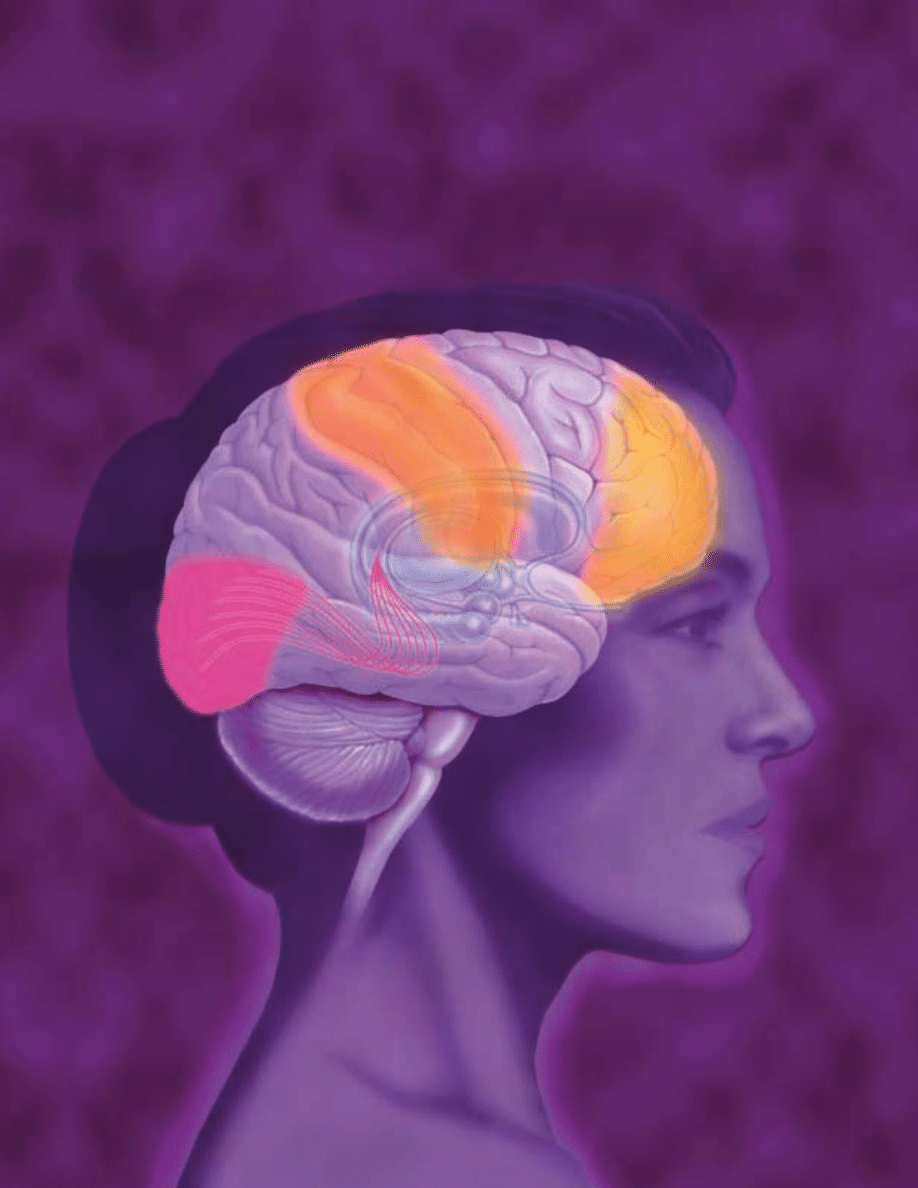
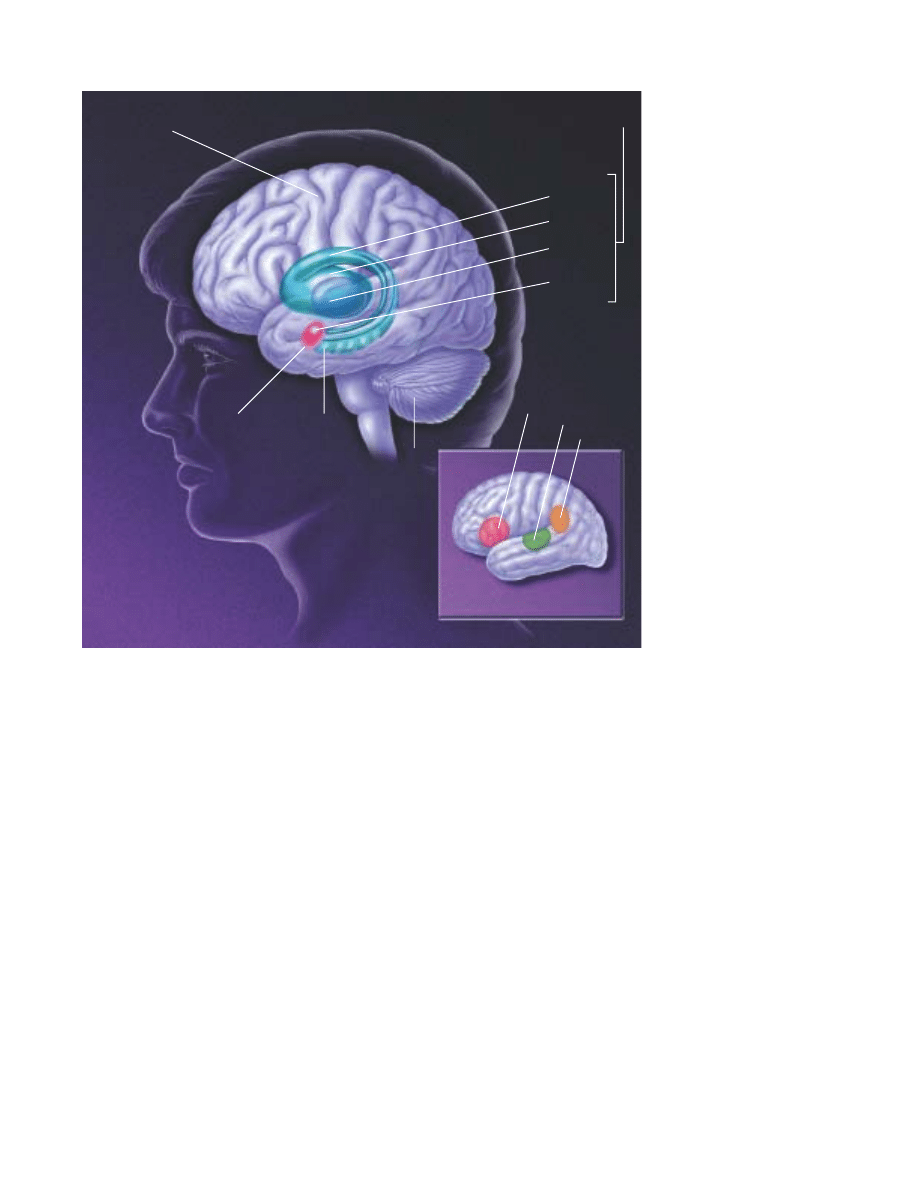
THE BRAIN. Cerebral cortex
(above). This part of the brain is
divided into four sections: the
occipital lobe, the temporal
lobe, the parietal lobe and the
frontal lobe. Functions, such as
vision, hearing and speech, are
distributed in selected regions.
Some regions are associated
with more than one function.
Major internal structures
(below). The (1) forebrain is
credited with the highest intel-
lectual functions—thinking,
planning and problem-solving.
The hippocampus is involved in
memory. The thalamus serves as
a relay station for almost all of
the information coming into the
brain. Neurons in the hypothala-
mus serve as relay stations for
internal regulatory systems by
monitoring information coming
in from the autonomic nervous
system and commanding the
body through those nerves and
the pituitary gland. On the
upper surface of the (2) mid-
brain are two pairs of small
hills, colliculi, collections of
cells that relay specific sensory
information from sense organs
to the brain. The (3) hindbrain
consists of the pons and
medulla oblongata, which help
control respiration and heart
rhythms, and the cerebellum,
which helps control movement
as well as cognitive processes
that require precise timing.
Frontal lobe
Motor cortex
Sensory cortex
Parietal lobe
Occipital lobe
Temporal lobe
Cerebrum
1 Forebrain
Amygdala
Hippocampus
Thalamus
Hypothalamus
2 Midbrain
3 Hindbrain
Pons
Cerebellum
Medulla
oblongata
Spinal cord

4
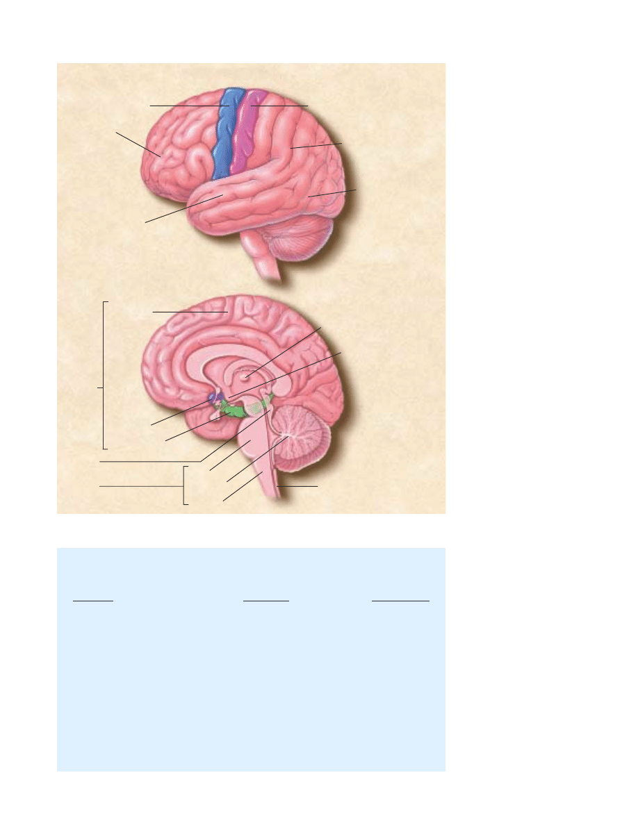
A
specialized cell designed to transmit infor-
mation to other nerve cells, muscle or gland
cells, the neuron is the basic working unit of
the brain. The brain is what it is because of
the structural and functional properties of
neurons. The brain contains between one bil-
lion and one trillion neurons.
The neuron consists of a cell body containing the nucleus
and an electricity-conducting fiber, the axon, which also gives
rise to many smaller axon branches before ending at nerve ter-
minals. Synapses, from the Greek words meaning to “clasp
together,” are the contact points where one neuron communi-
cates with another. Other cell processes, dendrites, Greek for
the branches of a tree, extend from the neuron cell body and
receive messages from other neurons. The dendrites and cell
body are covered with synapses formed by the ends of axons of
other neurons.
Neurons signal by transmitting electrical impulses along
their axons that can range in length from a tiny fraction of an
inch to three or more feet. Many axons are covered with a lay-
ered insulating myelin sheath, made of specialized cells, that
speeds the transmission of electrical signals along the axon.
Nerve impulses involve the opening and closing of ion chan-
nels, water-filled molecular tunnels that pass through the cell
membrane and allow ions—electrically charged atoms—or
small molecules to enter or leave the cell. The flow of these ions
creates an electrical current that produces tiny voltage changes
across the membrane.
The ability of a neuron to fire depends on a small dif-
ference in electrical charge between the inside and outside of
the cell. When a nerve impulse begins, a dramatic reversal
occurs at one point on the cell’s membrane. The change, called
an action potential, then passes along the membrane of the axon
at speeds up to several hundred miles an hour. In this way, a
neuron may be able to fire impulses scores or even hundreds
of times every second.
On reaching the ends of an axon, these voltage changes
trigger the release of neurotransmitters, chemical messengers.
Neurotransmitters are released at nerve ending terminals and
bind to receptors on the surface of the target neuron.
These receptors act as on and o∑ switches for the next cell.
Each receptor has a distinctly shaped part that exactly matches
a particular chemical messenger. A neurotransmitter fits into
this region in much the same way as a key fits into an automo-
bile ignition. And when it does, it alters the neuron’s outer
membrane and triggers a change, such as the contraction of a
muscle or increased activity of an enzyme in the cell.
Knowledge of neurotransmitters in the brain and the action
of drugs on these chemicals—gained largely through the study
of animals—is one of the largest fields in neuroscience. Armed
with this information, scientists hope to understand the circuits
responsible for disorders such as Alzheimer’s disease and Parkin-
son’s disease. Sorting out the various chemical circuits is vital
to understanding how the brain stores memories, why sex is such
a powerful motivation and what is the biological basis of men-
tal illness.
Neurotransmitters
Acetylcholine The first neurotransmitter to be identified 70
years ago, was acetylcholine (ACh). This chemical is released
by neurons connected to voluntary muscles (causing them to
contract) and by neurons that control the heartbeat. ACh also
serves as a transmitter in many regions of the brain.
ACh is formed at the axon terminals. When an action
potential arrives at the terminal, the electrically charged cal-
cium ion rushes in, and ACh is released into the synapse and
attaches to ACh receptors. In voluntary muscles, this opens
sodium channels and causes the muscle to contract. ACh is
then broken down and re-synthesized in the nerve terminal.
Antibodies that block the receptor for ACh cause myasthenia
gravis, a disease characterized by fatigue and muscle weakness.
Much less is known about ACh in the brain. Recent dis-
coveries suggest, however, that it may be critical for normal
attention, memory and sleep. Since ACh-releasing neurons die
in Alzheimer’s patients, finding ways to restore this neuro-
transmitter is one goal of current research.
Amino Acids Certain amino acids, widely distributed
throughout the body and the brain, serve as the building blocks
A
The Neuron
Nucleus
Myelin sheath
Dendrites
Direction
of impulse
Axon
terminals
Cell body
Axon
Neurotransmitters
Receptor molecules
Synapse
Dendrite
of receiving
neuron
Vesicle
Nerve impulse
Axon
5
of proteins. However, it is now apparent that certain amino
acids can also serve as neurotransmitters in the brain.
The neurotransmitters glutamate and aspartate act as exci-
tatory signals. Glycine and gamma-aminobutyric acid (GABA)
inhibit the firing of neurons. The activity of GABA is increased
by benzodiazepine (Valium) and by anticonvulsant drugs. In
Huntington’s disease, a hereditary disorder that begins during
mid-life, the GABA-producing neurons in the brain centers
coordinating movement degenerate, thereby causing incontrol-
lable movements.
Glutamate or aspartate activate N-methyl-D-aspartate
(NMDA) receptors, which have been implicated in activities
ranging from learning and memory to development and speci-
fication of nerve contacts in a developing animal. The stimula-
tion of NMDA receptors may promote beneficial changes in
the brain, whereas overstimulation can cause nerve cell damage
or cell death in trauma and stroke.
Key questions remain about this receptor’s precise structure,
regulation, location and function. For example, developing
drugs to block or stimulate activity at NMDA receptors holds
NEURON. A neuron fires by
transmitting electrical signals
along its axon. When signals
reach the end of the axon, they
trigger the release of neuro-
transmitters that are stored in
pouches called vesicles. Neuro-
transmitters bind to receptor
molecules that are present on
the surfaces of adjacent neu-
rons. The point of virtual contact
is known as the synapse.

6
promise for improving brain function and treating neurologi-
cal disorders. But this work is still in the early stage.
Catecholamines Dopamine and norepinephrine are widely
present in the brain and peripheral nervous system. Dopamine,
which is present in three circuits in the brain, controls move-
ment, causes psychiatric symptoms such as psychosis and reg-
ulates hormonal responses.
The dopamine circuit that regulates movement has been
directly related to disease. The brains of people with Parkinson’s
disease—with symptoms of muscle tremors, rigidity and
di≈culty in moving—have practically no dopamine. Thus,
medical scientists found that the administration of levodopa, a
substance from which dopamine is synthesized, is an e∑ective
treatment for Parkinson’s, allowing patients to walk and per-
form skilled movements successfully.
Another dopamine circuit is thought to be important for
cognition and emotion; abnormalities in this system have been
implicated in schizophrenia. Because drugs that block dopamine
receptors in the brain are helpful in diminishing psychotic
symptoms, learning more about dopamine is important to
understanding mental illness.
In a third circuit, dopamine regulates the endocrine sys-
tem. It directs the hypothalamus to manufacture hormones and
hold them in the pituitary gland for release into the blood-
stream, or to trigger the release of hormones held within cells
in the pituitary.
Nerve fibers containing norepinephrine are present through-
out the brain. Deficiencies in this transmitter occur in patients
with Alzheimer’s disease, Parkinson’s disease and those with
Korsako∑’s syndrome, a cognitive disorder associated with chronic
alcoholism. Thus, researchers believe norepinephrine may play
a role in both learning and memory. Norepinephrine also is
secreted by the sympathetic nervous system in the periphery to
regulate heart rate and blood pressure. Acute stress increases
the release of norepinephrine.
Serotonin This neurotransmitter is present in many tissues,
particularly blood platelets and the lining of the digestive tract
and the brain. Serotonin was first thought to be involved in
high blood pressure because it is present in blood and induces
a very powerful contraction of smooth muscles. In the brain, it
has been implicated in sleep, mood, depression and anxiety.
Because serotonin controls the di∑erent switches a∑ecting var-
ious emotional states, scientists believe these switches can be
manipulated by analogs, chemicals with molecular structures
similar to serotonin. Drugs that alter serotonin’s action, such as
fluoxetine (Prozac), have relieved symptoms of depression and
obsessive-compulsive disorder.
Peptides These chains of amino acids linked together, have
been studied as neurotransmitters only in recent years. Brain
peptides called opioids act like opium to kill pain or cause sleepi-
ness. (Peptides di∑er from proteins, which are much larger and
more complex combinations of amino acids.)
In 1973, scientists discovered receptors for opiates on neu-
rons in several regions in the brain that suggested the brain
must make substances very similar to opium. Shortly thereafter,
scientists made their first discovery of an opiate produced by
the brain that resembles morphine, an opium derivative used
medically to kill pain. They named it enkephalin, literally mean-
ing “in the head.” Subsequently, other opiates known as endor-
phins—from endogenous morphine—were discovered.
The precise role of the opioids in the body is unclear. A
plausible guess is that enkephalins are released by brain neurons
in times of stress to minimize pain and enhance adaptive behav-
ior. The presence of enkephalins may explain, for example, why
injuries received during the stress of combat often are not
noticed until hours later.
Opioids and their receptors are closely associated with path-
ways in the brain that are activated by painful or tissue-damag-
ing stimuli. These signals are transmitted to the central nervous
system—the brain and spinal cord—by special sensory nerves,
small myelinated fibers and tiny unmyelinated or C fibers.
Scientists have discovered that some C fibers contain a pep-
tide called substance P that causes the sensation of burning pain.
The active component of chili peppers, capsaicin, causes the
release of substance P.
Trophic factors Researchers have discovered several small
proteins in the brain that are necessary for the development,
function and survival of specific groups of neurons. These small
proteins are made in brain cells, released locally in the brain,
and bind to receptors expressed by specific neurons. Researchers
also have identified genes that code for receptors and are
involved in the signaling mechanisms of trophic factors. These
findings are expected to result in a greater understanding of
how trophic factors work in the brain. This information also
should prove useful for the design of new therapies for brain
disorders of development and for degenerative diseases, includ-
ing Alzheimer’s disease and Parkinson’s disease.
Hormones After the nervous system, the endocrine system
is the second great communication system of the body. The
pancreas, kidney, heart and adrenal gland are sources of hor-
mones. The endocrine system works in large part through the
pituitary that secretes hormones into the blood. Because endor-
phins are released from the pituitary gland into the blood-
stream, they might also function as endocrine hormones. Hor-
mones activate specific receptors in target organs that release
other hormones into the blood, which then act on other tissues,
the pituitary itself and the brain. This system is very important
for the activation and control of basic behavioral activities such
as sex, emotion, response to stress and the regulation of body
functions, such as growth, energy use and metabolism. Actions
of hormones show the brain to be very malleable and capable
of responding to environmental signals.

7
The brain contains receptors for both the thyroid hormone
and the six classes of steroid hormones—estrogens, androgens,
progestins, glucocorticoids, mineralocorticoids and vitamin D. The
receptors are found in selected populations of neurons in the
brain and relevant organs in the body. Thyroid and steroid hor-
mones bind to receptor proteins that in turn bind to the DNA
genetic material and regulate action of genes. This can result in
long-lasting changes in cellular structure and function.
In response to stress and changes in our biological clocks,
such as day-and-night cycles and jet-lag, hormones enter the
blood and travel to the brain and other organs. In the brain,
they alter the production of gene products that participate in
synaptic neurotransmission as well as the structure of brain
cells. As a result, the circuitry of the brain and its capacity for
neurotransmission are changed over a course of hours to days.
In this way, the brain adjusts its performance and control of
behavior in response to a changing environment. Hormones are
important agents of protection and adaptation, but stress and
stress hormones also can alter brain function, including learn-
ing. Severe and prolonged stress can cause permanent brain
damage.
Reproduction is a good example of a regular, cyclic process
driven by circulating hormones: The hypothalamus produces
gonadotropin-releasing hormone (GnRH), a peptide that acts on
cells in the pituitary. In both males and females, this causes two
hormones—the follicle-stimulating hormone (FSH) and the
luteinizing hormone (LH)—to be released into the bloodstream.
In males, these hormones are carried to receptors on cells in the
testes where they release the male hormone testosterone into
the bloodstream. In females, FSH and LH act on the ovaries
and cause the release of the female hormones estrogen and prog-
esterone. In turn, the increased levels of testosterone in males
and estrogen in females act back on the hypothalamus and pitu-
itary to decrease the release of FSH and LH. The increased lev-
els also induce changes in cell structure and chemistry that lead
to an increased capacity to engage in sexual behavior.
Scientists have found statistically and biologically signi-
ficant di∑erences between the brains of men and women that
are similar to sex di∑erences found in experimental animals.
These include di∑erences in the size and shape of brain struc-
tures in the hypothalamus and the arrangement of neurons in
the cortex and hippocampus. Some functions can be attributed
to these sex di∑erences, but much more must be learned in
terms of perception, memory and cognitive ability. Although
di∑erences exist, the brains of men and women are more sim-
ilar than they are di∑erent.
Recently, several teams of researchers have found anatom-
ical di∑erences between the brains of heterosexual and homo-
sexual men. Research suggests that hormones and genes act
early in life to shape the brain in terms of sex-related di∑erences
in structure and function, but scientists still do not have a firm
grip on all the pieces of this puzzle.
Gases Very recently, scientists identified a new class of neu-
rotransmitters that are gases. These molecules—nitric oxide and
carbon monoxide—do not obey the “laws” governing neuro-
transmitter behavior. Being gases, they cannot be stored in any
structure, certainly not in synaptic storage structures. Instead,
they are made by enzymes as they are needed. They are released
from neurons by di∑usion. And rather than acting at receptor
sites, they simply di∑use into adjacent neurons and act upon
chemical targets, which may be enzymes.
Though only recently characterized, nitric oxide has
already been shown to play important roles. For example, nitric
oxide neurotransmission governs erection in neurons of the
penis. In nerves of the intestine, it governs the relaxation that
contributes to normal movements of digestion. In the brain,
nitric oxide is the major regulator of the intracellular messen-
ger molecule—cyclic GMP. In conditions of excess glutamate
release, as occurs in stroke, neuronal damage following the
stroke may be attributable in part to nitric oxide. Exact func-
tions for carbon monoxide have not yet been shown.
Second messengers
Recently recognized substances that trigger biochemical com-
munication within cells, second messengers may be responsi-
ble for long-term changes in the nervous system. They convey
the chemical message of a neurotransmitter (the first messen-
ger) from the cell membrane to the cell’s internal biochemical
machinery. Second messengers take anywhere from a few milli-
seconds to minutes to transmit a message.
An example of the initial step in the activation of a second
messenger system involves adenosine triphosphate (ATP), the
chemical source of energy in cells. ATP is present throughout
the cell. For example, when norepinephrine binds to its recep-
tors on the surface of the neuron, the activated receptor binds
G-proteins on the inside of the membrane. The activated G-
protein causes the enzyme adenylyl cyclase to convert ATP to
cyclic adenosine monophosphate (cAMP). The second messenger,
cAMP, exerts a variety of influences on the cell, ranging from
changes in the function of ion channels in the membrane to
changes in the expression of genes in the nucleus, rather than
acting as a messenger between one neuron and another. cAMP
is called a second messenger because it acts after the first mes-
senger, the transmitter chemical, has crossed the synaptic space
and attached itself to a receptor.
Second messengers also are thought to play a role in the
manufacture and release of neurotransmitters, intracellular
movements, carbohydrate metabolism in the cerebrum—the
largest part of the brain consisting of two hemispheres—and
the processes of growth and development. Direct e∑ects of
these substances on the genetic material of cells may lead to
long-term alterations of behavior.

8
T
hree to four weeks after conception, one of the
two cell layers of the gelatin-like human embryo,
now about one-tenth of an inch long, starts to
thicken and build up along the middle. As this
flat neural plate grows, parallel ridges, similar to
the creases in a paper airplane, rise across its
surface. Within a few days, the ridges fold in toward each other
and fuse to form the hollow neural tube. The top of the tube
thickens into three bulges that form the hindbrain, midbrain
and forebrain. The first signs of the eyes and then the hemi-
spheres of the brain appear later.
How does all this happen? Although many of the mecha-
nisms of human brain development remain secrets, neurosci-
entists are beginning to uncover some of these complex steps
through studies of the roundworm, fruit fly, frog, zebrafish,
mouse, rat, chicken, cat and monkey.
Many initial steps in brain development are similar across
species, while later steps are different. By studying these simi-
larities and differences, scientists can learn how the human brain
develops and how brain abnormalities, such as mental retarda-
tion and other brain disorders, can be prevented or treated.
Neurons are initially produced along the central canal in
the neural tube. These neurons then migrate from their birth-
place to a final destination in the brain. They collect together
to form each of the various brain structures and acquire specific
ways of transmitting nerve messages. Their processes, or axons,
grow long distances to find and connect with appropriate part-
ners, forming elaborate and specific circuits. Finally, sculpting
action eliminates redundant or improper connections, honing
the specificity of the circuits that remain. The result is the cre-
ation of a precisely elaborated adult network of 100 billion neu-
rons capable of a body movement, a perception, an emotion or
a thought.
Knowing how the brain is put together is essential for
understanding its ability to reorganize in response to external
influences or to injury. These studies also shed light on brain
functions, such as learning and memory. Brain diseases, such as
schizophrenia and mental retardation, are thought to result
from a failure to construct proper connections during develop-
ment. Neuroscientists are beginning to discover some general
principles to understand the processes of development, many
of which overlap in time.
Birth of neurons and brain wiring
The embryo has three primary layers that undergo many inter-
actions in order to evolve into organ, bone, muscle, skin or
Brain development
BRAIN DEVELOPMENT. The human brain and nervous system begin to develop at three weeks’ gestation as the closing neural tube (left).
By four weeks, major regions of the human brain can be recognized in primitive form, including the forebrain, midbrain, hindbrain, and optic vesicle
(from which the eye develops). Irregular ridges, or convolutions, are clearly seen by six months.
T
Future
forebrain
Future
spinal
cord
Forebrain
Optic vesicle
Midbrain
Hindbrain
Hindbrain
Forebrain
Spinal cord
3 WEEKS
3 MONTHS
4 WEEKS
7 WEEKS
6 MONTHS
9 MONTHS
9
neural tissue. The skin and neural tissue arise from a single
layer, known as the ectoderm, in response to signals provided
by an adjacent layer, known as the mesoderm.
A number of molecules interact to determine whether the
ectoderm becomes neural tissue or develops in another way to
become skin. Studies of spinal cord development in frogs show
that one major mechanism depends on specific molecules that
inhibit the activity of various proteins. If nothing interrupts the
activity of such proteins, the tissue becomes skin. If other mol-
ecules, which are secreted from mesodermal tissue, block pro-
tein signaling, then the tissue becomes neural.
Once the ectodermal tissue has acquired its neural fate,
another series of signaling interactions determine the type of
neural cell to which it gives rise. The mature nervous system
contains a vast array of cell types, which can be divided into two
main categories: the neurons, primarily responsible for signal-
ing, and supporting cells called glial cells.
Researchers are finding that the destiny of neural tissue
depends on a number of factors, including position, that define
the environmental signals to which the cells are exposed. For
example, a key factor in spinal cord development is a secreted
protein called sonic hedgehog that is similar to a signaling pro-
tein found in flies. The protein, initially secreted from meso-
dermal tissue lying beneath the developing spinal cord, marks
young neural cells that are directly adjacent to become a spe-
cialized class of glial cells. Cells further away are exposed to
lower concentrations of sonic hedgehog protein, and they
become the motor neurons that control muscles. An even lower
concentration promotes the formation of interneurons that
relay messages to other neurons, not muscles.
A combination of signals also determines the type of chem-
ical messages, or neurotransmitters, that a neuron will use to
communicate with other cells. For some, such as motor neu-
rons, the choice is invariant, but for others it is a matter of
choice. Scientists found that when certain neurons are main-
tained in a dish without any other cell type, they produce the
neurotransmitter norepinephrine. In contrast, if the same neu-
rons are maintained with other cells, such as cardiac or heart
tissue cells, they produce the neurotransmitter acetylcholine.
Since all neurons have genes containing the information for the
production of these molecules, it is the turning on of a partic-
ular set of genes that begins the production of specific neuro-
transmitters. Many researchers believe that the signal to engage
the gene and, therefore, the final determination of the chemi-
cal messengers that a neuron produces, is influenced by factors
coming from the targets themselves.
As neurons are produced, they move from the neural tube’s
ventricular zone, or inner surface, to near the border of the mar-
ginal zone, or the outer surface. After neurons stop dividing,
they form an intermediate zone where they gradually accumu-
late as the brain develops.
The migration of neurons occurs in most structures of the
brain, but is particularly prominent in the formation of a large
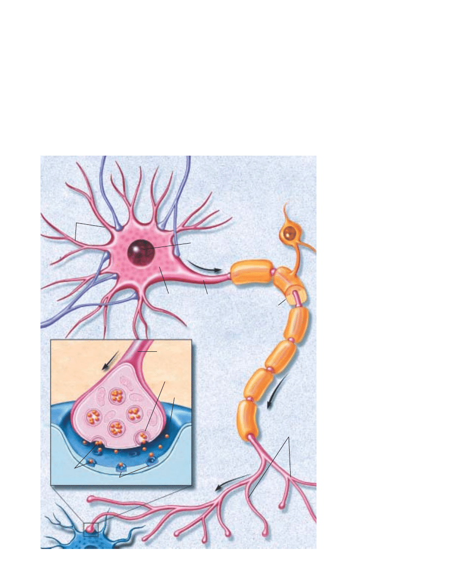
cerebral cortex in primates, including humans. In this structure,
NEURON MIGRATION. A cross-
sectional view of the occipital
lobe (which processes vision) of
a three-month-old monkey fetus
brain (center) shows immature
neurons migrating along glial
fibers. These neurons make
transient connections with other
neurons before reaching their
destination. A single migrating
neuron, shown about 2,500
times its actual size (right), uses
a glial fiber as a guiding
sca≈old. To move, it needs ad-
hesion molecules, which recog-
nize the pathway, and contrac-
tile proteins to propel it along.
Fetal
monkey
brain
Migrating
zone
Migrating
neuron
Glial
fiber
Outer surface
Inner surface

10
neurons slither from the place of origin near the ventricular sur-
face along nonneuronal fibers that form a trail to their proper
destination. Proper neuron migration requires multiple mech-
anisms, including the recognition of the proper path and the
ability to move long distances. One such mechanism for long
distance migration is the movement of neurons along elongated
fibers that form transient scaffolding in the fetal brain. Many
external forces, such as alcohol, cocaine or radiation, prevent
proper neuronal migration and result in misplacement of cells,
which may lead to mental retardation and epilepsy. Further-
more, mutations in genes that regulate migration have recently
been shown to cause some rare genetic forms of retardation and
epilepsy in humans.
Once the neurons reach their final location, they must make
the proper connections for a particular function, such as vision
or hearing, to occur. They do this through their axons. These
stalk-like appendages can stretch out a thousand times longer
than the cell body from which they arise. The journey of most
axons ends when they meet the branching areas, called den-
drites, on other neurons. These target neurons can be located
at a considerable distance, sometimes at opposite sides of the
brain. In the case of a motor neuron, the axon may travel from
the spinal cord all the way down to a foot muscle. The linkup
sites, called synapses, are where messages are transferred from
one neuron in a circuit to the next.
Axon growth is spearheaded by growth cones. These enlarge-
ments of the axon’s tip actively explore the environment as they
seek out their precise destinations. Researchers have discovered
that many special molecules help guide growth cones. Some
molecules lie on the cells that growth cones contact, while oth-
ers are released from sources found near the growth cone. The
growth cones, in turn, bear molecules that serve as receptors for
the environmental cues. The binding of particular signals with
its receptors tells the growth cone whether to move forward,
stop, recoil or change direction.
Recently researchers have identified some of the molecules
that serve as cues and receptors. These molecules include pro-
teins with names such as cadherin, netrin, semaphorin, ephrin,
neuropilin and plexin. In most cases, these are families of
related molecules; for example there are at least 15 semapo-
horins and at least 10 ephrins. Perhaps the most remarkable
result is that most of these are common to worms, insects and
mammals, including humans. Each family is smaller in flies
or worms than in mice or people, but their functions are quite
similar. It has therefore been possible to use the simpler ani-
mals to gain knowledge that can be directly applied to
humans. For example, the first netrin was discovered in a
worm and shown to guide neurons around the worm’s “nerve
ring.” Later, vertebrate netrins were found to guide axons
around the mammalian spinal cord. Worm receptors for
netrins were then found and proved invaluable in finding the
corresponding, and again related, human receptors.
Once axons reach their targets, they form synapses, which
permit electric signals in the axon to jump to the next cell, where
they can either provoke or prevent the generation of a new sig-
nal. The regulation of this transmission at synapses, and the inte-
gration of inputs from the thousands of synapses each neuron
receives, are responsible for the astounding information-
processing capabilities of the brain. For processing to occur prop-
erly, the connections must be highly specific. Some specificity
arises from the mechanisms that guide each axon to its proper
target area. Additional molecules mediate “target recognition”
whereby the axon chooses the proper neuron, and often the
proper part of the target, once it arrives at its destination. Few of
these molecules have been identified. There has been more suc-
cess, however, in identifying the ways in which the synapse forms
once the contact has been made. The tiny portion of the axon
that contacts the dendrite becomes specialized for the release of
neurotransmitters, and the tiny portion of the dendrite that
receives the contact becomes specialized to receive and respond
to the signal. Special molecules pass between the sending and
receiving cell to ensure that the contact is formed properly.
Paring back
Following the period of growth, the network is pared back to
create a more sturdy system. Only about one-half of the neu-
rons generated during development survive to function in the
adult. Entire populations of neurons are removed through
internal suicide programs initiated in the cells. The programs
are activated if a neuron loses its battle with other neurons to
receive life-sustaining nutrients called trophic factors. These
factors are produced in limited quantities by target tissues. Each
type of trophic factor supports the survival of a distinct group
of neurons. For example, nerve growth factor is important for
sensory neuron survival. It has recently become clear that the
internal suicide program is maintained into adulthood, and
constantly held in check. Based on this idea, researchers have
found that injuries and some neurodegenerative diseases kill
neurons not directly by the damage they inflict, but rather by
activating the death program. This discovery, and its implica-
tion that death need not inevitably follow insult, have led to
new avenues for therapy.
Brain cells also form too many connections at first. For
example, in primates, the projection from the two eyes to the
brain initially overlaps, and then sorts out to separate territo-
ries devoted only to one or the other eye. Furthermore, in the
young primate cerebral cortex, the connections between neu-
rons are greater in number and twice as dense as an adult pri-
mate. Communication between neurons with chemical and
electrical signals is necessary to weed out the connections. The
connections that are active and generating electrical currents
survive while those with little or no activity are lost.
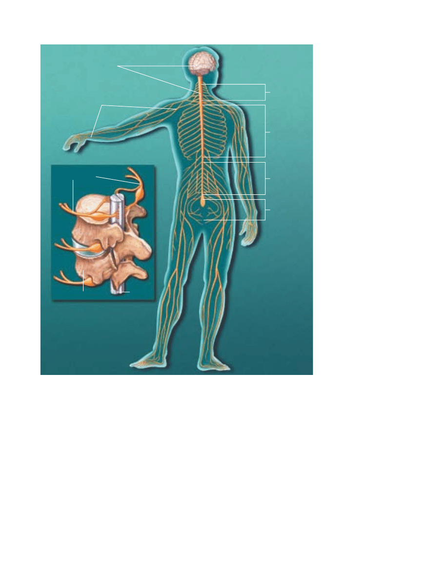
Vertebrae
Peripheral nerves
11
SPINAL CORD AND NERVES. The
mature central nervous system
(CNS) consists of the brain and
spinal cord. The brain sends
nerve signals to specific parts of
the body through peripheral
nerves, known as the peripheral
nervous system (PNS). Peripheral
nerves in the cervical region
serve the neck and arms; those in
the thoracic region serve the
trunk; those in the lumbar region
serve the legs; and those in the
sacral region serve the bowels
and bladder. The PNS consists of
the somatic nervous system that
connects voluntary skeletal mus-
cles with cells specialized to re-
spond to sensations, such as
touch and pain. The autonomic
nervous system is made of neu-
rons connecting the CNS with
internal organs. It is divided into
the sympathetic nervous system,
which mobilizes energy and
resources during times of stress
and arousal, and the parasympa-
thetic nervous system, which
conserves energy and resources
during relaxed states.
Critical periods
The brain’s refining and building of the network in mammals,
including humans, continues after birth. An organism’s interac-
tions with its surroundings fine-tune connections.
Changes occur during critical periods. These are windows of
time during development when the nervous system must obtain
certain critical experiences, such as sensory, movement or emo-
tional input, to develop properly. Following a critical period, con-
nections become diminished in number and less subject to
change, but the ones that remain are stronger, more reliable and
more precise. Injury, sensory or social deprivation occurring at a
certain stage of postnatal life may affect one aspect of develop-
ment, while the same injury at a different period may affect
another aspect. In one example, a monkey is raised from birth
up to six months of age with one eyelid closed. As a result of
diminished use, the animal permanently loses useful vision in
that eye. This gives cellular meaning to the saying “use it or lose
it.” Loss of vision is caused by the actual loss of functional con-
nections between that eye and neurons in the visual cortex. This
finding has led to earlier and better treatment of the eye disor-
ders congenital cataracts and “crossed-eyes” in children.
Research also shows that enriched environments can bolster
brain development during postnatal life. For example, studies
show that animals brought up in toy-filled surroundings have more
branches on their neurons and more connections than isolated ani-
mals. In one recent study, scientists found enriched environments
resulted in more neurons in a brain area involved in memory.
Scientists hope that new insights on development will lead
to treatments for those with learning disabilities, brain damage
and even neurodegenerative disorders or aging.
CENTRAL NERVOUS SYSTEM
Brain and spinal cord
PERIPHERAL NERVOUS SYSTEM
Nerves extending from spinal cord
Cervical region
Thoracic region
Lumbar region
Sacral region
Spinal cord

12
V
ision. This wonderful sense allows us to
image the world around us from the genius
of Michelangelo’s Sistine Chapel ceiling to
mist-filled vistas of a mountain range. Vision
is one of the most delicate and complicated
of all the senses.
It also is the most studied. About one-fourth of the brain
is involved in visual processing, more than for all other senses.
More is known about vision than any other vertebrate sensory
system, with most of the information derived from studies of
monkeys and cats.
Vision begins with the cornea, which does about three-
quarters of the focusing, and then the lens, which varies the
focus. Both help produce a clear image of the visual world on
the retina, the sheet of photoreceptors, which process vision,
and neurons lining the back of the eye.
As in a camera, the image on the retina is reversed: objects
to the right of center project images to the left part of the retina
and vice versa. Objects above the center project to the lower
part and vice versa. The shape of the lens is altered by the mus-
cles of the iris so near or far objects can be brought into focus
on the retina.
Visual receptors, about 125 million in each eye, are neurons
specialized to turn light into electrical signals. They occur in
two forms. Rods are most sensitive to dim light and do not con-
vey the sense of color. Cones work in bright light and are
responsible for acute detail, black and white and color vision.
The human eye contains three types of cones that are sensitive
to red, green and blue but in combination convey information
about all visible colors.
Primates, including humans, have well-developed vision
using two eyes. Visual signals pass from each eye along the mil-
lion or so fibers of the optic nerve to the optic chiasma where
some nerve fibers cross over, so both sides of the brain receive
signals from both eyes. Consequently, the left halves of both
retinae project to the left visual cortex and the right halves pro-
ject to the right visual cortex.
The e∑ect is that the left half of the scene you are watch-
ing registers in your right hemisphere. Conversely, the right half
of the scene you are watching registers in your left hemisphere.
A similar arrangement applies to movement and touch: each
half of the cerebrum is responsible for the opposite half of the
body.
Scientists know much about the way cells code visual infor-
mation in the retina, lateral geniculate nucleus—an intermedi-
ate point between the retina and visual cortex—and visual cor-
tex. These studies give us the best knowledge so far about how
the brain analyzes and processes information.
The retina contains three stages of neurons. The first, the
layer of rods and cones, sends its signals to the middle layer,
which relays signals to the third layer. Nerve fibers from the
third layer assemble to form the optic nerve. Each cell in the
middle or third layer receives input from many cells in the pre-
vious layer. Any cell in the third layer thus receives signals—
via the middle layer—from a cluster of many thousands of rods
and cones that cover about one-square millimeter (the size of
a thumb tack hole). This region is called the receptive field of
the third-layer cell.
About 50 years ago, scientists discovered that the receptive
field of such a cell is activated when light hits a tiny region in
its receptive field center and is inhibited when light hits the part
of the receptive field surrounding the center. If light covers the
entire receptive field, the cell reacts only weakly and perhaps
not at all.
Thus, the visual process begins with a comparison of the
amount of light striking any small region of the retina and the
amount of light around it. Located in the occipital lobe, the pri-
mary visual cortex—two millimeters thick (twice that of a
dime) and densely packed with cells in many layers—receives
messages from the lateral geniculate. In the middle layer, which
receives input from the lateral geniculate, scientists found pat-
terns of responsiveness similar to those observed in the retina
and lateral geniculate cells. Cells above and below this layer
responded di∑erently. They preferred stimuli in the shape of
bars or edges. Further studies showed that di∑erent cells pre-
ferred edges at particular angles, edges that moved or edges
moving in a particular direction.
Although the process is not yet completely understood,
V
Sensation and perception
13
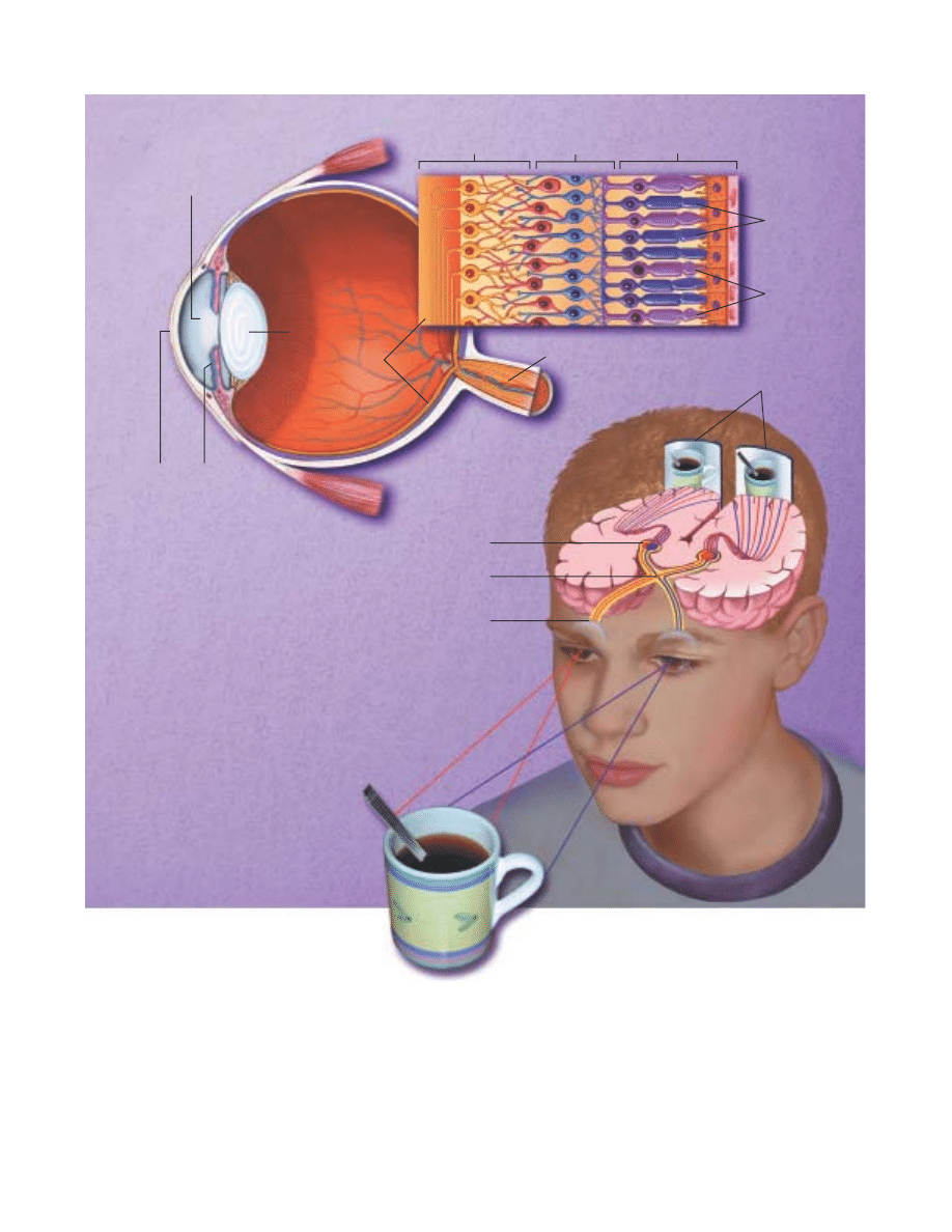
VISION. The cornea and lens help produce a clear image of the visual world on the retina, the sheet of photoreceptors and neurons lining the back
of the eye. As in a camera, the image on the retina is reversed: objects to the right of center project images to the left part of the retina and vice
versa. The eye’s 125 million visual receptors—composed of rods and cones—turn light into electrical signals. Rods are most sensitive to dim light
and do not convey the sense of color; cones work in bright light and are responsible for acute detail, black and white and color vision. The human
eye contains three types of cones that are sensitive to red, green and blue but, in combination, convey information about all visible colors. Rods and
cones connect with a middle cell layer and third cell layer (see inset, above). Light passes through these two layers before reaching the rods and
cones. The two layers then receive signals from rods and cones before transmitting the signals onto the optic nerve, optic chiasm, lateral geniculate
nucleus and, finally, the visual cortex.
Optic chiasm
Middle cell layer
Rods and Cones
Third cell layer
Pupil
Lens
Rods
Cones
Optic nerve
Retina
Iris
Cornea
Visual cortex
Right visual field
Left visual field
Optic nerve
Lateral geniculate nucleus
Modified from Jane Hurd
14
recent findings suggest that visual signals are fed into at least three separate processing systems.
One system appears to process information about shape; a second, color; and a third, movement,
location and spatial organization. These findings of separate processing systems come from mon-
key anatomical and physiological data. They are verified by human psychological studies showing
that the perception of movement, depth, perspective, the relative size of objects, the relative move-
ment of objects and shading and gradations in texture all depend primarily on contrasts in light
intensity rather than in color.
Why movement and depth perception should be carried by only one processing system may
be explained by a school of thought called Gestalt psychology. Perception requires various ele-
ments to be organized so that related ones are grouped together. This stems from the brain’s abil-
ity to group the parts of an image together and also to separate images from one another and from
their individual backgrounds.
How do all these systems produce the solid images you see? By extracting biologically rele-
vant information at each stage and associating firing patterns with past experience.
Vision studies also have led to better treatment for visual disorders. Information from research
in cats and monkeys has improved the therapy for strabismus, or squint, a term for “cross-eye” or
wall-eye. Children with strabismus initially have good vision in each eye. But because they can-
not fuse the images in the two eyes, they tend to favor using one eye and often lose useful vision
in the other eye.
Vision can be restored but only during infancy or early childhood. Beyond the age of six or
so, the blindness becomes permanent. But until a few decades ago, ophthalmologists waited until
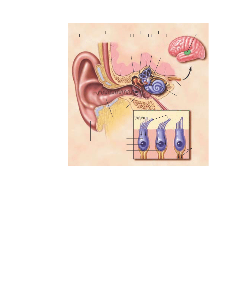
HEARING. From the chirping of
crickets to the roar of a rocket
engine, almost all of the thou-
sands of single tones processed
by the human ear are heard by a
mechanism known as air con-
duction. In this process, sound
waves are first funneled
through the external ear—the
pinna and the external auditory
canal—to the middle ear—the
tympanic membrane (eardrum)
that vibrates at di≈erent
speeds. The malleus (hammer),
which is attached to the tym-
panic membrane, transmits the
vibrations to the incus (anvil).
The vibrations are then passed
onto the stapes (stirrup) and
oval window that, in turn, pass
them onto the inner ear. In the
inner ear, the fluid-filled spiral
passage of the cochlea contains
cells with microscopic, hairlike
projections that respond to the
vibrations produced by sound.
The hair cells, in turn, excite the
28,000 fibers of the auditory
nerve that end in the medulla in
the brain. Auditory information
flows via the thalamus to the
temporal gyrus, the part of the
cerebral cortex involved in
receiving and perceiving sound.
Auditory area
External
auditory
canal
Pinna
Cochlea
Auditory nerve
External ear
Middle ear
Inner ear
Malleus Incus Stapes
Oval
window
To brain
BONES OF THE MIDDLE EAR
Released
chemicals
excite nerve
and send
impulses to
brain
Displacement of hair bundles
Tympanic
membrane
Transmitters
released
Hair cell
of cochlea
Nucleus
Soundwaves

15
children reached the age of four before operating to align the
eyes, or prescribe exercises or an eye patch. Now strabismus is
corrected very early in life—before age four—when normal
vision can still be restored.
Hearing
Often considered the most important sense for humans, hear-
ing allows us to communicate with each other by receiving
sounds and interpreting speech. It also gives us information
vital to survival. For example, the sound of an oncoming train
tells us to stay clear of the railroad track.
Like the visual system, our hearing system distinguishes sev-
eral qualities in the signal it detects. However, our hearing system
does not blend di∑erent sounds, as the visual system does when
two di∑erent wavelengths of
light are mixed to produce
color. We can follow the sep-
arate melodic lines of several
instruments as we listen to an
orchestra or rock band.
From the chirping of
crickets to the roar of a rocket
engine, most of the sounds
processed by the ear are heard
by a mechanism known as air conduction. In this process, sound
waves are first funneled through the externally visible part of the
ear, the pinna (or external ear) and the external auditory canal to
the tympanic membrane (eardrum) that vibrates at di∑erent
speeds. The malleus (hammer), which is attached to the tym-
panic membrane, transmits the vibrations to the incus (anvil).
This structure passes them onto the stapes (stirrup) which deliv-
ers them, through the oval window, to the inner ear.
The fluid-filled spiral passages of each cochlea contain
16,000 hair cells whose microscopic, hairlike projections
respond to the vibrations produced by sound. The hair cells, in
turn, excite the 28,000 fibers of the auditory nerve that termi-
nate in the medulla of the brain. Auditory information flows
via the thalamus to the temporal gyrus, the part of the cerebral
cortex involved in receiving and perceiving sound.
The brain’s analysis of auditory information follows a pat-
tern similar to that of the visual system. Adjacent neurons
respond to tones of similar frequency. Some neurons respond
to only a small range of frequencies, others react to a wide
range; some react only to the beginning of a sound, others only
respond to the end.
Speech sounds, however, may be processed di∑erently than
others. Our auditory system processes all the signals that it
receives in the same way until they reach the primary auditory
cortex in the temporal lobe of the brain. When speech sound
is perceived, the neural signal is funneled to the left hemisphere
for processing in language centers.
Taste and smell
Although di∑erent, the two sensory experiences of taste and
smell are intimately entwined. They are separate senses with
their own receptor organs. However, these two senses act
together to allow us to distinguish thousands of di∑erent
flavors. Alone, taste is a relatively focused sense concerned with
distinguishing among sweet, salty, sour and bitter. The interac-
tion between taste and smell explains why loss of the sense of
smell apparently causes a serious reduction in the overall taste
experience, which we call flavor.
Tastes are detected by taste buds, special structures of which
every human has some 5,000. Taste buds are embedded within
papillae, or protuberances, located mainly on the tongue, with
others found in the back of the mouth and on the palate. Taste
substances stimulate hairs pro-
jecting from the sensory cells.
Each taste bud consists of 50 to
100 sensory cells that respond
to salts, acidity, sweet sub-
stances and bitter compounds.
Some researchers add a fifth
category named umami, for the
taste of monosodium gluta-
mate and related substances.
Taste signals in the sensory cells are transferred by synapses
to the ends of nerve fibers, which send impulses along cranial
nerves to taste centers in the brain. From here, the impulses are
relayed to other brain stem centers responsible for the basic
responses of acceptance or rejection of the tastes, and to the
thalamus and on to the cerebral cortex for conscious perception
of taste.
Specialized smell receptor cells are located in a small patch
of mucus membrane lining the roof of the nose. Axons of these
sensory cells pass through perforations in the overlying bone
and enter two elongated olfactory bulbs lying on top of the bone.
The portion of the sensory cell that is exposed to odors pos-
sesses hair-like cilia. These cilia contain the receptor sites that
are stimulated by odors carried by airborne molecules. The odor
molecules dissolve in the mucus lining in order to stimulate
receptor molecules in the cilia to start the smell response. An
odor molecule acts on many receptors to di∑erent degrees. Sim-
ilarly, a receptor interacts with many di∑erent odor molecules
to di∑erent degrees.
Axons of the cells pass through perforations in the overly-
ing bone and enter two elongated olfactory bulbs lying on top
of the bone. The pattern of activity set up in the receptor cells
is projected to the olfactory bulb, where it forms a spatial image
of the odor. Impulses created by this stimulation pass to smell
centers, to give rise to conscious perceptions of odor in the
frontal lobe and emotional responses in the limbic system of
the brain.
Taste and smell are two separate senses with
their own sets of receptor organs, but they act
together to distinguish an enormous number of
di≈erent flavors.
16
Touch and pain
Touch is the sense by which we determine the characteristics of objects: size, shape and texture.
We do this through touch receptors in the skin. In hairy skin areas, some receptors consist of webs
of sensory nerve cell endings wrapped around the hair bulbs. They are remarkably sensitive, being
triggered when the hairs are moved. Other receptors are more common in non-hairy areas, such
as lips and fingertips, and consist of nerve cell endings that may be free or surrounded by bulb-
like structures.
Signals from touch receptors pass via sensory nerves to the spinal cord, then to the thalamus
and sensory cortex. The transmission of this information is highly topographic, meaning that the
body is represented in an orderly fashion at di∑erent levels of the nervous system. Larger areas of
the cortex are devoted to sensations from the hands and lips; much smaller cortical regions rep-
resent less sensitive parts of the body.
Di∑erent parts of the body vary in their sensitivity to touch discrimination and painful stim-
uli according to the number and distribution of receptors. The cornea is several hundred times
more sensitive to painful stimuli than are the soles of the feet. The fingertips are good at touch
discrimination but the chest and back are less sensitive.
Until recently, pain was thought to be a simple message by which neurons sent electrical
impulses from the site of injury directly to the brain.
Recent studies show that the process is more complicated. Nerve impulses from sites of injury
that persist for hours, days or longer lead to changes in the nervous system that result in an
amplification and increased duration of the pain. These changes involve dozens of chemical mes-
sengers and receptors.
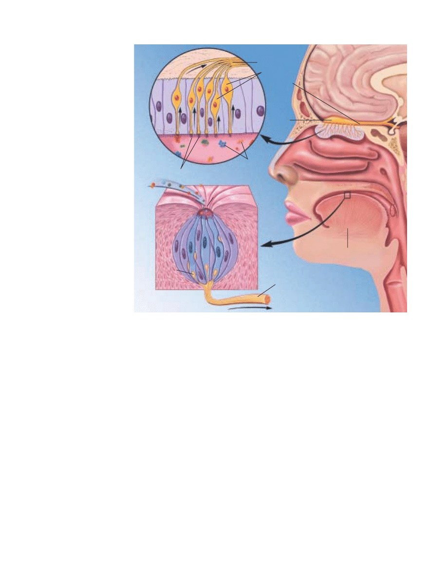
SMELL AND TASTE. Specialized
receptors for smell are located
in a patch of mucous membrane
lining the roof of the nose. Each
cell has several fine hairlike
cilia containing receptor pro-
teins, which are stimulated by
odor molecules in the air, and a
long fiber (axon), which passes
through perforations in the
overlying bone to enter the
olfactory bulb. Stimulated cells
give rise to impulses in the
fibers, which set up patterns in
the olfactory bulb that are
relayed to the brain’s frontal
lobe to give rise to smell per-
ception, and to the limbic sys-
tem to elicit emotional
responses. Tastes are detected
by special structures, taste
buds, of which every human has
some 10,000. Taste buds are
embedded within papillae (pro-
tuberances) mainly on the
tongue, with a few located in
the back of the mouth and on
the palate. Each taste bud con-
sists of about 100 receptors that
respond to the four types of
stimuli—sweet, salty, sour and
bitter—from which all tastes are
formed. A substance is tasted
when chemicals in foods dis-
solve in saliva, enter the pores
on the tongue and come in con-
tact with taste buds. Here they
stimulate hairs projecting from
the receptor cells and cause sig-
nals to be sent from the cells,
via synapses, to cranial nerves
and taste centers in the brain.
Olfactory tract
Olfactory
bulb
Nerve fibers to brain
Receptor cells
Cilia
Airborne odors
Food
chemicals
Taste bud pore
Synapse
Taste (gustatory) nerve to brain
Tongue
17
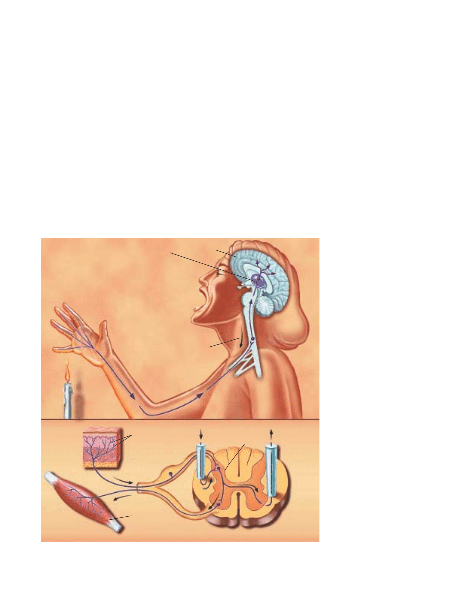
At the point of injury, nociceptors, special receptors, respond
to tissue-damaging stimuli. Injury results in the release of
numerous chemicals at the site of damage and inflammation.
One such chemical, prostaglandin, enhances the sensitivity of
receptors to tissue damage and ultimately can result in more
intense pain sensations. It also contributes to the clinical con-
dition in which innocuous stimuli can produce pain (such as in
sunburned skin) because the threshold of the nociceptor is
significantly reduced.
Pain messages are transmitted to the spinal cord via small
myelinated fibers and C fibers—very small unmyelinated fibers.
Myelin is a covering around nerve fibers that helps them send
their messages more rapidly.
In the ascending system, the impulses are relayed from the
spinal cord to several brain structures, including the thalamus
and cerebral cortex, which are involved in the process by which
“pain” messages become conscious experience.
Pain messages can also be suppressed by a system of neu-
rons that originate within the gray matter in the brainstem of
the midbrain. This descending system sends messages to the dor-
sal horn of the spinal cord where it suppresses the transmission
of pain signals to the higher brain centers. Some of these
descending systems use naturally occurring chemicals similar to
opioids. The three major families of opioids—enkephalins,
endorphins and dynorphins—identified in the brain originate
from three precursor proteins coded by three di∑erent genes.
They act at multiple opioid receptors in the brain and spinal
cord. This knowledge has led to new treatments for pain: Opiate-
like drugs injected into the space above the spinal cord provide
long-lasting pain relief.
Scientists are now using modern tools for imaging brain
structures in humans to determine the role of the higher cen-
ters of the brain in pain experience and how signals in these
structures change with long-lasting pain.
PAIN. Messages about tissue
damage are picked up by recep-
tors and transmitted to the
spinal cord via small, myeli-
nated fibers and very small
unmyelinated fibers. From the
spinal cord, the impulses are
carried to the brainstem, thala-
mus and cerebral cortex and
ultimately perceived as pain.
These messages can be sup-
pressed by a system of neurons
that originates in the gray
matter of the midbrain. This
descending pathway sends mes-
sages to the spinal cord where it
suppresses the transmission of
tissue damage signals to the
higher brain centers. Some of
these descending pathways use
naturally occurring, opiate-like
chemicals called endorphins.
Message is received in the thalamus and cerebral cortex
Tissue-damaging stimulus
activates nociceptors
Message carried
to spinal cord
Descending pathway
Nociceptors
From brain
To brain
Dorsal horn
Muscle fiber

18
T
he conscious memory of a patient known as
H.M. is limited almost entirely to events that
occurred years before his surgery, which
removed part of the medial temporal lobe of his
brain to relieve epilepsy. H.M. can remember
recent events for only a few minutes. Talk with
him awhile and then leave the room. When you return, he has
no recollection of ever having seen you before.
The medial temporal lobe, which includes the hippocam-
pus and adjacent brain areas, seems to play a role in converting
memory from a short-term to a long-term, permanent form.
The fact that H.M. retains memories for events that are remote
to his surgery is evidence that the medial temporal region is not
the site of permanent storage but that it plays a role in the for-
mation of new memories. Other patients like H.M. have also
been described.
Additional evidence comes from patients undergoing elec-
troconvulsive therapy (ECT) for depression. ECT is thought to
temporarily disrupt the function of the hippocampus and
related structures. These patients typically su∑er di≈culty with
new learning and have amnesia for events that occurred during
the several years before treatment. Memory of earlier events is
unimpaired. As time passes after treatment, much of the lost
part of memory becomes available once again.
The hippocampus and the medial temporal region are con-
nected with widespread areas of the cerebral cortex, especially
the vast regions responsible for thinking and language. Whereas
the medial temporal region is important for forming and orga-
nizing memory, cortical areas are important for the long-term
storage of knowledge about facts and events and for how these
are used in everyday situations.
Working memory, a type of transient memory that enables
us to retain what someone has said just long enough to reply,
depends in part on the prefrontal cortex. Researchers discov-
ered that certain neurons in this area are influenced by neurons
releasing dopamine and other neurons releasing glutamate.
While much is unknown about learning and memory, scien-
tists can recognize certain pieces of the process. For example, the
brain appears to process di∑erent kinds of information in sepa-
rate ways and then store it di∑erently. Procedural knowledge, the
knowledge of how to do something, is expressed in skilled behav-
ior and learned habits. Declarative knowledge provides an explicit,
consciously accessible record of individual previous experiences
and a sense of familiarity about those experiences. Declarative
knowledge requires processing in the medial temporal region and
parts of the thalamus, while procedural knowledge requires pro-
cessing by the basal ganglia. Other kinds of memory depend on
the amygdala (emotional aspects of memory) and the cerebellum
(motor learning where precise timing is involved).
An important factor that influences what is stored and how
strongly it is stored is whether the action is followed by reward-
ing or punishing consequences. This is an important principle
in determining what behaviors an organism will learn and
remember. The amygdala appears to play an important role in
these memory events.
How exactly does memory occur? After years of study, there
is much support for the idea that memory involves a persistent
change in the relationship between neurons. In animal studies,
scientists found that this occurs through biochemical events in
the short term that a∑ect the strength of the relevant synapses.
The stability of long-term memory is conferred by structural
modifications within neurons that change the strength and
number of synapses. For example, researchers can correlate
specific chemical and structural changes in the relevant cells
with several simple forms of behavioral change exhibited by the
sea slug Aplysia.
Another important model for the study of memory is the
phenomenon of long-term potentiation (LTP), a long-lasting
increase in the strength of a synaptic response following stim-
ulation. LTP occurs prominently in the hippocampus, as well
as in other brain areas. Studies of rats suggest LTP occurs by
changes in synaptic strength at contacts involving NMDA
receptors. It is now possible to study LTP and learning in
genetically modified mice that have abnormalities of specific
genes. Abnormal gene expression can be limited to particular
brain areas and time periods, such as during learning.
Scientists believe that no single brain center stores mem-
ory. It most likely is stored in the same, distributed collection
Learning and memory
T
19
of cortical processing systems involved in the perception, pro-
cessing and analysis of the material being learned. In short,
each part of the brain most likely contributes di∑erently to per-
manent memory storage.
One of the most prominent intellectual activities depen-
dent on memory is language. While the neural basis of lan-
guage is not fully understood, scientists have learned much
about this feature of the brain from studies of patients who have
lost speech and language abilities due to stroke, and from behav-
ioral and functional neuroimaging studies of normal people.
A prominent and influential model, based on studies of
these patients, proposes that the underlying structure of speech
comprehension arises in Wernicke’s area, a portion of the left
hemisphere of the brain. This temporal lobe region is connected
with Broca’s area in the frontal lobe where a program for vocal
expression is created. This program is then transmitted to a
nearby area of the motor cortex that activates the mouth,
tongue and larynx.
This same model proposes that, when we read a word, the
information is transmitted from the primary visual cortex to the
angular gyrus where the message is somehow matched with the
sounds of the words when spoken. The auditory form of the
word is then processed for comprehension in Wernicke’s area
as if the word had been heard. Writing in response to an oral
instruction requires information to be passed along the same
pathways in the opposite direction—from the auditory cortex
to Wernicke’s area to the angular gyrus. This model accounts
for much of the data from patients, and is the most widely used
model for clinical diagnosis and prognosis. However, some
refinements to this model may be necessary due to both recent
studies with patients and functional neuroimaging studies in
normal people.
For example, using an imaging technique called positron
emission tomography (PET), scientists have demonstrated that
some reading tasks performed by normal people activated nei-
ther Wernicke’s area nor the angular gyrus. These results sug-
gest that there is a direct reading route that does not involve
speech sound recoding of the visual stimulus before the pro-
cessing of either meaning or speaking. Other studies with
patients also have indicated that it is likely that familiar words
need not be recoded into sound before they can be understood.
Although the understanding of how language is imple-
mented in the brain is far from complete, there are now several
techniques that may be used to gain important insights.
LEARNING AND MEMORY,
SPEECH AND LANGUAGE.
Structures believed to be impor-
tant for various kinds of learning
and memory include the cere-
bral cortex, amygdala, hip-
pocampus, cerebellum and
basal ganglia. Areas of the left
hemisphere (inset) are known to
be active in speech and lan-
guage. The form and meaning of
an utterance is believed to arise
in Wernicke’s area and then
Broca’s area, which is related to
vocalization. Wernicke’s area is
also important for language
comprehension.
Cerebral cortex
Wernicke’s area
Broca’s area
AREAS OF SPEECH
AND LANGUAGE
Amygdala
Hippocampus
Cerebellum
BASAL GANGLIA
Caudate
nucleus
Putamen
Globus
pallidus
Amygdaloid
nucleus
Angular
gyrus

20
F
rom the stands, we marvel at the perfectly placed
serves of professional tennis players and lightning-
fast double plays executed by big league infielders.
But in fact, every one of us in our daily lives per-
forms highly skilled movements, such as walking
upright, speaking and writing, that are no less
remarkable. A finely tuned and highly complex central nervous
system controls the action of hundreds of muscles in accom-
plishing these everyday marvels.
In order to understand how the nervous system performs
this trick, we have to start with muscles. Most muscles attach
to points on the skeleton that cross one or more joints. Acti-
vation of a given muscle, the agonist, can open or close the
joints that it spans or act to sti∑en them, depending on the
forces acting on those joints from the environment or other
muscles that oppose the agonist, the antagonists. Relatively few
muscles act on soft tissue. Examples include the muscles that
move the eyes and tongue, and the muscles that control facial
expression.
A muscle is made up of thousands of individual muscle
fibers, each of which is controlled by one alpha motor neuron in
either the brain or spinal cord. On the other hand, a single
alpha neuron can control hundreds of muscle fibers, forming a
motor unit. These motor neurons are the critical link between
the brain and muscles. When these neurons die, a person is no
longer able to move.
The simplest movements are reflexes—fixed muscle
responses to particular stimuli. Studies show sensory stretch
receptors—called muscle spindles, which include small, special-
ized muscle fibers and are located in most muscles—send infor-
mation about muscles directly to alpha motor neurons.
Sudden muscle stretch (such as when a doctor taps a mus-
cle tendon to test your reflexes) sends a barrage of impulses into
the spinal cord along the muscle spindle sensory fibers. This,
in turn, activates motor neurons in the stretched muscle, caus-
ing a contraction which is called the stretch reflex. The same
sensory stimulus causes inactivation, or inhibition, in the motor
neurons of the antagonist muscles through connecting neurons,
called inhibitory neurons, within the spinal cord.
The sensitivity of the muscle spindle organs is controlled
by the brain through a separate set of gamma motor neurons that
control the specialized spindle muscle fibers and allow the brain
to fine-tune the system for di∑erent movement tasks. Other
muscle sense organs signal muscle force that a∑ects motor neu-
rons through separate sets of spinal neurons. We now know that
this complex system responds di∑erently for tasks that require
precise control of position (holding a full teacup), as opposed
to those that require rapid, strong movement (throwing a ball).
You can experience such changes in motor strategy when you
compare walking down an illuminated staircase with the same
task done in the dark.
Another useful reflex is the flexion withdrawal that occurs
if your bare foot encounters a sharp object. Your leg is imme-
diately lifted from the source of potential injury (flexion) but
the opposite leg responds with increased extension in order to
maintain your balance. The latter event is called the crossed
extension reflex. These responses occur very rapidly and without
your attention because they are built into systems of neurons
located within the spinal cord itself.
It seems likely that the same systems of spinal neurons also
participate in controlling the alternating action of the legs dur-
ing normal walking. In fact, the basic patterns of muscle acti-
vation that produce coordinated walking can be generated in
four-footed animals within the spinal cord itself. It seems likely
that these spinal mechanisms, which evolved in primitive ver-
tebrates, are probably still present in the human spinal cord.
The most complex movements that we perform, including
voluntary ones that require conscious planning, involve control
of the spinal mechanisms by the brain. Scientists are only
beginning to understand the complex interactions that take
place between di∑erent brain regions during voluntary move-
ments, mostly through careful experiments on animals. One
important area is the motor cortex, which exerts powerful con-
trol of the spinal cord neurons and has direct control of some
motor neurons in monkeys and humans. Some neurons in the
motor cortex appear to specify the coordinated action of many
muscles, so as to produce organized movement of the limb to
a particular place in space.
F
Movement
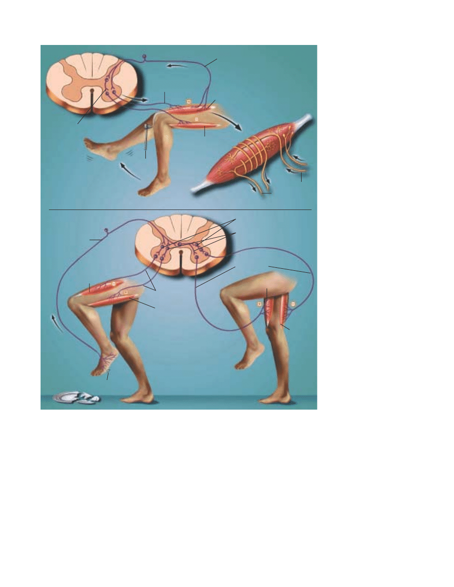
21
In addition to the motor cortex, movement control also
involves the interaction of many other brain regions, including
the basal ganglia and thalamus, the cerebellum and a large
number of neuron groups located within the midbrain and
brainstem—regions that connect cerebral hemispheres with the
spinal cord.
Scientists know that the basal ganglia and thalamus have
widespread connections with sensory and motor areas of the
cerebral cortex. Loss of regulation of the basal ganglia by
dopamine depletion can cause serious movement disorders,
such as Parkinson’s disease. Loss of dopamine neurons in the
substantia nigra on the midbrain, which connects with the basal
ganglia, is a major factor in Parkinson’s.
The cerebellum is critically involved in the control of all
skilled movements. Loss of cerebellar function leads to poor
coordination of muscle control and disorders of balance. The
cerebellum receives direct and powerful sensory information
from the muscle receptors, and the sense organs of the inner
ear, which signal head position and movement, as well as sig-
nals from the cerebral cortex. It apparently acts to integrate all
this information to ensure smooth coordination of muscle
action, enabling us to perform skilled movements more or less
automatically. There is evidence that, as we learn to walk, speak
or play a musical instrument, the necessary detailed control
information is stored within the cerebellum where it can be
called upon by commands from the cerebral cortex.
MOVEMENT. The stretch reflex
(above) occurs when a doctor taps
a muscle tendon to test your
reflexes. This sends a barrage of
impulses into the spinal cord
along muscle spindle sensory
fibers and activates motor neu-
rons to the stretched muscle to
cause contraction (stretch reflex).
The same sensory stimulus
causes inactivation, or inhibition,
of the motor neurons to the antag-
onist muscles through connection
neurons, called inhibitory neu-
rons, within the spinal cord.
A≈erent nerves carry messages
from sense organs to the spinal
cord; e≈erent nerves carry motor
commands from the spinal cord to
muscles. Flexion withdrawal
(below) can occur when your bare
foot encounters a sharp object.
Your leg is immediately lifted
(flexion) from the source of poten-
tial injury, but the opposite leg
responds with increased exten-
sion in order to maintain your bal-
ance. The latter event is called the
crossed extension reflex. These
responses occur very rapidly and
without your attention because
they are built into systems of neu-
rons located within the spinal
cord itself.
Inhibitory neuron
Alpha motor neuron
Sensory neuron
Extensor muscles activated
Flexor muscles inhibited
Response
Stimulus
Afferent nerves
Muscle
spindle
Efferent nerves
Sensory neuron
Motor
neurons
Motor neurons
Inhibitory neurons
Excitatory neurons
Extensor muscles activated
Flexor
muscles
inhibited
Flexor
muscles
activated
Stimulus
Right leg extends to
balance body
Extensor
muscles
inhibited
-
22
S
leep remains one of the great mysteries of mod-
ern neuroscience. We spend nearly one-third
of our lives asleep, but the function of sleep still
is not known. Fortunately, over the last few
years researchers have made great headway in
understanding some of the brain circuitry that
controls wake-sleep states.
Scientists now recognize that sleep consists of several
di∑erent stages; that the choreography of a night’s sleep
involves the interplay of these stages, a process that depends
upon a complex switching mechanism; and that the sleep stages
are accompanied by daily rhythms in bodily hormones, body
temperature and other functions.
Sleep disorders are among the nation’s most common
health problems, a∑ecting up to 70 million people, most of
whom are undiagnosed and untreated. These disorders are one
of the least recognized sources of disease, disability and even
death, costing an estimated $100 billion annually in lost pro-
ductivity, medical bills and industrial accidents. Research holds
the promise for devising new treatments to allow millions of
people to get a good night’s sleep.
The stu≈ of sleep
Sleep appears to be a passive and restful time when the brain is
less active. In fact, this state actually involves a highly active
and well-scripted interplay of brain circuits to produce the
stages of sleeping.
The stages of sleep were discovered in the 1950s in experi-
ments examining the human brain waves or electroencephalo-
gram (EEG) during sleep. Researchers also measured move-
ments of the eyes and the limbs during sleep. They found that
over the course of the first hour or so of sleep each night, the
brain progresses through a series of stages during which the
brain waves progressively slow down. The period of slow wave
sleep is accompanied by relaxation of the muscles and the eyes.
Heart rate, blood pressure and body temperature all fall. If
awakened at this time, most people recall only a feeling or
image, not an active dream.
Sleep
SLEEP PATTERNS. During a night of sleep, the brain waves of a young adult recorded by the electroencephalogram (EEG) gradually slow down and
become larger as the individual passes into deeper stages of slow wave sleep. After about an hour, the brain re-emerges through the same series of
stages, and there is usually a brief period of REM sleep (on dark areas of graph), during which the EEG is similar to wakefulness. The body is com-
pletely relaxed, the person is deeply unresponsive and usually is dreaming. The cycle repeats over the course of the night, with more REM sleep,
and less time spent in the deeper stages of slow wave sleep as the night progresses.
S
Awake
Stage 1
Stage 2
Stage 3
Stage 4
Awake
Stage 1
Stage 2
Stage 3
Stage 4
1 2 3 4 6 7
Hours
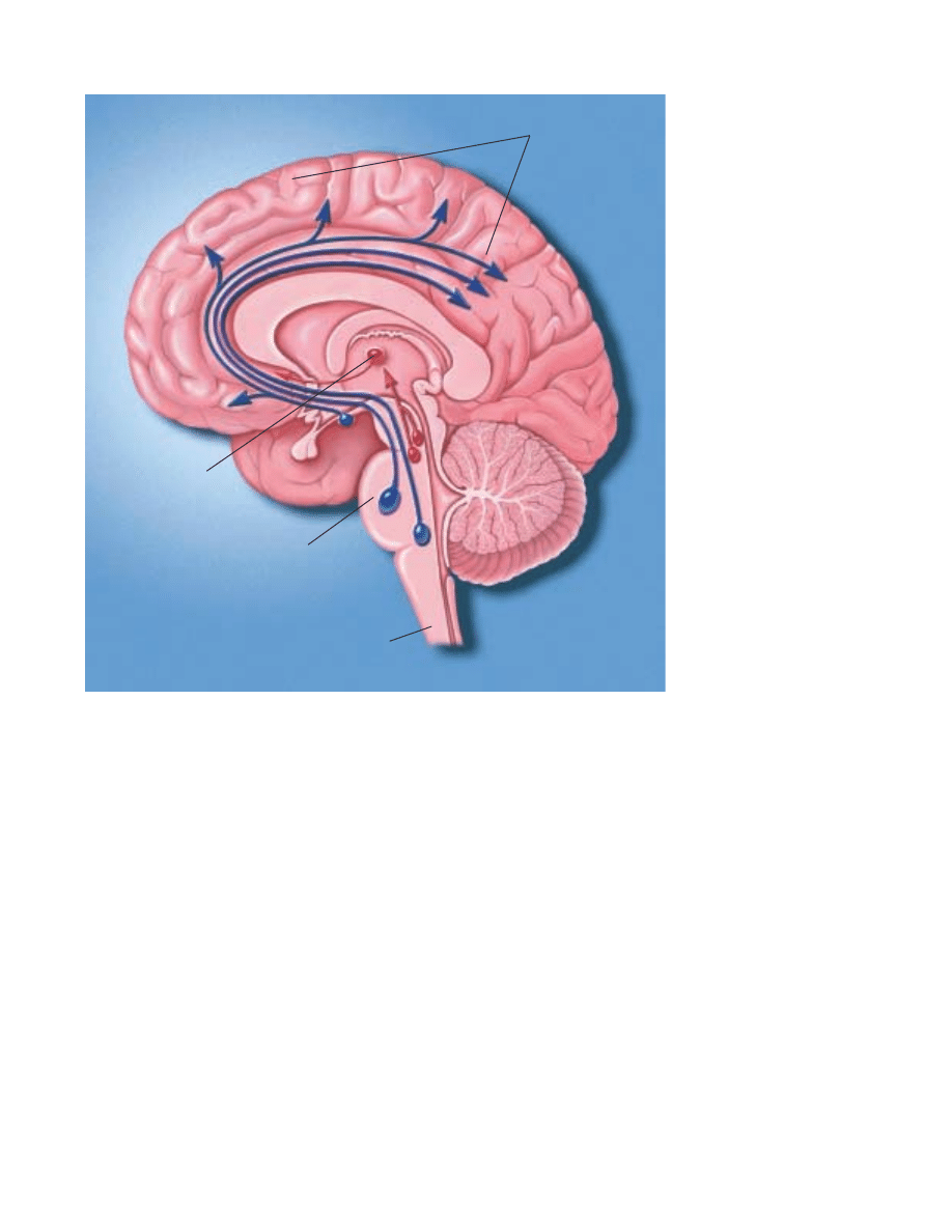
23
THE WAKING AND SLEEPING
BRAIN. Wakefulness is main-
tained by activity in two systems
of brainstem neurons. Nerve
cells that make the neurotrans-
mitter acetylcholine stimulate
the thalamus, which activates
the cerebral cortex (red path-
way). Full wakefulness also
requires cortical activation by
other neurons that make
monoamine neurotransmitters
such as norepinephrine, sero-
tonin and histamine (blue path-
way). During slow wave sleep,
when the brain becomes less
active, neuron activity in both
pathways slows down. During
rapid eye movement sleep, in
which dreaming occurs, the neu-
rons using acetylcholine fire
rapidly, producing a dreaming
state, but the monoamine cells
stop firing altogether.
Over the next half hour or so, the brain emerges from the
deep slow wave sleep as the EEG waves become progressively
faster. Similar to during waking, rapid eye movements emerge,
but the body’s muscles become almost completely paralyzed
(only the muscles that allow breathing remain active). This state
is often called rapid eye movement (REM) sleep. During REM
sleep, there is active dreaming. Heart rate, blood pressure and
body temperature become much more variable. Men often have
erections during this stage of sleep. The first REM period usu-
ally lasts ten to 15 minutes.
Over the course of the night, these alternative cycles of slow
wave and REM sleep alternate, with the slow wave sleep
becoming less deep, and the REM periods more prolonged,
until waking occurs.
Over the course of a lifetime, the pattern of sleep cycles
changes. Infants sleep up to 18 hours per day, and they spend
much more time in deep slow wave sleep. As children mature,
they spend less time asleep, and less time in deep slow wave
sleep. Older adults may sleep only six to seven hours per night,
often complain of early wakening that they cannot avoid, and
spend very little time in slow wave sleep.
Sleep disorders
The most common sleep disorder, and the one most people are
familiar with, is insomnia. Some people have di≈culty falling
asleep initially, but other people fall asleep, and then awaken
part way through the night, and cannot fall asleep again.
Although there are a variety of short-acting sedatives and
sedating antidepressant drugs available to help, none of these
produces a truly natural and restful sleep state because they tend
to suppress the deeper stages of slow wave sleep.
Excessive daytime sleepiness may have many causes. The
most common are disorders that disrupt sleep and result in
inadequate amounts of sleep, particularly the deeper stages.
These are usually diagnosed in the sleep laboratory. Here, the
EEG, eye movements and muscle tone are monitored electri-
cally as the individual sleeps. In addition, the heart, breathing,
and oxygen content of the blood can be monitored.
Obstructive sleep apnea causes the airway muscles in the
throat to collapse as sleep deepens. This prevents breathing,
which causes arousal, and prevents the su∑erer from entering
the deeper stages of slow wave sleep. This condition can also
cause high blood pressure and may increase the risk of heart
Cerebral
cortex
Thalamus
Pons
Spinal cord

24
attack. There is also an increased risk of daytime accident, espe-
cially automobile accidents, which may prevent driving. Treat-
ment is complex and may include a variety of attempts to reduce
airway collapse during sleep. While simple things like losing
weight, avoiding alcohol and sedating drugs prior to sleep, and
avoiding sleeping on one’s back can sometimes help, most peo-
ple with sleep apnea require positive airway pressure to keep the
airway open. This can be provided by fitting a small mask over
the nose that provides an air stream under pressure during sleep.
In some cases, surgery is needed to correct the airway anatomy.
Periodic limb movements of sleep are intermittent jerks of the
legs or arms, which occur as the individual enters slow wave
sleep, and can cause arousal from sleep. Other people have
episodes in which their muscles fail to be paralyzed during
REM sleep, and they act out their dreams. This REM behavior
disorder can also be very disruptive to a normal nights’ sleep.
Both disorders are more common in people with Parkinson’s
disease, and both can be treated with drugs that treat Parkin-
son’s, or with an anti-epileptic drug called clonazepam.
Narcolepsy is a relatively uncommon condition (one case per
2,500 people) in which the switching mechanism for REM
sleep does not work properly. Narcoleptics have sleep attacks
during the day, in which they suddenly fall asleep. This is
socially disruptive, as well as dangerous, for example, if they are
driving. They tend to enter REM sleep very quickly as well, and
may even enter a dreaming state while still awake, a condition
known as hypnagogic hallucinations. They also have attacks dur-
ing which they lose muscle tone, similar to what occurs during
REM sleep, but while they are awake. Often, this occurs while
they are falling asleep or just waking up, but attacks of paraly-
sis known as cataplexy can be triggered by an emotional expe-
rience or even hearing a funny joke.
Recently, insights into the mechanism of narcolepsy have
given major insights into the processes that control these mys-
terious transitions between waking, slow wave and REM sleep
states.
How is sleep regulated?
During wakefulness, the brain is kept in an alert state by the
interactions of two major systems of nerve cells. Nerve cells in
the upper part of the pons and in the midbrain, which make
acetylcholine as their neurotransmitter, send inputs to the thal-
amus, to activate it. When the thalamus is activated, it in turn
activates the cerebral cortex, and produces a waking EEG pat-
tern. However, that is not all there is to wakefulness. As dur-
ing REM sleep, the cholinergic nerve cells and the thalamus
and cortex are in a condition similar to wakefulness, but the
brain is in REM sleep, and is not very responsive to external
stimuli.
The di∑erence is supplied by three sets of nerve cells in the
upper part of the brainstem: nerve cells in the locus coeruleus
that contain the neurotransmitter norepinephrine; in the dor-
sal and median raphe groups that contain serotonin; and in the
tuberomammillary cell group that contains histamine. These
monoamine neurons fire most rapidly during wakefulness, but
they slow down during slow wave sleep, and they stop during
REM sleep.
The brainstem cell groups that control arousal are in turn
regulated by two groups of nerve cells in the hypothalamus, part
of the brain that controls basic body cycles. One group of nerve
cells, in the ventrolateral preoptic nucleus, contain inhibitory
neurotransmitters, galanin and GABA. When the ventrolateral
preoptic neurons fire, they are thought to turn o∑ the arousal
systems, causing sleep. Damage to the ventrolateral preoptic
nucleus produces irreversible insomnia.
A second group of nerve cells in the lateral hypothalamus
act as an activating switch. They contain the neurotransmitters
orexin and dynorphin, which provide an excitatory signal to the
arousal system, particularly to the monoamine neurons. In
experiments in which the gene for the neurotransmitter orexin
was experimentally removed in mice, the animals became nar-
coleptic. Similarly, in two dog strains with naturally occurring
narcolepsy, an abnormality was discovered in the gene for the
type 2 orexin receptor. Recent studies show that in humans with
narcolepsy, the orexin levels in the brain and spinal fluid are
abnormally low. Thus, orexin appears to play a critical role in
activating the monoamine system, and preventing abnormal
transitions, particularly into REM sleep.
Two main signals control this circuitry. First, there is home-
ostasis, or the body’s need to seek a natural equilibrium. There
is an intrinsic need for a certain amount of sleep each day. The
mechanism for accumulating sleep need is not yet clear. Some
people think that a chemical called adenosine may accumulate
in the brain during prolonged wakefulness, and that it may
drive sleep homeostasis. Interestingly, the drug ca∑eine, which
is widely used to prevent sleepiness, acts as an adenosine
blocker, to prevent its e∑ects.
If an individual does not get enough sleep, the sleep debt
progressively accumulates, and leads to a degradation of men-
tal function. When the opportunity comes to sleep again, the
individual will sleep much more, to “repay” the debt, and the
slow wave sleep debt is usually “paid o∑” first.
The other major influence on sleep cycles is the body’s cir-
cadian clock, the suprachiasmatic nucleus. This small group of
nerve cells in the hypothalamus contains clock genes, which go
through a biochemical cycle of almost exactly 24 hours, setting
the pace for daily cycles of activity, sleep, hormones and other
bodily functions. The suprachiasmatic nucleus also receives an
input directly from the retina, and the clock can be reset by
light, so that it remains linked to the outside world’s day-night
cycle. The suprachiasmatic nucleus provides a signal to the ven-
trolateral preoptic nucleus and probably the orexin neurons.

25
T
he urge to act in the presence of stress has been
with us since our ancient ancestors. In response
to impending danger, muscles are primed,
attention is focused and nerves are readied for
action—fight or flight. But in today’s corpora-
tion-dominated world, this response to stress is
simply inappropriate and may be a contributor to heart disease,
accidents and aging.
Indeed, nearly two-thirds of ailments seen in doctors’
o≈ces are commonly thought to be stress-induced or related to
stress in some way. Surveys indicate that 60 percent of Amer-
icans feel they are under a great deal of stress at least once a
week. Costs due to stress from absenteeism, medical expenses
and lost productivity are estimated at $300 billion annually.
Only recently admitted into the medical vocabulary, stress
is di≈cult to define because its e∑ects vary with each individ-
ual. Dr. Hans Selye, a founder of stress research, called it “the
rate of wear and tear in the body.” Other specialists now define
stress as any external stimulus that threatens homeostasis—the
normal equilibrium of body function. Among the most power-
ful stressors are psychological and psychosocial stressors that
exist between members of the same species. Lack or loss of con-
trol is a particularly important feature of severe psychological
stress that can have physiological consequences.
During the last six decades, researchers using animals found
that stress both helps and harms the body. When confronted
with a crucial challenge, properly controlled stress responses
can provide the extra strength and energy needed to cope.
Moreover, the acute physiological response to stress protects
the body and brain and helps to re-establish or maintain home-
ostasis. But stress that continues for prolonged periods of time
can repeatedly elevate the physiological stress responses or fail
to shut them o∑ when not needed. When this occurs, these
same physiological mechanisims can badly upset the body’s bio-
chemical balance and accelerate disease.
Scientists also believe that the individual variation in
responding to stress is somewhat dependent on a person’s per-
ception of external events. This perception ultimately shapes
his or her internal physiological response. Thus, by controlling
your perception of events, you can do much to avoid the harm-
ful consequences of stress.
The immediate response
A stressful situation activates three major communication sys-
tems in the brain that regulate bodily functions. Scientists have
come to understand these complex systems through experi-
ments primarily with rats, mice and nonhuman primates, such
as monkeys. Scientists then verified the action of these systems
in humans.
The first of these systems is the voluntary nervous system,
which sends messages to muscles so that we may respond to
sensory information. For example, the sight of a growling bear
on a trail in Yellowstone National Park prompts you to run as
quickly as possible.
The second communication system is the autonomic nervous
system. It combines the sympathetic or emergency branch,
which gets us going in emergencies, and the parasympathetic or
calming branch, which keeps the body’s maintenance systems,
such as digestion, in order and calms the body’s responses to
the emergency branch.
Each of these systems has a specific task. The emergency
branch causes arteries supplying blood to the muscles to relax
in order to deliver more blood, allowing greater capacity to act.
At the same time, the emergency system reduces blood flow to
the skin, kidney and digestive tract and increases blood flow to
the muscles. In contrast, the calming branch helps to regulate
bodily functions and soothe the body, preventing it from
remaining too long in a state of mobilization. Remaining mobi-
lized and left unchecked, these body functions could lead to
disease. Some actions of the calming branch appear to reduce
the harmful e∑ects of the emergency branch’s response to stress.
The brain’s third major communication process is the neu-
roendocrine system, which also maintains the body’s internal
functioning. Various “stress hormones” travel through the blood
and stimulate the release of other hormones, which a∑ect bod-
ily processes, such as metabolic rate and sexual functions.
Major stress hormones are epinephrine (also known as
adrenaline) and cortisol. When the body is exposed to stressors,
Stress
T
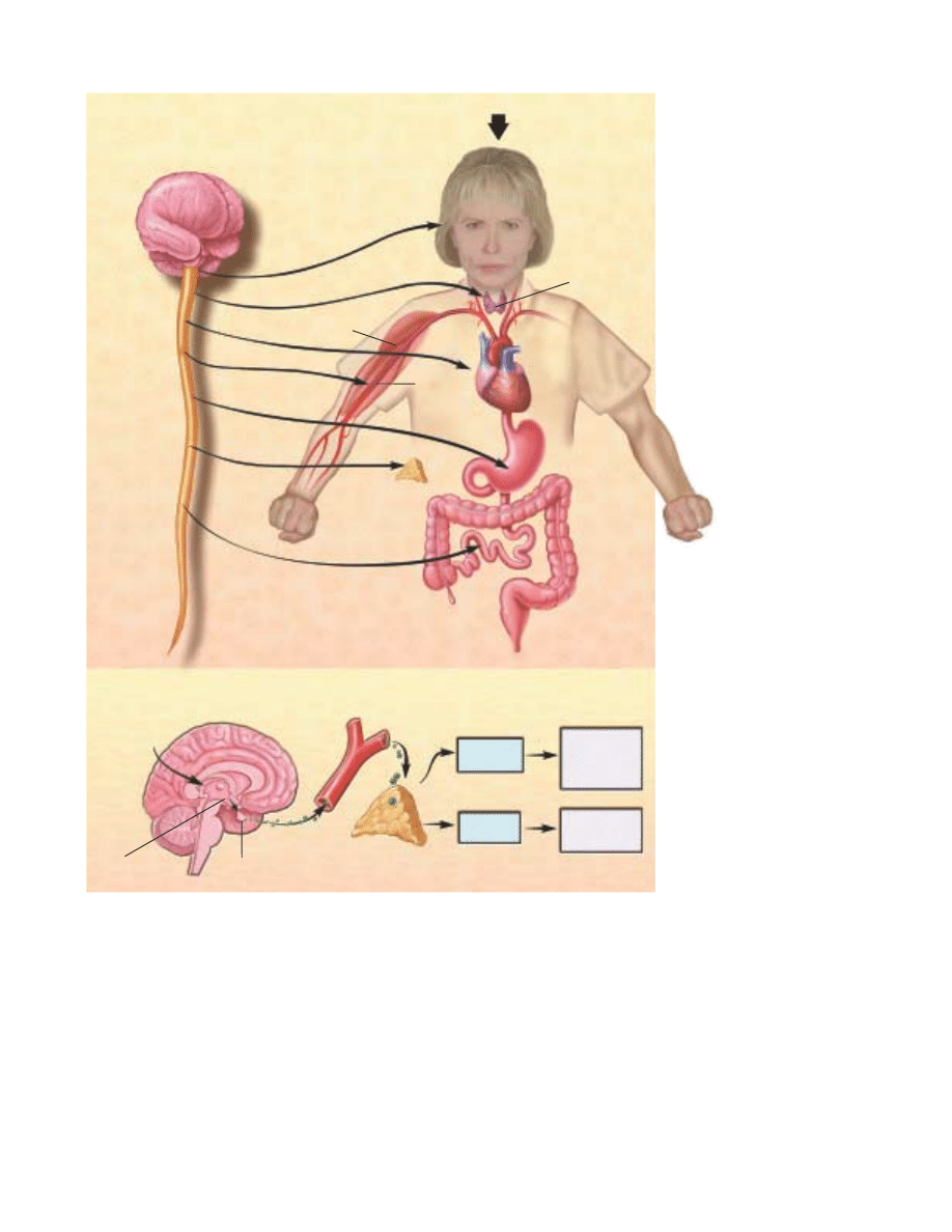
THE STRESS REACTION. When
stress occurs, the sympathetic
nervous system is triggered.
Norepinephrine is released by
nerves, and epinephrine is
secreted by the adrenal glands.
By activating receptors in blood
vessels and other structures,
these substances ready the
heart and working muscles for
action.
In the parasympathetic ner-
vous system, acetylcholine is
released, producing calming
e≈ects. The digestive tract is
stimulated to digest a meal, the
heart rate slows and the pupils
of the eye become smaller.
The neuroendocrine system
also maintains the body’s
normal internal function-
ing. Corticotrophin-releas-
ing factor (CRF), a peptide
formed by chains of amino acids,
is released from the hypothala-
mus, a collection of cells at the
base of the brain that acts as a
control center for the neuroen-
docrine system. CRF travels to
the pituitary gland where it trig-
gers the release of adrenocorti-
cotropic hormone (ACTH). ACTH
travels in the blood to the
adrenal glands where it stimu-
lates the release of cortisol.
26
NEUROENDOCRINE SYSTEM
AUTONOMIC
NERVOUS SYSTEM
Intestines
Stomach
Heart
Thymus and
Immune System
Muscle
Eyes
STRESS
Blood
vessels
Adrenal gland
Blood
stream
Epinephrine
Cortisol
Prepares body
for immediate
response
Re-establishes
homeostatis
Pituitary
Hypothalamus
ACTH
CRF
STRESS
epinephrine is quickly released into the bloodstream to put the
body into a general state of arousal and enable it to cope with
a challenge.
The secretion by the adrenal glands of cortisol—known as
a glucocorticoid because it a∑ects the metabolism of glucose, a
source of energy—starts about five minutes later. Some of its
actions help to mediate the stress-response, while some of its
other, slower ones, counteract the primary response to stress
and help re-establish homeostasis. Over the short run, cortisol
mobilizes energy and delivers it to muscles for the body’s
response. With prolonged exposure, cortisol enhances feeding
and helps the body recover from energy mobilization.
Acute stress also increases activity of the immune system
and helps protect the body from disease pathogens. The two
major stress hormones, cortisol and adrenaline, facilitate the
movement of immune cells from the bloodstream and storage
organs such as the spleen into tissue where they are needed to
defend against an infection.
Glucocorticoids also a∑ect food intake during the sleep-
wake cycle. Cortisol levels peak in the body in the early morn-
ing hours just before waking. This hormone acts as a wake-up
signal and helps to turn on appetite and physical activity. This

27
e∑ect of glucocorticoids may help to explain disorders, such as
jet-lag, which result when the light-dark cycle is altered by jet
travel over long distances, causing the body’s biological clock
to reset itself more slowly. Until that clock is reset, cortisol
secretion and hunger, as well as sleepiness and wakefulness,
occur at inappropriate times of day in the new location.
Glucocorticoids do more than help the body respond to
stress. In fact, they are an integral part of daily life and the
adaptation to environmental change. The adrenal glands help
protect us from stress and are essential for survival.
Chronic stress
When glucocorticoids or epinephrine are secreted in response
to the prolonged psychological stress commonly encountered
by humans, the results are not
ideal. Normally, bodily sys-
tems gear up under stress, and
hormones are released to
enhance muscular activity and
restore homeostasis. If you are
not fighting or fleeing—but
standing frustrated in a super-
market check-out line or sit-
ting in a tra≈c jam—you are
not engaging in muscular exer-
cise. Yet these systems continue to be stimulated.
Overexposure to cortisol also can lead to weakened mus-
cles and the suppression of major bodily systems. Elevated epi-
nephrine production increases blood pressure. Together, ele-
vated cortisol and epinephrine can contribute to chronic
hypertension (high blood pressure), abdominal obesity and ath-
erosclerosis (hardening of the arteries).
Scientists have identified a variety of stress-related disor-
ders, including colitis, high blood pressure, clogged arteries,
impotency and loss of sex drive in males, irregular menstrual
cycles in females, adult-onset diabetes and possibly cancer.
Aging rats show impairment of neuronal function in the hip-
pocampus—an area of the brain important for learning, mem-
ory and emotion—as a result of cortisol secretion throughout
their lifetimes. Overexposure to glucocorticoids also increases the
number of neurons damaged by stroke. Moreover, prolonged
exposure before or immediately after birth can cause a decrease
in the normal number of brain neurons and smaller brain size.
The immune system, which receives messages from the ner-
vous system, also is sensitive to many of the circulating hor-
mones of the body, including stress hormones. Moderate to
high levels of glucocorticoids act to suppress immune function,
although acute elevations of stress hormones actually facilitate
immune function.
Although acute stress-induced immunoenhancement can
be protective against disease pathogens, the glucocorticoid-
induced immunosuppression can also be beneficial. It reduces
inflammation and counteracts allergic reactions and autoim-
mune responses that occur when the body’s defenses turn against
body tissue. Synthetic glucocorticoids like hydrocortisone and
prednisone are used all the time to decrease inflammation and
autoimmunity. But glucocorticoids may be harmful in the case
of increased tumor growth associated with stress in experiments
on animals—an area of intense research yet to yield any final
conclusions.
One important determinant of the immune system’s resis-
tance or susceptibility to disease may be a person’s sense of
control as opposed to a feeling of helplessness. This phenom-
enon may help explain large individual variations in response
to disease. Scientists are trying to identify how the percep-
tion of control or helpless-
ness influences physiologi-
cal processes that regulate
immune function.
The cardiovascular system
receives many messages from
the autonomic nervous sys-
tem, and stressful experiences
have an immediate and direct
e∑ect on heart rate and blood
pressure. In the short run,
these changes help in response to stressors. But when stressors
are chronic and psychological, the e∑ect can be harmful and
result in accelerated atherosclerosis and increased risk for heart
attack. Research supports the idea that people holding jobs that
carry high demands and low control, such as telephone opera-
tors, waiters and cashiers, have higher rates of heart disease
than people who can dictate the pace and style of their work-
ing lives.
Behavioral type a∑ects a person’s susceptibility to heart
attack. People at greatest risk are hostile, irritated by trivial
things and exhibit signs of struggle against time and other
challenges.
Researchers found that two groups of men—one with high
hostility scores and the other with low hostility scores—exhibited
similar increases in blood pressure and blood muscle flow when
performing a lab test. This confirmed that hostility scores do
not predict the biological response to simple mental tasks.
Then the researchers added harassment to the test by lead-
ing the subjects to believe that their performances were being
unfairly criticized. Men with high hostility scores showed much
larger increases in muscle blood flow and blood pressure, and
slower recovery than those with low hostility scores. Scientists
found that harassed men with high hostility scores have larger
increases in levels of stress hormones. Thus, if you are a hostile
person, learning to reduce or avoid anger could be important to
avoid cardiovascular damage.
Stressful experiences have a direct e≈ect on
heart rate and blood pressure. When stressors
are chronic and psychological, the e≈ect can be
extremely harmful and result in an increased
risk for heart attack.

28
P
ablo Picasso, Georgia O’Keefe and Grandma
Moses, artists. Louise Nevelson, sculptor. Albert
Einstein, physicist. Giuseppe Verdi, musician.
Robert Frost, poet. Each of these great minds
worked di∑erently, but they all shared an out-
standing trait: They were creative and produc-
tive in old age. They defied the popular notion that aging always
leads to a pronounced decline and loss of cognitive ability.
Neuroscientists now believe that the brain can remain rel-
atively healthy and fully functioning as it ages, and that diseases
are the causes of the most severe decline in memory, intelligence,
verbal fluency and other tasks. Researchers are investigating the
normal changes that occur over time and the e∑ect that these
changes have on reasoning and other intellectual activities.
It appears that the e∑ects of age on brain function vary
widely. The vast majority of people get only a bit forgetful in
old age, particularly in forming memories of recent events. For
example, once you reach your 70s, you may start to forget names
or phone numbers, or respond more slowly to conflicting infor-
mation. This is not disease. However, other individuals develop
senile dementia, the progressive and severe impairment in men-
tal function that interferes with daily living. The senile demen-
tias include Alzheimer’s and cerebrovascular diseases and a∑ect
about one percent of people younger than age 65, with the inci-
dence increasing to nearly 50 percent in those older than 85. In
a small, third group, that includes the Picassos, Nevelsons and
others, mental functioning seems una∑ected by age. Many peo-
ple do well throughout life and continue to do well even when
they are old. The oldest human, Jeanne Calment, was consid-
ered to have all her wits during her 122-year lifespan.
It’s important to understand that scientific studies measure
trends and reflect what happens to the norm. They don’t tell what
happens to everybody. Some people in their 70s and 80s func-
tion as well as those in their 30s and 40s. The wisdom and expe-
rience of older people often make up for deficits in performance.
The belief that pronounced and progressive mental decline
is inevitable was and still is popular for several reasons. For one,
until the 20th century, few people lived to healthy old ages. In
1900, when life expectancy was about 47 years, three million
people, or four percent of the population, were older than age
65, and typically they were ill. In 1990, when life expectancy was
more than 75 years, 30 million people, or 12 percent of the pop-
ulation, were older than age 65. A generation ago, frailty was
seen among people in their 60s; today it is more typical among
those in their 80s. Moreover, few people challenged the notion
that aging meant inevitable brain decline because scientists
knew little about the brain or the aging process. Today’s under-
standing of how the normal brain ages comes from studies of
the nervous system that began decades ago and are just now
bearing results. Modern technologies now make it possible to
explore the structure and functions of the brain in more depth
than ever before and to ask questions about what actually hap-
pens in its aging cells.
Thus, neuroscientists are increasingly able to distinguish
between the processes of normal aging and disease. While some
changes do occur in normal aging, they are not as severe as sci-
entists once thought.
All human behavior is determined by how well the brain’s
communication systems work. Often a failure in the cascade of
one of these systems results in a disturbance of normal func-
tions. Such a failure may be caused by an abnormal biochemi-
cal process or by a loss of neurons.
The cause of brain aging still remains a mystery. Dozens of
theories abound. One says that specific “aging genes” are
switched on at a certain time of life. Another points to genetic
mutations or deletions. Other theories implicate hormonal
influences, an immune system gone awry and the accumulation
of damage caused by cell byproducts that destroy fats and pro-
teins vital to normal cell function.
Aging neurons
The brain reaches its maximum weight near age 20 and slowly
loses about 10 percent of its weight over a lifetime. Subtle
changes in the chemistry and structure of the brain begin at
midlife in most people. During a lifetime, the brain is at risk
for losing some of its neurons, but neuron loss is not a normal
process of aging. Brain tissue can respond to damage or loss of
neurons in Alzheimer’s disease or after stroke by expanding
Aging
P
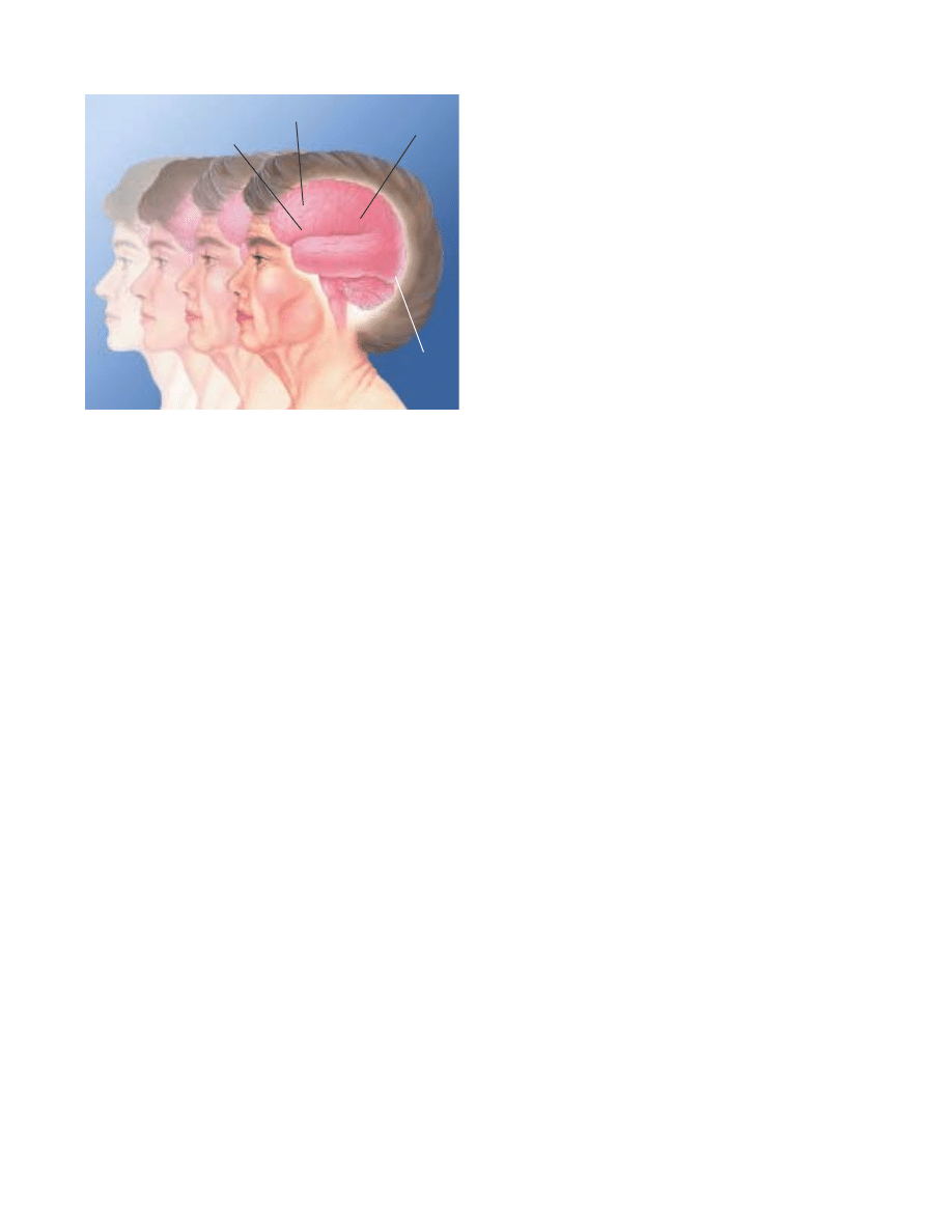
29
dendrites and refining connections between neurons. A dam-
aged brain neuron can readjust to damage only if its cell body
remains intact. If it does, regrowth can occur in dendrites and
axons. When neurons are completely destroyed, nearby surviv-
ing neurons can compensate, in part, by growing new dendrites
and connections.
Intellectual capacity
In the first large studies to follow the same group of normal
healthy humans for many years, scientists have uncovered unex-
pected results. They report declines in some mental functions
and improvements in others. In one study, the speed of carry-
ing out certain tasks became slower, but vocabulary improved.
Several studies found less severe declines in the type of intelli-
gence relying on learned or stored information, compared with
the type that uses the ability to deal with new information.
This research is supported by animal studies in which sci-
entists found that changes in mental function are subtle. For
example, in rodents and primates in which only minor brain
abnormalities can be detected, certain spatial tasks, such as nav-
igating to find food, tend to become more di≈cult with age.
The aging brain is only as resilient as its circuitry. Scien-
tists debate whether this circuitry is changed only by neuron
atrophy or whether some neuron loss over time also is in-
evitable. In any event, when the circuitry begins to break down,
remaining neurons can respond by expanding their roles.
Learning conditions may dictate what happens to brain
cells. Studies of rats shed light on some of the changes that
occur in brain cells when the animals live in challenging and
stimulating environments. In tests of middle-aged rats exposed
to these environments, researchers found that dendrites in the
cerebral cortex, which is responsible for all conscious activity,
developed more and longer branches when compared with rats
housed in isolated conditions. Another study showed that brain
cells in rats given acrobatic training had greater numbers of
synapses per cell than rats given only physical exercise or rats
who were inactive. The scientists concluded that motor learn-
ing generates new synapses. Physical exercise, however, improved
blood circulation in the brain.
Other scientists report that rats reared in a stimulating envi-
ronment made significantly fewer errors on a maze test than
did similar rats kept in an isolated environment. Moreover, the
stimulated rats showed an increase in brain weight and corti-
cal thickness when compared with the control animals.
Older rats tend to form new dendrites and synapses as do
younger animals in response to enriched environments. But the
response is more sluggish and not as large. Compared to younger
rats, the older rats have less growth of the new blood vessels
that nourish neurons.
While much has been learned about the aging brain, many
questions remain to be answered. For instance, does the pro-
duction of proteins decline with age in all brain neurons? In a
given neuron, does atrophy cause a higher likelihood of death?
How does aging a∑ect gene expression in the brain—the organ
with the greatest number of active genes? Are there gender
di∑erences in brain aging that may be due to hormonal changes
at menopause?
Neuroscientists speculate that certain genes may be linked to
events leading to death in the nervous system. By understanding
the biology of the proteins produced by genes, scientists hope to
be able to influence the survival and degeneration of neurons.
THE AGING BRAIN. Studies of people who have died contradict the
popular belief that adults lose an enormous number of neurons every
day. In fact, many areas of the brain, primarily in the cortex, maintain
most of their neurons. Examples include the parietal cortex, which
plays a role in sensory processes and language, and the striate cortex,
which processes visual information. However, neurons in regions far
below the cortex—such as the nucleus basalis, the principal compo-
nent of the basal forebrain—decrease in number with age, and cell
processes, such as axons and synapses, also change. The nucleus
basalis sends connections to the cortex that produce acetylcholine,
a chemical important for memory. Thus, these abnormalities may con-
tribute to the mental declines that occur in some elderly individuals.
Parietal cortex
Frontal cortex
Basal forebrain
Striate cortex

30
arkinson’s disease. This neurologic disorder af-
flicts one million individuals in the U.S., the
majority of whom are older than 50. Parkinson’s
is characterized by symptoms of slowness of
movement, muscular rigidity and tremor.
The discoveries in the late 1950s that the level of
dopamine was decreased in the brains of patients was followed
in the 1960s by the successful treatment of this disorder by
administration of the drug levodopa. This drug is changed to
dopamine in the brain. The successful treatment of Parkinson’s
by replacement therapy is one of the greatest success stories in
all of neurology. Levodopa is now combined with another drug,
carbidopa, that reduces the peripheral breakdown of levodopa,
thus allowing greater levels to reach the brain and reducing side
e∑ects. Also playing an important role are newer drugs such as
pergolide that act directly upon dopamine receptors and other
inhibitors of dopamine breakdown.
Genetic studies have demonstrated several inheritable gene
abnormalities in certain families, but the vast majority of cases
of Parkinson’s occur sporadically. It is believed that heredity fac-
tors may render some individuals more vulnerable to environ-
mental factors such as pesticides. The discovery in the late 1970s
that a chemical substance, MPTP, can cause parkinsonism in
drug addicts stimulated intensive research on the causes of the
disorder. MPTP was accidently synthesized by illicit drug
designers seeking to produce a heroin-like compound. MPTP
was found to be converted in the brain to a substance that
destroys dopamine neurons. Parkinson’s is now being inten-
sively studied in a primate MPTP model.
In the past several decades, scientists have shown in a pri-
mate model of Parkinson’s that specific regions in the basal gan-
glia, the collections of cell bodies deep in the brain, are abnor-
mally overactive. Most importantly, they found that surgical
destruction of these overactive nuclei—the pallidum and sub-
thalamic nucleus—can greatly reduce symptoms. The past
decade has witnessed a resurgence in this surgical procedure,
pallidotomy, and more recently chronic deep brain stimulation.
These techniques are highly successful for treating patients who
have experienced significant worsening of symptoms and are
troubled by the development of drug-related involuntary move-
ments. The past decade has also seen further attempts to treat
such patients with surgical implantation of cells, such as fetal
cells, capable of producing dopamine. Replacement therapy
with stem cells also is being explored.
Pain
If there is a universal experience, pain is it. Each year, more than
97 million Americans su∑er chronic, debilitating headaches or
a bout with a bad back or the pain of arthritis—all at a total
cost of some $100 billion. But it need not be that way. New dis-
coveries about how chemicals in the body transmit and inter-
cept pain have paved the way for new treatments for both
chronic and acute pain.
Until the middle of the 19th century, pain relief during
surgery relied on natural substances, such as opium, alcohol and
cannabis. All were inadequate and short-lived. Not until 1846
did doctors discover the anesthetic properties of ether, first in
animals and then in humans. Soon afterwards, the usefulness
of chloroform and nitrous oxide became known and heralded a
new era in surgery. The dozens of drugs used today during
surgery abolish pain, relax muscles and induce unconsciousness.
Other agents reverse these e∑ects.
Local anesthesia is used in a limited area of a person’s body
to prevent pain during examinations, diagnostic procedures,
treatments and surgical operations. The most famous of these
agents, which temporarily interrupt the action of pain-carrying
nerve fibers, is Novocain. Until recently, Novocain was used as
a local anesthetic by dentists; lidocaine is more popular today.
Analgesia produces loss of pain sensation without loss of sen-
sitivity to touch. The two main types of analgesics are nonopioids
(aspirin and related non-steroidal anti-inflammatory drugs such
as ibuprofen, naproxen and acetaminophen) and opioids (mor-
phine, codeine). Nonopioid analgesics are useful for treating
mild or moderate pain, such as headache or toothache. Mod-
erate pain also can be treated by combining a mild opioid, such
as codeine with aspirin. Opioids are the most potent pain-
killers and are used for severe pain, such as that occurring after
major chest or abdominal surgery.
Advances
P
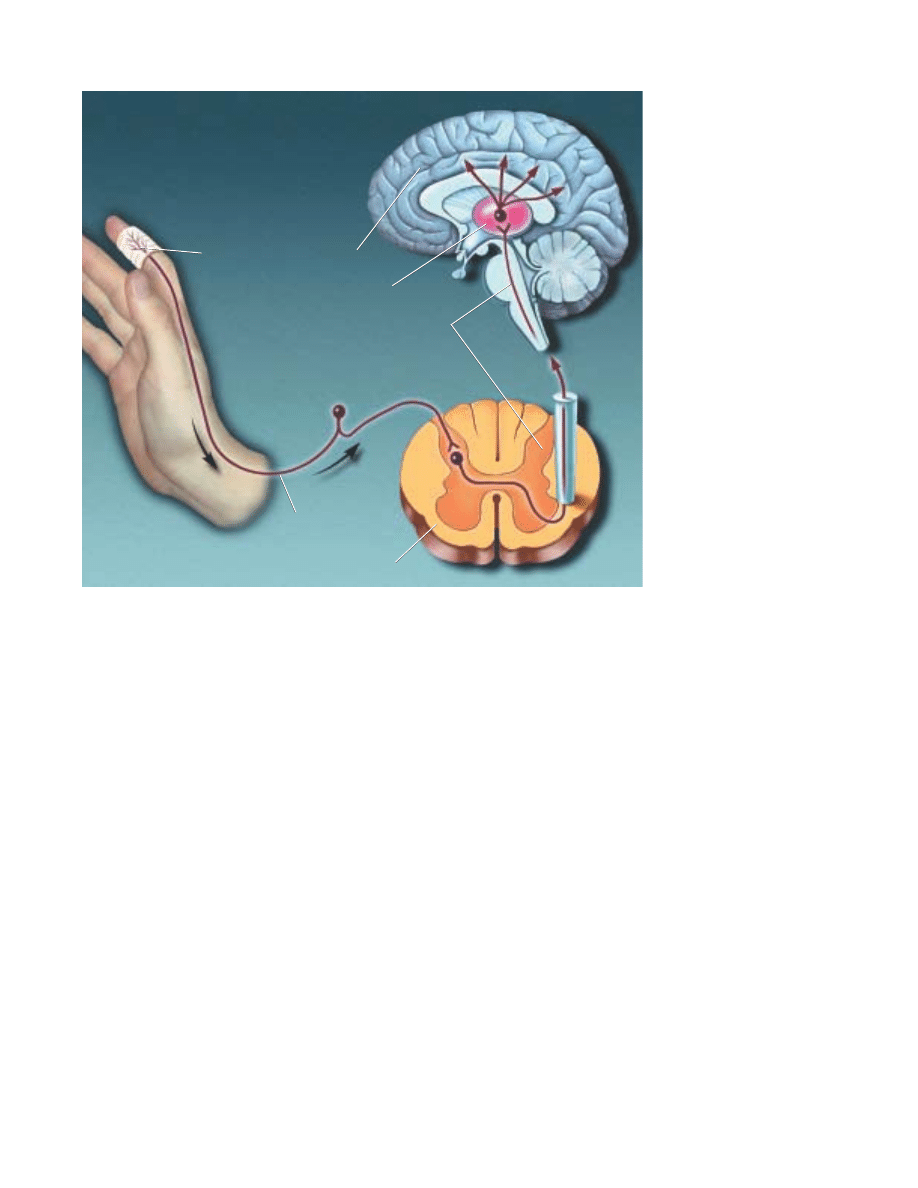
31
Insights into the body’s own pain-control system mediated
by naturally occurring opioids led to the use of injections of
morphine and endorphins, and other opioids, into the cere-
brospinal fluid in which the spinal cord is bathed without caus-
ing paralysis, numbness or other severe side e∑ects. This tech-
nique came about through experiments with animals that first
showed that injecting opioids into the spinal cord could pro-
duce profound pain control. This technique is now commonly
used in humans to treat pain after surgery.
New knowledge about other receptors and chemical medi-
ators involved in the transmission of pain are leading to the
development of new approaches to managing pain. These
include drugs that intercept pain messages at receptors that
bind glutamate, the major excitatory neurotransmitter in pain
pathways. Other studies are using molecular biology techniques
to identify specialized receptors and ion channels in nerve end-
ings that signal tissue damage of the skin, muscle or viscera.
These studies have the promise of leading to new classes of
analgesic agents in the future.
Epilepsy
A chronic neurological disorder characterized by sudden, dis-
orderly discharge of brain cells, epilepsy is marked by recurrent
seizures that temporarily alter one or more brain functions. The
disorder a∑ects approximately one percent of the population.
Many di∑erent forms of epilepsy have been recognized.
Epilepsy, which can start at any age, can result from inheriting
a mutant gene. It also can result from a wide variety of diseases
or injuries (including head injury), birth trauma, brain infec-
tion (such as meningitis), brain tumors, stroke, drug intoxica-
tion, drug or alcohol withdrawal states and metabolic disorders.
More than a dozen mutant genes that cause human epilepsy
have been identified during the past decade. In 70 percent of
cases, however, the cause is unknown.
Seizures are of two types. Generalized seizures, which result
in loss of consciousness, can cause several behavioral changes
including convulsions or sudden changes in muscle tone and
arise when there is excessive electrical activity over a wide area
of the brain. Partial seizures may occur in full consciousness or
with altered awareness, and can cause behavioral changes. They
can range from visual, auditory and sensory disturbances to
uncontrolled movements, and arise from excessive electrical
activity in a limited area of the brain.
The drug phenytoin was a major advance in the treatment
of epilepsy because it illustrated that antiseizure medications
need not cause sedation (as does phenobarbital, an older drug
for epilepsy) and encouraged the search for other drugs. Today
more than a dozen medications, approximately half of which
were introduced in the last several years, are used to prevent
seizures. The principal targets of antiseizure drugs are voltage-
HOW PAIN KILLERS WORK.
At the site of injury, the body
produces prostaglandins that
increase pain sensitivity.
Aspirin, which acts primarily in
the periphery, prevents the pro-
duction of prostaglandins.
Acetaminophen is believed to
block pain impulses in the brain
itself. Local anesthetics inter-
cept pain signals traveling up
the nerve. Opiate drugs, which
act primarily in the central ner-
vous system, block the transfer
of pain signals from the spinal
cord to the brain.
Aspirin acts here
Spinal cord
Local anesthetics
act here
Opiate drugs
act here
Cerebral cortex
Thalamus
P A I
N

32
gated ion channels permeable to sodium or calcium and synapses
using the transmitter GABA, a naturally occurring substance
in the brain that acts to inhibit electrical discharge. Identi-
fication of the mutant genes underlying human epilepsy is pro-
viding new targets for the next generation of antiseizure drugs.
In many instances, epilepsy can be controlled with a single
antiseizure drug that lessens the frequency of seizures. Some-
times a combination of drugs is necessary. Complete control of
seizures can be achieved in more than 50 percent of patients, and
another 25 percent can be improved significantly. It is hoped
that the newly available antiseizure drugs will provide complete
control in additional patients.
Surgery, considered for the patients who do not respond to
drugs, should be performed only at specialized medical centers
qualified to handle epilepsy surgery. One type of surgery requires
precise location and removal of the area of the brain where the
seizures originate. About 90 percent of properly selected patients
experience striking improvement or complete remission of
seizures. Another type of surgery separates the left and right
hemispheres of the brain to control a type of seizure that origi-
nates in one hemisphere and spreads to involve the whole brain.
A new form of epilepsy treatment, electrical stimulation
therapy, was introduced during the mid-1990s as another option
for hard-to-control seizures. The implantable pacemaker-like
device delivers small bursts of electrical energy to the brain via
the vagus nerve on the side of the neck.
Major depression
This aΩiction, with its harrowing feelings of sadness, hope-
lessness, pessimism, loss of interest in life and reduced emo-
tional wellbeing, is one of the most common and debilitating
mental disorders. Depression is as disabling as coronary disease
or arthritis. Depressed individuals are 18 times more likely to
attempt suicide than people with no mental illness.
Annually, major depression a∑ects five percent of the pop-
ulation or 9.8 million Americans aged 18 years and older. For-
tunately, 80 percent of patients respond to drugs, psychother-
apy or a combination of the two. Some severely depressed
patients can be helped with electroconvulsive therapy.
Depression arises from many causes: biological (including
genetic), psychological, environmental or a combination of
these. Stroke, hormonal disorders, antihypertensives and birth
control pills also can play a part.
Physical symptoms—disturbances of sleep, sex drive,
appetite and digestion—are common. Some of these symptoms
may reflect the fact that the disorder a∑ects the delicate hor-
monal feedback system linking the hypothalamus, the pituitary
gland and the adrenal glands. For example, many depressed
patients secrete excess cortisol, a stress hormone, and do not
respond appropriately to a hormone that should counter corti-
sol suppression. When tested in sleep laboratories, depressed
patients’ electroencephalograms (EEGs) often exhibit abnor-
malities in their sleep patterns.
The modern era of drug treatment for depression began in
the late 1950s. Most antidepressants a∑ect norepinephrine and
serotonin in the brain, apparently by correcting the abnormal
excess or inhibition of the signals that control mood, thoughts,
pain and other sensations. The tricyclic antidepressants pri-
marily block the reabsorption and inactivation of serotonin and
norepinephrine to varying degrees.
Another class of antidepressant medications is the mono-
amine oxidase inhibitors (MAOIs). MAOIs are thought to be
more complicated than tricyclics. These agents inhibit mon-
amine oxidase, an enzyme that breaks down serotonin, norepi-
nephrine and dopamine, allowing these chemicals to remain
active. During the 1950s, the first of the MAOIs, iproniazid,
was found to make experimental animals hyperalert and hyper-
active. By 1957, scientists had proven iproniazid’s benefit in
patients. Later, other MAOIs were developed. Today, three are
available for use: isocarboxazid, phenelzine and tranylcypromine.
The popular medication fluoxetine (Prozac) is the first of a
new class of drugs, serotonin reuptake inhibitors. Fluoxetine
blocks the reabsorption and inactivation of serotonin and keeps
it active in certain brain circuits. This seems to restore overall
serotonin activity to a more normal state and ease depression.
Manic-depressive illness
Patients with manic-depressive illness, also known as bipolar
disorder, usually experience episodes of deep depression and
manic highs, with a return to relatively normal functioning in
between. They also have an increased risk of suicide. Manic
depression a∑ects 1.2 percent of Americans age 18 or older
annually, or 2.2 million individuals. Approximately equal num-
bers of men and women su∑er from this disorder.
Manic-depressive disorder tends to be chronic, and episodes
can become more frequent without treatment. Because manic
depression runs in families, e∑orts are under way to identify the
responsible gene or genes.
However, manic-depressive patients also can benefit from
a broad array of treatments. One of these is lithium. During the
1940s, researchers showed that lithium injections into guinea
pigs made them placid, which implied mood-stabilizing e∑ects.
When given to manic patients, lithium calmed them and
enabled them to return to work and live relatively normal lives.
Regarded as both safe and e∑ective, lithium is often used to
prevent recurrent episodes.
Other useful medications include anticonvulsants, such as
valproate or carbamazepine, which can have mood-stabilizing
e∑ects and may be especially useful for di≈cult-to-treat bipo-
lar episodes. Newer anticonvulsant medications, are being
studied to determine how well they work in stabilizing mood
cycles.

33
A
ddiction. Drug abuse is one of the nation’s
most serious health problems. Indeed, six per-
cent of Americans, roughly 15 million people,
abuse drugs on a regular basis. Recent esti-
mates show that the abuse of drugs, includ-
ing alcohol and nicotine from tobacco, costs
the nation more than $276 billion each year.
If continued long enough, drug abuse—often defined as
harmful drug use—can eventually alter the very structure of the
brain, producing a true brain disorder. This disorder is called
drug addiction or drug dependence. Drug addiction is defined as
having lost much control over drug taking, even in the face of
adverse physical, personal or social consequences.
People abuse drugs for a simple reason: Drugs produce feel-
ings of pleasure, or they remove feelings of stress and emotional
pain. Neuroscientists have found that almost all abused drugs
produce pleasure by activating a specific network of neurons
called the brain reward system. The circuit is normally involved
in an important type of learning that helps us to stay alive. It is
activated when we fulfill survival functions, such as eating when
we are hungry or drinking when we are thirsty. In turn, our
brain rewards us with pleasurable feelings that teach us to
repeat the task. Because drugs inappropriately turn on this
reward circuit, people want to repeat drug use.
Neuroscientists have also learned specifically how drugs
a∑ect neurons to exert their actions. Neurons release special
chemicals, called neurotransmitters, to communicate with each
other. Drugs of abuse alter the ways in which neurotransmitters
carry their messages from neuron to neuron. Some drugs mimic
neurotransmitters while others block them. Still others alter the
way that the neurotransmitters are released or inactivated. The
brain reward system is inappropriately activated because drugs
alter the chemical messages sent among neurons in this circuit.
Finally, neuroscientists also have learned that addiction
requires more than the activation of the brain reward system.
The process of becoming addicted appears to be influenced by
many factors. Motivation for drug use is an important one. For
example, people who take drugs to get high may get addicted,
but people who use them properly as medicine rarely do. Also
genetic susceptibility or environmental factors, like stress, may
alter the way that people respond to drugs. In addition, the
development of tolerance—the progressive need that accompa-
nies chronic use for a higher drug dose to achieve the same
e∑ect—varies in di∑erent people. So does drug dependence—
the adaptive physiological state that results in withdrawal
symptoms when drug use stops. While tolerance and depen-
dence are standard responses of the brain and body to the pres-
ence of drugs, addiction requires that these occur while a moti-
vational form of dependence—the feeling that a person can’t live
without a drug, accompanied by negative a∑ective states—is
also developing. Together these insights on abuse and addic-
tion are leading to new therapies.
Nicotine Some 57 million Americans were current smokers
in 1999, and another 7.6 million used smokeless tobacco, mak-
ing nicotine one of the most widely abused substances. Tobacco
kills more than 430,000 U.S. citizens each year—more than
alcohol, cocaine, heroin, homicide, suicide, car accidents, fire,
and AIDS combined. Tobacco use is the leading preventable
cause of death in the United States. Smoking is responsible for
approximately seven percent of total U.S. health care costs, an
estimated $80 billion each year. The direct and indirect costs of
smoking are estimated at more than $138 billion per year.
Nicotine acts through the well known cholinergic nicotinic
receptor. This drug can act as both a stimulant and a sedative.
Immediately after exposure to nicotine, there is a “kick” caused
in part by the drug’s stimulation of the adrenal glands and
resulting discharge of epinephrine. The rush of adrenaline stim-
ulates the body and causes a sudden release of glucose as well
as an increase in blood pressure, respiration and heart rate.
Nicotine also suppresses insulin output from the pancreas,
which means that smokers are always slightly hyperglycemic.
In addition, nicotine indirectly causes a release of dopamine in
the brain regions that control pleasure and motivation. This is
thought to underlie the pleasurable sensations experienced by
many smokers.
Much better understanding of addiction, coupled with the
identification of nicotine as an addictive drug, has been instru-
mental in developing treatments. Nicotine gum, the transder-
A
Challenges
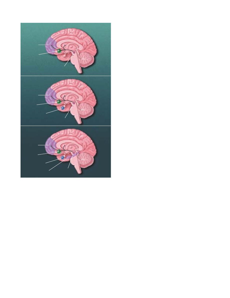
34
mal patch, nasal spray and inhaler all appear to be equally
e∑ective in successfully treating more than one million people
addicted to nicotine. These techniques are used to relieve with-
drawal symptoms, produce less severe physiological alterations
than tobacco-based systems and generally provide users with
lower overall nicotine levels than they receive with tobacco. The
first non-nicotine prescription drug, bupropion, an antidepres-
sant marketed as Zyban, has been approved for use as a phar-
macological treatment for nicotine addiction. Behavioral treat-
ments are important for helping an individual learn coping
skills for both short- and long-term prevention of relapse.
Psychostimulants In 1997, 1.5 million Americans were cur-
rent cocaine users. A popular, chemically altered form of
cocaine, crack, is smoked. It enters the brain in seconds, pro-
ducing a rush of euphoria and feelings of power and self-
confidence. The key biochemical factor that underlies the rein-
forcing e∑ects of psychostimulants is the brain chemical
dopamine. We feel pleasure-like e∑ects when dopamine-
containing neurons release dopamine into specific brain areas
that include a special portion of the nucleus accumbens.
Cocaine and amphetamines produce their intense feelings of
euphoria by increasing the amount of dopamine that is avail-
able to send messages within the brain reward system.
Cocaine users often go on binges, consuming a large
amount of the drug in just a few days. A “crash” occurs after
this period of intense drug-taking and includes symptoms of
emotional and physical exhaustion and depression. These
symptoms may result from an actual crash in dopamine func-
tion and the activity of another brain chemical, serotonin, as
well as an increase in the response of the brain systems that
react to stress. Vaccines to produce antibodies to cocaine in the
bloodstream are in clinical trials.
Opiates Humans have used opiate drugs, such as mor-
phine, for thousands of years. Monkeys and rats readily self-
administer heroin or morphine and, like humans, will become
tolerant and physically dependent with unlimited access. With-
drawal symptoms range from mild flu-like discomfort to major
physical ailments, including severe muscle pain, stomach
cramps, diarrhea and unpleasant mood.
Opiates, like psychostimulants, increase the amount of
dopamine released in the brain reward system and mimic the
e∑ects of endogenous opioids such as opioid peptides. Heroin
injected into a vein reaches the brain in 15 to 20 seconds and
binds to opiate receptors found in many brain regions, includ-
ing the reward system. Activation of the receptors in the reward
circuits causes a brief rush of intense euphoria, followed by a
couple of hours of a relaxed, contented state.
Opiates create e∑ects like those elicited by the naturally
occurring opioid peptides. They relieve pain, depress breath-
ing, cause nausea and vomiting, and stop diarrhea—important
medical uses. In large doses, heroin can make breathing shal-
BRAIN DRUG REWARD SYSTEMS. Scientists are not certain about
all the structures involved in the human brain reward system. How-
ever, studies of rat and monkey brains, and brain imaging studies in
humans, have provided many clues. These illustrations show what
areas are most likely part of the reward systems in the human brain.
A central group of structures is common to the actions of all drugs.
These structures include a collection of dopamine-containing neurons
found in the ventral tegmental area. These neurons are connected to
the nucleus accumbens and other areas, such as the prefrontal cor-
tex. Cocaine exerts its e≈ects mainly through this system. Opiates act
in this system and many other brain regions, including the amygdala,
that normally use opioid peptides. Opioids are naturally occurring
brain chemicals that induce the same actions as drugs, such as heroin
and morphine. Alcohol activates the core reward system and addi-
tional structures throughout the brain because it acts where GABA
and glutamate are used as neurotransmitters. GABA and glutamate
are widely distributed in the brain, including the cortex, hippocam-
pus, amygdala and nucleus accumbens.
COCAINE AND AMPHETAMINES
OPIATES
ALCOHOL
Hippocampus
Amygdala
Prefrontal
cortex
Nucleus
accumbens
Ventral
tegmental area
Amygdala
Prefrontal
cortex
Nucleus
accumbens
Ventral
tegmental area
Prefrontal
cortex
Nucleus
accumbens
Ventral
tegmental area
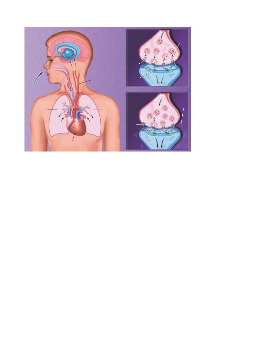
Dopamine
transporter
35
low or stop altogether—the cause of death in thousands of people who have died of a heroin
overdose.
A standard treatment for opiate addiction involves methadone, a long-acting oral opiate that
helps keep craving, withdrawal and relapse under control. Methadone helps opiate addicts reha-
bilitate themselves by preventing withdrawal symptoms that are powerful motivators of drug use.
A synthetic opiate, known as LAAM, can block the e∑ects of heroin for up to 72 hours with min-
imal side e∑ects when taken orally. In 1993 the Food and Drug Administration approved the use
of LAAM for treating patients addicted to heroin. Its long duration of action permits dosing just
three times per week, thereby eliminating the need for daily dosing. LAAM will be increasingly
available in clinics that already dispense methadone. Naloxone and naltrexone are medications
that also block the e∑ects of morphine, heroin and other opiates. As antagonists, they are espe-
cially useful as antidotes. Another medication to treat heroin addiction, buprenorphine, causes
weaker opiate e∑ects and is less likely to cause overdose problems.
Alcohol Although legal, alcohol is highly addictive. Alcohol abuse and alcohol addiction
(sometimes referred to as alcoholism or alcohol dependence) are the nation’s major drug problem,
with some people being more susceptible to them than others. Nearly 14 million abuse alcohol or
are alcoholic. Fetal alcohol syndrome is the leading preventable cause of mental retardation. It a∑ects
about 0.5 to 3 of every 1,000 babies born in the United States. Chronic liver diseases, including
cirrhosis—the main chronic health problem associated with alcohol addiction—are responsible
for more than 25,000 deaths each year. The annual cost of alcohol abuse and addiction is estimated
at $185 billion.
Genetic and environmental factors contribute to alcoholism, but no single factor or combi-
nation of factors enables doctors to predict who will become an alcoholic.
Ethanol, the active ingredient in alcoholic beverages, reduces anxiety, tension and inhibitions.
In low doses it may act as a stimulant, whereas at higher doses, it acts as a depressant. In both
cases, it significantly alters mood and behavior. It can also cause heat loss and dehydration.
The drug, which is easily absorbed into the bloodstream and the brain, a∑ects several neuro-
transmitter systems. For example, alcohol’s interaction with the GABA receptor can calm anxi-
ety, impair muscle control and delay reaction time. At higher doses, alcohol also decreases the
HOW CRACK COCAINE AFFECTS
THE BRAIN. Crack cocaine takes
the same route as nicotine by
entering the bloodstream
through the lungs. Within sec-
onds, it is carried by the blood
to the brain. The basis for
increased pleasure occurs at the
gap where the impulses that
represent neural messages are
passed from one neuron to
another. This gap is called a
synapse. Dopamine-containing
neurons normally relay their
signals by releasing dopamine
into many synapses. Dopamine
crosses the synapse and fits
into receptors on the surface of
the receiving cell. This triggers
an electrical signal that is
relayed through the receiver.
Then, to end the signal,
dopamine molecules break
away from the receptors and are
pumped back into the nerve ter-
minals that released them.
Cocaine molecules block the
pump or “transporter,” causing
more dopamine to accumulate in
the synapse. Pleasure circuits
are stimulated again and again,
producing euphoria.
NORMAL
COCAINE HIGH
Right
lung
Left lung
Dopamine
recycled
Dopamine
transporter
Dopamine
Dopamine
receptor site
Transmitter
cell
Receiver
cell
Crack
cocaine
inhaled
into
lungs
Blood carries cocaine
to brain in seconds
Heart
Cocaine
blocks
trans-
porter
Dopamine
not
recycled

36
function of NMDA receptors that recognize the neurotrans-
mitter glutamate. This interaction can cloud thinking and even-
tually lead to a coma.
Researchers are developing treatments, which interfere with
molecules, such as the opioid peptides, and trigger alcohol’s
positive reinforcing e∑ects. One such drug, naltrexone, recently
has been approved for treating alcoholism.
Marijuana This drug can distort perception, and alter the
sense of time, space and self. In certain situations, marijuana
can produce intense anxiety.
In radioactive tracing studies, scientists found that tetrahy-
drocannabinol (THC), the active ingredient in marijuana, binds
to specific receptors, many of which coordinate movement. This
may explain why people who drive after they smoke marijuana
are impaired. The hippocampus, a structure involved with
memory storage and learning, also contains many THC recep-
tors. This may explain why heavy users or those intoxicated on
marijuana have poor short-term memory and problems pro-
cessing complex information. Scientists recently discovered that
these receptors normally bind to a natural internal chemical
called anandamide, and are now working to see how this nat-
ural marijuana a∑ects brain function.
Club Drugs Ecstasy, Herbal Ecstasy, Rohypnol, GHB and
ketamine are among the drugs used by some teens and young
adults as part of rave and trance events which are generally
night-long dances, often held in warehouses. The drugs are
rumored to increase stamina and to produce intoxicating highs
that are said to deepen the rave or trance experience. Recent
hard science, however, is showing serious damage to several
parts of the brain from use of some of these drugs.
Many users tend to experiment with a variety of club drugs
in combination. This creates a larger problem because combi-
nations of any of these drugs, particularly with alcohol, can lead
to unexpected adverse reactions and even death after high
doses. Physical exhaustion can enhance some toxicities and
problems.
MDMA, called “Adam,” “Ecstasy,” or “XTC,” on the street,
is a synthetic, psychoactive drug with hallucinogenic and
amphetamine-like properties. Users encounter problems similar
to those found with the use of amphetamines and cocaine.
Recent research also links Ecstasy use to long-term damage to
those parts of the brain critical to thought, memory and pleasure.
Rohypnol, GHB, and ketamine are predominantly central
nervous system depressants. Because they are often colorless,
tasteless and odorless, they can be easily added to beverages and
ingested unknowingly. These drugs have emerged as the so-
called “date rape” drugs. When mixed with alcohol, Rohypnol
can incapacitate a victim and prevent them from resisting sex-
ual assault. Also, Rohypnol may be lethal when mixed with
alcohol and other depressants. Since about 1990, GHB (gamma
hydroxy-butyrate) has been abused in the U.S. for euphoric, seda-
tive and anabolic (body building) e∑ects. It, too, has been asso-
ciated with sexual assault. Ketamine is another central nervous
system depressant abused as a “date rape” drug. Ketamine, or
“Special K,” is a rapid-acting general anesthetic. It has sedative-
hypnotic, analgesic and hallucinogenic properties. It is marketed
in the U.S. and a number of foreign countries for use as a gen-
eral anesthetic in both human and veterinary medical practice.
Alzheimer’s disease
One of the most frightening and devastating of all neurologi-
cal disorders, is the dementia that occurs in the elderly. The
most common cause of this illness is Alzheimer’s disease (AD).
Rare before age 60 but increasingly prevalent in each decade
thereafter, AD a∑ects an estimated 4 to 5 million Americans.
By the year, 2040, it is predicted to a∑ect approximately 14 mil-
lion individuals in the U.S.
The earliest symptoms are forgetfulness and memory loss;
disorientation to time or place; and di≈culty with concentra-
tion, calculation, language and judgment. Some patients have
severe behavioral disturbances and may even become psychotic.
The illness is progressive. In the final stages, the a∑ected indi-
vidual is incapable of self-care. Unfortunately, no e∑ective treat-
ments exist, and patients usually die from pneumonia or some
other complication. AD, which kills 100,000 people a year, is
one of the leading causes of death in the U.S.
In the earliest stages, the clinical diagnosis of possible or
probable AD can be made with greater than 80 percent accu-
racy. As the course of the disease progresses, the accuracy of
diagnosis at Alzheimer’s research centers exceeds 90 percent.
The diagnosis depends on medical history, physical and neuro-
logical examinations, psychological testing, laboratory tests and
brain imaging studies. At present, final confirmation of the
diagnosis requires examination of brain tissue, usually obtained
at autopsy.
The causes and mechanisms of the brain abnormalities are
not yet fully understood, but great progress has been made
through genetics, biochemistry, cell biology and experimental
treatments. Microscopic examination of AD brain tissue shows
abnormal accumulations of a small fibrillar peptide, termed a
beta amyloid, in the spaces around synapses (neuritic plaques),
and by abnormal accumulations of a modified form of the pro-
tein tau in the cell bodies of neurons (neurofibrillary tangles).
The plaques and tangles are mostly in brain regions important
for memory and intellectual functions.
In cases of AD, there are reductions in levels of markers for
several neurotransmitters, including acetylcholine, somato-
statin, monoamine and glutamate, that allow cells to commu-
nicate with one another. Damage to these neural systems,
which are critical for attention, memory, learning and higher
cognitive abilities, is believed to cause the clinical symptoms.
Approximately five percent to 10 percent of individuals with

37
AD have an inherited form of the disease. These patients often
have early-onset illness. Recently, scientists have identified
mutations in AD-linked genes on three chromosomes. The gene
encoding the amyloid precursor protein gene is on chromosome
21. In other families with early-onset AD, mutations have been
identified in the presenilin 1 and 2 genes, which are on chro-
mosomes 14 and 1, respectively. Apolipoprotein E (apoE), a chro-
mosome 19 gene, which influences susceptibility in late life, exists
in three forms, with apoE4 clearly associated with enhanced risk.
Treatments are available mostly only for some symptoms of
AD, such as agitation, anxiety, unpredictable behavior, sleep
disturbances and depression. Three drugs treat cognitive symp-
toms in patients with mild to moderate Alzheimer’s. These
agents improve memory deficits temporarily and modestly in
20 percent to 30 percent of patients. Several other approaches,
such as antioxidants, anti-inflammatories and estrogens, are
being tested.
An exciting new area of research is the use of approaches
in which genes are introduced in mice. These transgenic mice
carrying mutant genes linked to inherited AD develop behav-
ioral abnormalities and some of the cellular changes that occur
in humans. It is anticipated that these mice models will prove
very useful for studying the mechanisms of AD and testing
novel therapies.
Moreover, researchers have begun to knock out genes play-
ing critical roles in the production of amyloid. These enzymes,
termed beta and gamma secretase, which cleave the amyloid
peptide from the precursor, are clearly targets for development
of drugs to block amyloid.
Learning disorders
An estimated 10 percent of the population, as many as 25 mil-
lion Americans, have some form of learning disability involv-
ing di≈culties in the acquisition and use of listening, speaking,
reading, writing, reasoning or mathematical abilities. They
often occur in people with normal or high intelligence.
Dyslexia, or specific reading disability, is the most common
and most carefully studied of the learning disabilities. It a∑ects
80 percent of all of those identified as learning-disabled.
Dyslexia is characterized by an unexpected di≈culty in reading
in children and adults who otherwise possess the intelligence,
motivation and schooling considered necessary for accurate and
fluent reading.
Previously, it was believed that dyslexia a∑ected boys pri-
marily. However, more recent data indicate similar numbers of
boys and girls are affected. Studies indicate that dyslexia is a
persistent, chronic condition. It does not represent a tran-
sient “developmental lag.”
There is now a strong consensus that the central di≈culty
in dyslexia reflects a deficit within the language system, and
more specifically, in a component of the language system called
phonology. This is illustrated in di≈culty transforming the let-
ters on the page to the sound structure of the language.
As children approach adolescence, a manifestation of
dyslexia may be a very slow reading rate. Children may learn to
read words accurately but they will not be fluent or automatic,
reflecting the lingering e∑ects of a phonologic deficit. Because
they are able to read words accurately (albeit very slowly), dyslexic
adolescents and young adults may mistakenly be assumed to
have “outgrown” their dyslexia. The ability to read aloud accu-
rately, rapidly and with good expression as well as facility with
spelling may be most useful clinically in distinguishing students
who are average from those who are poor readers.
A range of investigations indicates that there are di∑erences
in the temporo-parieto-occipital brain regions between dyslexic
and non-impaired readers. Recent data using functional brain
imaging indicate that dyslexic readers demonstrate a functional
disruption in an extensive system in the posterior portion of the
brain. The disruption occurs within the neural systems linking
visual representations of the letters to the phonologic structures
they represent. The specific cause of the disruption in neuronal
systems in dyslexia is not entirely understood. However, it is
clear that dyslexia runs in families and can be inherited.
Interventions to help children with dyslexia focus on teach-
ing the child that words can be segmented into smaller units of
sound and that these sounds are linked with specific letters and
letter patterns. In addition, children with dyslexia require prac-
tice in reading stories, both to allow them to apply their newly
acquired decoding skills to reading words in context and to
experience reading for meaning.
Stroke
Until recently, if you or a loved one had a stroke, your doctor
would tell your family there was no treatment. In all likelihood,
the patient would live out the remaining months or years with
severe neurological impairment.
This dismal scenario has now been radically altered. For
one, use of the clot-dissolving bioengineered drug, tissue plas-
minogen activator (tPA), is now a standard treatment in many
hospitals. This approach rapidly opens blocked vessels to
restore circulation before oxygen loss causes permanent dam-
age. Given within three hours of a stroke, it often can help in
returning patients to normal.
Also, attitudes about the nation’s third leading cause of
death are changing rapidly. Much of this has come from new
and better understandings of the mechanisms that lead to the
death of neurons following stroke and devising ways to protect
these neurons. A variety of chemicals appear to play a role,
including calcium, potassium and zinc, and may be important
in devising new treatments.
Stroke a∑ects roughly 700,000 Americans a year — 150,000
of whom die; total annual costs are estimated at $43 billion.
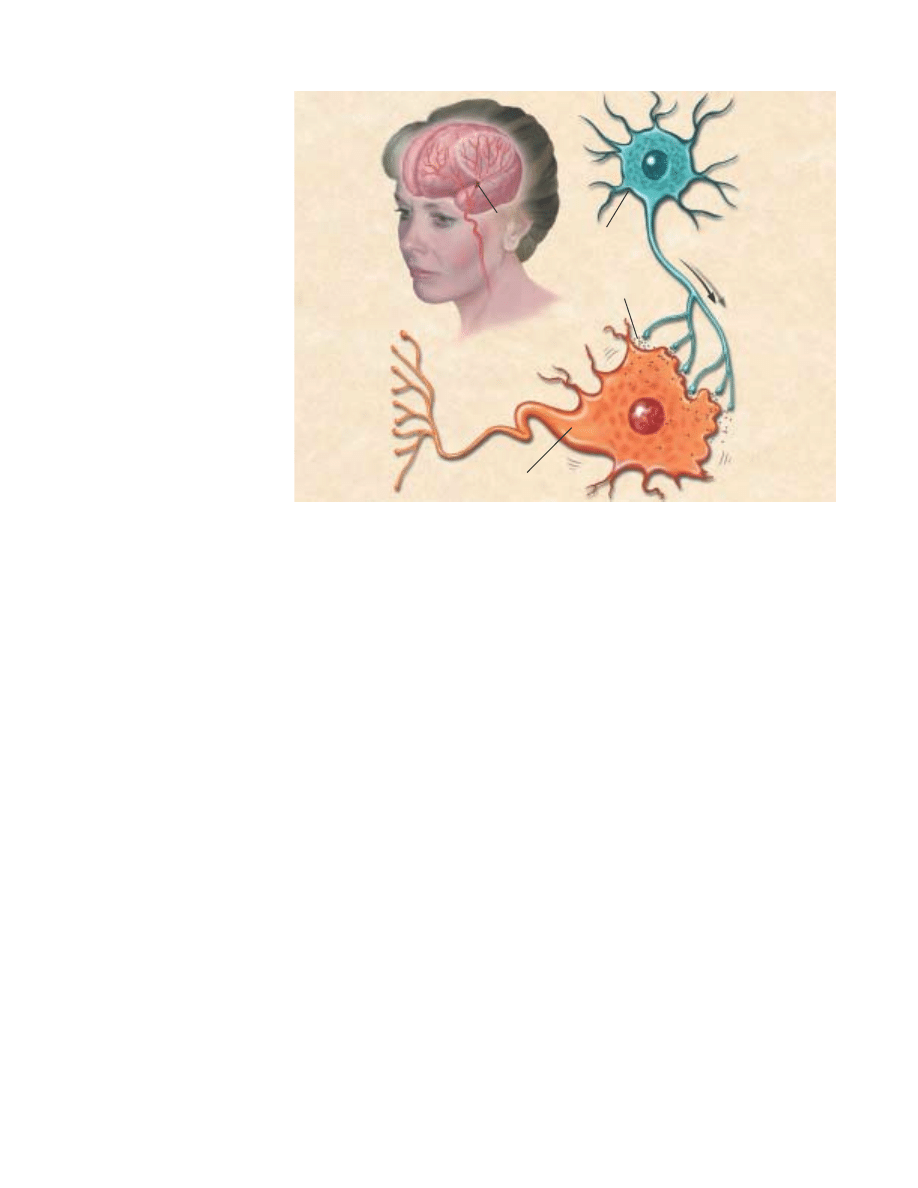
38
A stroke occurs when a blood vessel bringing oxygen and
nutrients to the brain bursts or is clogged by a blood clot or
some other particle. This deprives the brain of blood, causing
the death of neurons within minutes. Depending on its loca-
tion, a stroke can cause many permanent disorders, such as
paralysis on one side of the body and loss of speech.
Stroke often occurs in individuals over 65 years of age, yet
a third are younger. Stroke tends to occur more in males and
blacks and in those with diabetes, high blood pressure, heart
disease, obesity, high cholesterol and a family history of stroke.
In addition to tPA, increased use of preventive measures are
battling the disorder. Controlling risk factors such as obesity,
blood pressure, diabetes and high cholesterol can help prevent
stroke. Other specific treatments involving surgery can clear
clogs in the arteries of the neck region and help prevent a cut-
o∑ of blood supply.
Treatments that target the heart’s blood flow can prevent
stroke. Surgery can help repair damaged heart valves. Drugs
can reduce the chance of clots forming, traveling to the brain
and causing a stroke.
Other experimental therapies under investigation may lead
to even bigger payo∑s for patients in the future. Some strate-
gies target mechanisms inside the neuron. In this way, the
vicious cycle of local damage followed by a widening fringe of
biochemical-induced neuronal death can be slowed. A number
of classes of drugs have been shown to be e∑ective in animal
studies.
Another promising possibility is the use of neural stem
cells. Some animal studies have shown that an injection of stem
cells aids recovery even if administered as long as a day after
the injury. Stem cells administered along with a growth factor
resulted in greater improvement than with either treatment
alone. The double regimen resulted in behavioral improvement
as well as decreased stroke-induced brain loss.
Neurological trauma
A magic bullet has not been found, but doctors have discovered
several methods to stave o∑ severe neurological damage caused
by head and spinal cord injuries. These treatments include bet-
ter emergency care, a drug to help reduce damage and improved
rehabilitation techniques.
Some 500,000 people su∑er traumatic head injuries requir-
ing hospitalization each year, and roughly 100,000 die—many
before reaching the hospital. Economic costs approach $25 bil-
lion annually.
Greater use of computed tomography (CT) and magnetic res-
onance imaging (MRI) allows doctors more readily to see poten-
tially life-threatening swelling and act immediately more read-
ily. Doctors can bore a small hole in the skull and insert a tube
attached to a pressure monitor. When the intracranial pressure
is above safe levels, the patient is put on a ventilator to increase
the breathing rate. The more breaths a patient takes, the more
carbon dioxide is blown o∑, helping to shrink cerebral blood
vessels and thus reduce intracranial pressure. Drugs, such as
mannitol, help draw water away from the brain.
Doctors also now can identify blood clots with CT and
MRI scans and remove the clots before a patient completely
deteriorates.
STROKE. A stroke occurs when a
blood vessel bringing oxygen and
nutrients to the brain bursts or is
clogged by a blood clot. This lack of
blood can cause cell death within
minutes. One theory is that the
overexcited dying nerve cells re-
lease neurotransmitters, especially
glutamate, onto nearby nerve cells.
These nearby nerve cells become
overexcited and overloaded with
calcium and die. This is one of the
places where scientists think they
may be able to intervene to stop the
process of cell death. Depending on
its location, a stroke can have
di≈erent symptoms. They include
paralysis on one side of the body
or a loss of speech. The e≈ects of
stroke are often permanent because
dead brain cells are not replaced.
Glutamate released
Overexcited injured cell
Cell lacking
blood flow
Blockage

39
An estimated 250,000 individuals are living with spinal
cord injury in the U.S. Some 11,000 new injuries are reported
annually and are caused mostly by motor vehicle accidents, vio-
lence and falls. Economic costs approach $10 billion a year.
Researchers have found that people who su∑er spinal cord
injuries become less severely paralyzed if they receive high
intravenous doses of a commonly used steroid drug within eight
hours after injury. The drug methylprednisolone appears to help
regardless of how severely the spinal cord is injured, and in
some cases, makes the di∑erence between a patient being
confined to a wheelchair and being able to walk. Building on
this knowledge, researchers hope to decipher the precise order
of chemical reactions that leads to damage.
Scientists have known that following a complete injury to
the spinal cord, animals can regain the ability to bear their
weight and walk at various speeds on a treadmill belt. More
recently, scientists have recognized that the level of this recov-
ery is dependent to a large degree on whether these tasks are
practiced—that is, trained—after injury. It appears that humans
with spinal cord injury also respond to training interventions.
Anxiety disorders
The most widespread mental illnesses, anxiety disorders annu-
ally a∑ect an estimated 12.6 percent of the adult population, or
24.8 million Americans. They include phobias, panic disorder
and agoraphobia and obsessive-compulsive disorder (OCD).
Some can keep people completely housebound or, as in the case
of panic disorder, contribute to suicide.
In OCD, people become trapped, often for many years, in
repetitive thoughts and behaviors, which they recognize as
groundless but cannot stop, such as repeatedly washing hands,
or checking doors or stoves. The illness is estimated to a∑ect
3.8 million Americans annually. Social learning and genetics
may play a role in developing the disorder. But positron emis-
sion tomography (PET) scans reveal abnormalities in both cor-
tical and deep areas of the brain, suggesting a biological com-
ponent as well.
Scientists recently discovered that certain breeds of large
dogs that develop acral lick syndrome, severely sore paws through
compulsive licking, respond to the serotonergic antidepressant
clomipramine, which was the first e∑ective treatment developed
for OCD in people.
Serotonergic antidepressants, especially the tricyclics,
clomipramine and serotonin reuptake inhibitors are e∑ective in
treating OCD. A specialized type of behavioral intervention,
exposure and response prevention, is also e∑ective in many
patients.
Panic disorder, which a∑ects 2.4 million Americans annu-
ally, usually starts “out of the blue.” Patients experience an over-
whelming sense of impending doom, accompanied by sweat-
ing, weakness, dizziness and shortness of breath. With repeated
attacks, patients may develop anxiety in anticipation of another
attack and avoid public settings where attacks might occur.
Untreated, their lives may constrict until they develop agora-
phobia, or the fear of crowds.
The recent discovery of brain receptors for the benzo-
diazepine anti-anxiety drugs has sparked research to identify
the brain’s own anti-anxiety chemical messengers. This finding
may lead to ways to regulate this brain system and correct its
possible defects in panic disorder. PET scans reveal that dur-
ing such attacks, the tip of the brain’s temporal lobe is unusu-
ally active compared with controls. When normal people expect
to receive a shock to the finger, the same general area is acti-
vated.
The serotonin reuptake inhibitors, cognitive behavior ther-
apy, or a combination are now the first choice treatments of
panic disorder. Tricyclic antidepressants, MAO inhibitors and
high-potency benzodiazepines also are e∑ective.
Schizophrenia
Marked by disturbances in thinking, emotional reactions and
social behavior, schizophrenia usually results in chronic illness
and personality change. Delusions, hallucinations and thought
disorder are common.
A∑ecting about one percent of the population or 2 million
Americans a year, schizophrenia is disabling and costly. On a
given day, these patients occupy up to 100,000 hospital beds.
Annual costs total about $32.5 billion.
Schizophrenia is thought to reflect changes in the brain,
possibly caused by disease or injury at the time of birth, or a
genetic disposition that may be exacerbated by environmental
stress. Brain systems using dopamine appear to be particularly
involved. Brain scans and postmortem studies show abnormal-
ities in some people with schizophrenia, such as enlarged ven-
tricles (fluid-filled spaces) and reduced size of certain brain
regions. PET scans taken during intellectual tasks show
abnormal functioning in specific brain areas of persons with this
illness.
The disorder usually begins in persons between the ages of
15 and 25. Some patients fully recover following treatment, but
most continue to have moderate or severe symptoms, particu-
larly in response to stress. About 15 percent of patients return
to normal life after a single episode; 60 percent will have inter-
mittent episodes throughout their lives; another 25 percent will
not recover their ability to live as independent adults.
After a long search for an e∑ective antipsychotic medica-
tion, scientists synthesized the drug chlorpromazine during the
late 1940s. By the 1950s, it was found useful for treating psy-
chotic states and later became a mainstay of drug treatment.
Since then a large number of agents similar to chlorpro-
mazine have been developed. When given as long-acting injec-
tions, these drugs reduce some symptoms and aid patients’

40
readiness for adjustment back into the community. However,
chronic use may cause abnormal muscle movements and
tremors in some patients. Safer treatments are being sought.
Thus far, most drugs are successful in treating hallucina-
tions and thought disorder. Clozapine, acts somewhat dif-
ferently from other antipsychotics. It treats the approximately
30 percent of patients who are not helped by conventional med-
ications. However, the drug can induce a potentially fatal blood
disorder, agranulocytosis, in about one percent of patients. To
prevent this disorder, patients must take regular weekly to
biweekly blood tests, a precaution that makes the use of the
drug very costly. Several new antipsychotics—risperidone, olan-
zapine and sertindole—are now available. They do not involve
risk of angranulocytosis but may have other side e∑ects.
Neurological AIDS
By the end of 2000, some 448,000 deaths and up to 774,000
infections from acquired immune deficiency syndrome (AIDS)
had occurred in the U.S. This is dwarfed by the more than 21.8
million deaths and 58 million infections identified worldwide.
While the principal target of human immunodeficiency virus
(HIV) is the immune system, the nervous system also may be
profoundly a∑ected. Some 20 percent to 40 percent of patients
with full-blown AIDS also develop clinically significant
dementia that includes movement impairment. Those a∑ected
have mental problems ranging from mild di≈culty with con-
centration or coordination to progressive, fatal dementia.
Despite advances in treating other aspects of the disease,
AIDS dementia remains a mystery. Most current hypotheses
center on an indirect e∑ect of HIV infection related to secreted
viral products or cell-coded signal molecules called cytokines.
Nonetheless, HIV infection appears to be the prime mover in
this disorder since antiviral treatment may prevent or reverse
this condition in some patients.
Experts believe that serious neurologic symptoms are
uncommon early in AIDS infection. But later, patients develop
di≈culty with concentration and memory and experience gen-
eral slowing of their mental processes. At the same time, patients
may develop leg weakness and a loss of balance. Imaging tech-
niques, such as CT and MRI, show that the brains in these
patients have undergone some shrinkage. The examination of
brain cells under a microscope suggests that abnormalities are
present principally in subcortical areas. Neurons in the cortex
also may be altered.
Recent studies indicate that highly active combination anti-
retroviral treatment (‘cocktails’ of three or more drugs active
against HIV) is e∑ective in reducing the incidence of AIDS
dementia. Such treatment also can e∑ectively reverse the cog-
nitive abnormalities attributed to brain HIV infection.
Despite this remarkable progress, some patients develop
these problems and fail to respond to treatment, thus requiring
additional approaches to prevention and treatment of these
symptoms as well as the common peripheral neuropathy that
can aΩict those with AIDS.
Multiple sclerosis
The most common central nervous system disease of young
adults after epilepsy, multiple sclerosis (MS) is a life-long ail-
ment of unknown origin that a∑ects more than 300,000 Amer-
icans. MS is diagnosed in individuals who are mainly between
the ages of 20 and 50, with two of three cases occurring in
women. MS results in earning losses of about $2 billion annu-
ally for families with MS.
Although a cause has yet to be found, MS is thought to be
an autoimmune disease in which the body’s natural defenses act
against the myelin in the central nervous system as though it
were foreign tissue. In MS, myelin is destroyed and replaced by
scars of hardened “sclerotic” patches of tissue. Such lesions are
called “plaques,” and appear in multiple places within the cen-
tral nervous system. This can be compared to a loss of insulat-
ing material around an electrical wire, which interferes with the
transmission of signals. Some nerve fibers are actually cut in
association with the loss of myelin.
Siblings of people with MS are 10 to 15 times more likely
than others to be aΩicted by the disorder. In addition, the dis-
ease is five times more prevalent in temperate zones, such as the
Northern United States and Northern Europe, than it is in the
tropics. Thus, genetic and environmental factors are probably
involved in the cause. An infection acquired during the first
15 years of life may be responsible for triggering the disease in
a genetically susceptible individual.
The most common symptoms are blurred vision, awkward
gait, numbness and fatigue. These can occur singly or in com-
bination, vary in intensity and last from several weeks to months.
In some patients, symptoms include slurred speech, weakness,
loss of coordination, uncontrollable tremors, loss of bladder con-
trol, memory problems, depression and paralysis. Muscle spas-
ticity can a∑ect balance and coordination, causing pain and
involuntary jerking movement—and, if untreated, can create
contractures or the “freezing” of a joint that prevents movement.
MS cannot be cured at present, but several medications
control relapsing forms of MS. A wide range of medications
and therapies are available to control symptoms such as spas-
ticity, pain, fatigue, mood swings and bladder, bowel or sexual
dysfunctions. Steroids, which have been used in MS for three
decades, e∑ectively shorten attacks and speed recovery from
MS-related optic nerve inflammation. Promising new agents to
control MS or to alleviate its symptoms are in clinical trials.
Down syndrome
Down syndrome, the most frequently occurring chromosomal
abnormality, appears in one out of every 800 to 1,000 babies

41
born. It occurs when an extra copy of chromosome 21 or part of
its long arm is present in the egg or, less commonly, the sperm,
at the time of conception. It is not known why this error in cell
division occurs. It is not linked to any environmental or behav-
ioral factors, either before or during pregnancy.
This disorder is associated with approximately 50 physical
and developmental characteristics. An individual with Down
syndrome is likely to possess, to varying degrees, some of these
characteristics. They include mild to moderate mental retarda-
tion, low muscle tone, an upward slant to the eyes, a flat facial
profile, an enlarged tongue and an increased risk of congenital
heart defects, respiratory problems and obstructed digestive
tracts.
The risk of having a child with this syndrome increases with
the age of the mother. At age 35,
the risk is about one in 365
births. At age 40, it is one in 110.
However, it is important to note
that the average age of women
who give birth to children with
Down syndrome is 28. This is
because younger women are giv-
ing birth more often.
Prenatal screening tests, such as the Triple Screen and
Alpha-fetaprotein Plus, can accurately detect about 60 percent
of fetuses with Down syndrome.
Babies with Down syndrome will develop much like typi-
cal children, but at a somewhat slower rate. They will learn to
sit, walk, talk and toilet train, just like their peers. Early inter-
vention programs can begin shortly after birth and can help fos-
ter an infant’s development.
Down syndrome patients have been able to have longer and
fuller lives, thanks to medical advances and a greater under-
standing of the potential of those with this condition. Individ-
uals with Down syndrome are being educated in their neigh-
borhood schools, participating in community activities and
finding rewarding employment and relationships.
Although there is no cure or means of preventing Down
syndrome, scientists are moving closer to understanding the role
that the genes on chromosome 21 play in a person’s develop-
ment. Once this mystery is understood, they hope to decode the
biochemical processes that occur in Down syndrome and treat
or cure this disorder.
Huntington’s disease
A∑ecting some 30,000 Americans and placing another 150,000
at risk, Huntington’s disease (HD) is now considered one of the
most common hereditary brain disorders. The disease that killed
folk singer Woody Guthrie in 1967 progresses slowly over a ten
to 20-year period and eventually robs the a∑ected individual of
the ability to walk, talk, think and reason. HD usually appears
between the ages of 30 and 50. It a∑ects both the basal ganglia
that controls coordination and the brain cortex, which serves
as the center for thought, perception and memory.
The most recognizable symptoms include involuntary jerk-
ing movements of the limbs, torso and facial muscles. These are
often accompanied by mood swings, depression, irritability,
slurred speech and clumsiness. As the disease progresses, com-
mon symptoms include di≈culty swallowing, unsteady gait, loss
of balance, impaired reasoning and memory problems. Even-
tually, the individual becomes totally dependent on others for
care, with death often due to pneumonia, heart failure or
another complication.
Diagnosis consists of a detailed clinical examination and
family history. Brain scans may be helpful. The identification
in 1993 of the gene that causes
HD has simplified genetic test-
ing, which can be used to help
confirm a diagnosis. However,
HD researchers and genetic
counselors have established
specific protocols for predictive
testing to ensure that the psy-
chological and social consequences of a positive or negative
result are understood. Predictive testing is available only for
adults, though children under 18 may be tested to confirm a
diagnosis of juvenile onset HD. Prenatal testing may be per-
formed. The ethical issues of testing must be considered and
the individual adequately informed, because there is no e∑ective
treatment or cure.
The HD mutation is an expanded triplet repeat in the HD
gene—a kind of molecular stutter in the DNA. This abnormal
gene codes for an abnormal protein called huntingtin. The
huntingtin protein, whose normal function is still obscure, is
widely distributed in the brain and appears to be associated
with the intracellular machinery involved in the transport of
proteins. But the cause of HD probably involves a gain of a new
and toxic function. Cell and transgenic animal models can
replicate many features of the disease and are now being used
to test new theories and therapies. Many researchers hope that
transplanted or resident stem cells may one day be able to
replace the neurons that have been lost to the disease.
Tourette syndrome
One of the most common and least understood neurobiological
disorders, Tourette syndrome (TS) is a genetic condition that
a∑ects an estimated one in 500 Americans, roughly 200,000 peo-
ple. Males are a∑ected three to four times as frequently as
females.
Symptoms usually appear between the ages of four and
eight, but in rare cases may emerge as late as age 18. The symp-
toms include motor and vocal tics that are repetitive, involuntary
Scientists are moving closer to understand-
ing the role that the genes on chromosome
21 play in Down syndrome.

movements or utterances that are rapid and sudden. The types
of tics may change frequently, and increase or decrease in sever-
ity over time. Generally, this disorder lasts a lifetime, but one-
third of patients may experience a remission or decrease in
symptoms as they get older. Most people with TS do not
require medication; their symptoms are mild and do not a∑ect
functioning.
The disorder seems to result from a hypersensitivity of
dopamine receptors. Another neurotransmitter, serotonin, also
has been implicated. The most e∑ective drugs for control of
movements, such as haloperidol, act by blocking the overactive
system. Other symptoms, such as obsessive-compulsive traits
and attention deficit disorder, often require treatment with
other classes of drugs that act on serotonin.
The neuroleptic drugs haloperidol and pimozide have been
the mainstays of treatment. They are not perfect medications,
however, because they can cause disturbing side e∑ects—
abnormal involuntary movements, sti∑ness of the face and
limbs, or sedation and weight gain in some patients. Recently,
newer medications have been found e∑ective in some patients.
Brain tumors
Although brain tumors are not always malignant—a condition
that spreads and becomes potentially lethal—these growths are
always serious because they can cause pressure in the brain and
compression of nearby structures, interfering with normal brain
activity.
Primary brain tumors arise within the brain while sec-
ondary brain tumors spread from other parts of the body
through the bloodstream. For tumors starting in the brain,
about 60 percent of which are malignant, the cause is unknown.
Tumors that begin as cancer elsewhere and spread to the brain
are always malignant.
The incidence of primary brain tumors is about 12 per
100,000 population. About 36,000 new cases occur in the
United States annually. Because of di≈culties diagnosing and
classifying brain tumors, exact statistics on secondary tumors
are unknown.
Symptoms vary according to location and size. The com-
pression of brain tissue or nerve tracts, as well as expansion of
the tumor, can cause symptoms such as seizures, headaches,
muscle weakness, loss of vision or other sensory problems and
speech di≈culties. An expanding tumor can increase pressure
within the skull, causing headache, vomiting, visual distur-
bances and impaired mental functioning. Brain tumors are
diagnosed with MRI and CT scanning.
Surgery is a common treatment if the tumor is accessible
and vital structures will not be disturbed. Radiation is used to
stop a tumor’s growth or cause it to shrink. Chemotherapy
destroys tumor cells that may remain after surgery and radia-
tion. Steroid drugs relieve swelling and other symptoms.
Immunotherapy uses the body’s own immune system against the
tumor. Promising areas of research include bioengineered genes,
monoclonal antibodies that attach to specifically targeted cells;
growth factors; angiogenesis inhibitors and di∑erentiation thera-
pies; targeted toxins; and tumor vaccines.
Amyotrophic lateral sclerosis
This fatal disorder strikes 5,000 Americans annually with 50
percent of patients dying within three to five years of diagno-
sis. It is the most common disorder within a group of diseases
a∑ecting movement and costs Americans some $300 million
annually.
Commonly known as Lou Gehrig’s disease, amyotrophic
lateral sclerosis (ALS) destroys neurons that control voluntary
muscle movements, such as walking. For reasons that are not
understood, brain and spinal motor neurons in the spinal cord
begin to disintegrate. Because signals from the brain are not
carried by these damaged nerves to the body, the muscles begin
to weaken and deteriorate from the lack of stimulation and use.
The first signs of progressive paralysis are usually seen in
the hands and feet. They include leg weakness, walking
di≈culty, and clumsiness of the hands when washing and dress-
ing. Eventually, almost all muscles under voluntary control,
including those of the respiratory system, are a∑ected. Despite
the paralysis, the mind and the senses remain intact. Death is
usually caused by respiratory failure or pneumonia.
No specific test identifies ALS; but muscle biopsies, blood
studies, electrical tests of muscle activity, CT and MRI scans
and X-rays of the spinal cord help identify the disease and rule
out other disorders. Still, diagnosis is often di≈cult because its
causes remain unknown. Potential causes include glutamate
toxicity, oxidative stress, factors in the environment and an
autoimmune response in which the body’s defenses turn against
body tissue.
In about 90 percent of cases, ALS is sporadic, arising in
individuals with no known family history of the disorder. In the
other 10 percent of cases, it is familial—transmitted to family
members because of a gene defect.
Scientists recently found a gene responsible for one form of
ALS. Mutations in the gene that codes for super oxide dismu-
tase located on chromosome 21 were linked to the presence of
this disorder. Scientists believe that whatever they learn from
studying the gene will have relevance for understanding other
forms of motor neuron disease.
Once diagnosed, physical therapy and rehabilitation meth-
ods help strengthen unused muscles. Various drugs can ease
specific problems like twitching and muscle weakness, but there
is no cure. An antiglutamate drug modestly slows down the dis-
ease. Additional drugs are now under study. Protecting or
regenerating motor neurons using nerve growth factors and
stem cells may someday provide significant hope for patients.
42

43
any of the recent advances in understand-
ing the brain are due to the development
of techniques that allow scientists to di-
rectly monitor neurons throughout the
body.
Electrophysiological recordings trace
brain electrical activity in response to a specific external stim-
ulus. In this method, electrodes placed in specific parts of the
brain—depending on which sensory system is being tested—
make recordings that are then processed by a computer. The
computer makes an analysis based on the time lapse between
stimulus and response. It then extracts this information from
background activity.
Following the discovery that material is transported within
neurons, methods have been developed to visualize activity and
precisely track fiber connections within the nervous system.
This can be done by injecting a radioactive amino acid into the
brain of an experimental animal; the animal is killed a few
hours later; and then the presence of radioactive cells is visual-
ized on film. In another technique, the enzyme horseradish per-
oxidase is injected and taken up by nerve fibers that can be later
identified under a microscope.
These and other methods have resulted in many advances
in knowledge about the workings of the nervous system and are
still useful today. New methods, safely applicable to humans,
promise to give even more precise information, particularly
about the point of origin of disorders such as epilepsy.
Imaging techniques
Positron emission tomography (PET) This method of measur-
ing brain function is based on the detection of radioactivity
emitted when positrons, positively charged particles, undergo
radioactive decay in the brain. Substances labeled with
positron-emitting radionuclides are used to produce three-
dimensional PET images that reflect blood flow as well as
metabolic and chemical activity in the brain.
So far, PET studies have helped scientists understand more
about how drugs a∑ect the brain and what happens during
learning, language and certain brain disorders—such as stroke
and Parkinson’s disease. Within the next few years, PET could
enable scientists to identify the biochemical nature of neuro-
logical and mental disorders and determine how well therapy
is working in patients. For instance, depression produces very
marked changes in the brain as seen by PET. Knowing the
location of these changes helps researchers understand better
the causes of depression and monitor the e∑ectiveness of
specific treatments.
Another technique, single photon emission computed tomog-
raphy (SPECT), is similar to PET but its pictures are not as
detailed. SPECT is much less expensive than PET because the
tracers it uses have a longer half-life and do not require a nearby
accelerator to produce them.
Magnetic resonance imaging (MRI) Providing a high-
quality, three-dimensional image of organs and structures
inside the body without X-rays or other radiation, MRI images
are unsurpassed in anatomical detail and may reveal minute
changes that occur with time.
MRI is expected to tell scientists when structural abnor-
malities first appear in the course of a disease, how they a∑ect
subsequent development and precisely how their progression
correlates with mental and emotional aspects of a disorder.
During the 15-minute MRI imaging procedure, a patient
lies inside a massive, hollow, cylindrical magnet and is exposed
to a powerful, steady magnetic field. The protons of the body’s
hydrogen atoms, especially those in water and fat, normally
point randomly in di∑erent directions, but in a very strong
magnetic field (many times the earth’s magnetic field) they line
up parallel to each other like rows of tiny bar magnets. If the
hydrogen nuclei are then knocked out of alignment by a strong
pulse of radio waves, they produce a detectable radio signal as
they fall back into alignment.
Magnetic coils in the machine detect these signals and a
computer changes them into an image based on di∑erent types
of body tissue. Tissue that contains a lot of water and fat pro-
duces a bright image; tissue that contains little or no water, such
as bone, appears black. (The image is similar to that produced
by CT scanning, but MRI generally gives much greater con-
trast between normal and abnormal tissues.)
New diagnostic methods
M

44
MRI allows images to be constructed in any plane and is
particularly valuable in studying the brain and spinal cord. It
reveals tumors rapidly and vividly, indicating their precise extent.
MRI provides early evidence of potential damage from stroke,
thus allowing physicians to administer proper treatments early.
Magnetic resonance spectroscopy (MRS), a technique related to
MRI which uses the same machinery but examines molecular
composition and metabolic processes, rather than anatomy, also
holds great promise to provide insights into how the brain
works. By measuring the molecular and metabolic changes that
occur in the brain, MRS has already provided new information
on brain development and aging, Alzheimer’s disease, schizo-
phrenia, autism and stroke. Because this method is noninvasive,
it is ideally suited to study the natural course of a disease or its
response to treatment.
Functional magnetic resonance imaging (fMRI) Another
exciting recent development in imaging is fMRI. This technique
measures brain activity under resting and activated conditions.
It combines the high spatial resolution, noninvasive imaging of
brain anatomy o∑ered by standard MRI with a strategy for
detecting changes in blood oxygenation levels driven by neuronal
activity. This technique allows for more detailed maps of brain
areas underlying human mental activities in health and disease.
To date, fMRI has been applied to the study of various functions
of the brain ranging from primary sensory responses to cognitive
activities. While the exact origin of the signal changes found in
fMRI is still under debate, the success of fMRI in numerous
studies has clearly demonstrated its great potential.
Magnetoencephalography (MEG) One of the latest advances
in scanners, MEG reveals the source of weak magnetic fields
emitted by neurons. An array of cylinder-shaped sensors mon-
itor the magnetic field pattern near the patient’s head to deter-
mine the positions and strengths of activity in various regions of
the brain. In contrast with other imaging techniques, MEG can
characterize rapidly changing patterns of neural activity with
millisecond resolution and provide a quantitative measure of its
strength for individual subjects. Moreover, by presenting stimuli
at various rates, it is possible to determine how long the neural
activation is sustained in the diverse brain areas that respond.
One of the most exciting developments in imaging is the
combined use of information from fMRI and MEG. The for-
mer provides detailed information about the areas of brain activ-
ity in a particular task whereas MEG tells researcher and physi-
cian when they become active. The combined use of this
information allows a much more precise understanding of how
the brain works in health and disease.
Gene diagnosis
The inherited blueprint for all human characteristics, genes
consist of short sections of deoxyribonucleic acid (DNA), the
long, spiraling, helix structure found on the 23 pairs of chromo-
somes in the nucleus of every human cell.
New gene diagnosis techniques now make it possible to find
the chromosomal location of genes responsible for neurologic
and psychiatric diseases and to identify structural changes in
these genes that are responsible for causing disease.
This information is useful for identifying individuals who
carry faulty genes and thereby improving diagnosis; for under-
standing the precise cause of diseases in order to improve meth-
ods of prevention and treatment; and for evaluating the malig-
nancy and susceptibility of certain tumors.
So far, scientists have identified defective genes for more
than 50 neurological disorders and the chromosomal location of
the defect in up to 100. Prenatal or carrier tests exist for many
of the most prevalent of these illnesses.
Scientists have tracked down the gene on chromosome 4
that goes awry in Huntington’s patients. The defect is an expan-
sion of a CAG repeat. CAG is the genetic code for the amino
acid glutamine, and the expanded repeat results in a long string
of glutamines within the protein. This expansion appears to
alter the protein’s function. Scientists have found that the size
of the expanded repeat in an individual is predictive of Hunt-
ington’s disease. Other neurodegenerative disorders have been
identified as due to expanded CAG repeats in other genes. The
mechanisms by which these expansions caused adult onset neuro-
degeneration is the focus of intense research.
Sometimes patients with single gene disorders are found to
have a chromosomal abnormality—a deletion or break in the
DNA sequence of the gene—that can lead scientists to a more
accurate position of the disease gene. This is the case with some
abnormalities found on the X-chromosome in patients with
Duchenne muscular dystrophy and on chromosome 13 in patients
with inherited retinoblastoma, a rare childhood eye tumor that
can lead to blindness and other cancers.
Gene mapping has led to the localization on chromosome
21 of the gene coding the beta amyloid precursor protein that is
abnormally cut to form the smaller peptide, beta amyloid. It is
this peptide that accumulates in the senile plaques that clog the
brains of patients with Alzheimer’s disease. This discovery shed
light on the reason why individuals with Down syndrome (Tri-
somy 21) invariably accumulate amyloid deposits: they make too
much amyloid as a consequence of having three copies instead
of two copies of this gene. Mutations in this gene have recently
been shown to underlie Alzheimer’s in a distinct subset of these
patients.
Several other genetic factors have been identified in
Alzheimer’s disease, including genes for two proteins, presenilin
1 and presenilin 2, located on chromosomes 14 and 1. A risk fac-
tor for late onset Alzheimer’s is the gene for the apolipoprotein
E protein located on chromosome 19.
Gene mapping has enabled doctors to diagnose Fragile X
mental retardation, the most common cause of inherited mental
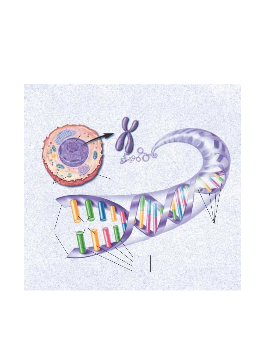
45
retardation. Scientists now believe they have identified this gene.
Several groups of scientists are investigating whether there
are genetic components to schizophrenia, manic depression and
alcoholism, but their findings are not yet conclusive.
Overall, the characterizations of the structure and function
of individual genes causing diseases of the brain and nervous
system are in the early stages. Factors that determine variations
in the genetic expression of a single-gene abnormality—such
as what contributes to the early or late start or severity of a dis-
order—are still unknown.
Scientists also are studying the genes in mitochondria, struc-
tures found outside the cell nucleus that have their own genome
and are responsible for the production of energy used by the
cell. Recently, di∑erent mutations in mitochondrial genes were
found to cause several rare neurological disorders. Some scien-
tists speculate that an inheritable variation in mitochondrial
DNA may play a role in diseases such as Alzheimer’s, Parkin-
son’s and some childhood diseases of the nervous system.
ANATOMY OF A GENE. Within the nucleus of every human cell, two long, threadlike strands of DNA encode the instructions for making all the pro-
teins necessary for life. Each cell holds more than 50,000 di≈erent genes found on 23 paired chromosomes of tightly coiled DNA. Each strand of
DNA bears four types of coding molecules—adenine (A), cytosine (C), guanine (G), and thymine (T). The sequence of coding molecules in a gene
(segment of DNA) is the code for protein manufacture.
CHROMOSOME
DNA DOUBLE HELIX
THE CELL
Cytosine (C)
Adenine (A)
Guanine (G)
Thymine (T)
DNA chains
Paired
bases
Bases
Linked
sequence pairs
Cell membrane
Mitochondria
Nucleus

46
N
ew drugs. Most drugs used today were devel-
oped using trial-and-error techniques that often
do not reveal why a drug produces a particular
e∑ect. But the expanding knowledge gained
from the new methods of molecular biology—
the ability to make a receptor gene and deter-
mine its structure—make it possible to design safer and more
e∑ective drugs.
In a test tube, the potency of an agent can be determined
by how well it binds to a receptor. A scientist then can vary the
drug’s structure to enhance its action on the receptor. Thus,
subsequent generations of drugs can be designed to interact
with the receptors more e≈ciently, producing higher potency
and fewer side e∑ects.
While this “rational drug design” holds promise for devel-
oping drugs for conditions ranging from stroke and migraine
headaches to depression and anxiety, it will take considerable
e∑ort to clarify the role of the di∑erent receptors in these dis-
orders.
Trophic factors
One result of basic neuroscience research involves the discov-
ery of numerous survival or trophic factors found in the brain that
control the development and survival of specific groups of neu-
rons. Once the specific actions of these molecules and their
receptors are identified and their genes cloned, procedures can
be developed to modify trophic factor-regulated function in ways
that might be useful in the treatment of neurological disorders.
Already, researchers have demonstrated the possible value
of at least one of these factors, nerve growth factor (NGF).
Infused into the brains of rats, NGF prevented cell death and
stimulated the regeneration and sprouting of damaged neurons
that are known to die in Alzheimer’s disease. When aged ani-
mals with learning and memory impairments were treated with
NGF, scientists found that these animals were able to remem-
ber a maze task as well as healthy aged rats.
Recently, several new factors have been identified and are
beginning to be studied. They are potentially useful for ther-
apy, but scientists must first understand how they may influence
neurons. Alzheimer’s, Parkinson’s and Lou Gehrig’s diseases
may be treated in the future with trophic factors or their genes.
Because the destruction of neurons that use acetylcholine
is one feature of Alzheimer’s disease, any substance that can
prevent this destruction is an important topic of research. NGF,
which can do this, also holds promise for slowing the memory
deficits associated with normal aging.
Once a trophic factor for a particular cell is found, copies
of the factor can be genetically targeted to the area of the brain
where this type of cell has died. The treatment may not cure a
disease but could improve symptoms and delay progression.
In an interesting twist on growth factor therapy, researchers
for the first time demonstrated that a neutralization of
inhibitory molecules can help repair damaged nerve fiber tracts
in the spinal cord. Using antibodies to Nogo-A, a protein that
inhibits nerve regeneration, Swiss researchers succeeded in get-
ting nerves of damaged spinal cords in rats to regrow. Treated
rats showed large improvements in their ability to walk after
spinal cord damage.
In these experiments, scientists cut one of the major groups
of nerve fiber tracts in the spinal cord that connect the spinal
cord and the brain. When an antibody, directed against the fac-
tor Nogo-A, was administered to the spinal cords or the brains
of adult rats, “massive sprouting” of nerve fibers occurred where
the spinal cord had been cut. Within two to three weeks, neu-
rons grew to the lower level of the spinal cord and in some ani-
mals along its whole length. In untreated spinal cord-injured
rats, the maximum distance of nerve regrowth rarely exceeded
one-tenth of an inch. This research could eventually have clin-
ical implications for spinal cord or brain-damaged people.
Cell and gene therapy
Researchers throughout the world are pursuing a variety of new
ways to repair or replace healthy neurons and other cells in the
brain. Most of the experimental approaches are still being worked
out in animals and cannot be considered real therapies at this
time.
Scientists have identified an embryonic neuronal stem cell—
an unspecialized cell that gives rise to cells with specific func-
N
Potential therapies
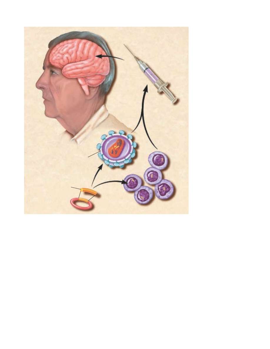
47
tions. They have located this type of cell in the brain and spinal
cord of embryonic and adult mice that can be stimulated to
divide by known proteins, epidermal growth factor and fibrob-
last growth factor. The stem cells can continuously produce all
three major cell types of the brain—neurons; astrocytes, the cells
that nourish and protect neurons; and oligodendrocytes, the cells
that surround axons and allow them to conduct their signals
e≈ciently. Someday their production abilities may become use-
ful for replacing missing neurons. A very similar stem cell also
has been discovered in the adult nervous system in various
kinds of tissue, raising the possibility that these stem cells can
be pharmacologically directed to replace damaged neurons.
In other work, researchers are studying a variety of viruses
that may ultimately be used to act as “Trojan horses” to carry
therapeutic genes to the brain to correct nervous system dis-
eases. The viruses include herpes simplex type 1 virus (HSV), ade-
novirus, lentivirus, adeno-associated virus and others naturally
attracted to neurons. All have been found to be capable of being
modified to carry new genes to cells in tissue culture and in the
rodent central nervous system. HSV and adenovirus vectors
have also been evaluated in early-stage human trials for treat-
ing brain tumors.
In one gene therapy experiment, scientists created an ani-
mal model of Parkinson’s disease (PD) in rhesus monkeys. One
week later, these monkeys received injections of the glial cell-
derived neurotrophic factor (GDNF) gene into the striatum
and substantia nigra using a lentiviral vector system. The
nigrostriatal system is the main brain area a∑ected by PD. The
injections reversed the motor deficits seen on a clinical rating
scale and a hand-reach task for up to three months. PET scans
showed that these animals displayed marked increases in mea-
sures of dopamine, a chemical that is deficient in patients. Post-
mortem studies revealed a comprehensive protection in striatal
dopamine as well as the number of nigrostriatal neurons. The
results support the concept that lentiviral delivery of GDNF
may provide neuroprotection for patients with early PD.
CELL AND GENE THERAPY. In
potential therapy techniques,
scientists plan to insert genetic
material for a beneficial neuro-
transmitter or trophic factor into
stem cells or a virus. The cells or
virus are then put into a syringe
and injected into the patient
where they will produce the
beneficial molecule and, it is
hoped, improve symptoms.
Virus
Stem cells
Patient with
a neurological
disease
New genetic
material

48
ACETYLCHOLINE
A neurotransmitter in both the brain, where it
regulates memory, and in the peripheral nervous system,
where it controls the actions of skeletal and smooth muscle.
ACTION POTENTIAL
This occurs when a neuron is activated and
temporarily reverses the electrical state of its interior mem-
brane from negative to positive. This electrical charge travels
along the axon to the neuron’s terminal where it triggers the
release of a neurotransmitter.
ADRENAL CORTEX
An endocrine organ that secretes corticos-
teroids for metabolic functions; for example, in response to
stress.
ADRENAL MEDULLA
An endocrine organ that secretes epineph-
rine and norepinephrine in concert with the activation of the
sympathetic nervous system; for example, in response to stress.
AGONIST
A neurotransmitter, a drug or other molecule that
stimulates receptors to produce a desired reaction.
ALZHEIMER’S DISEASE
The major cause of dementia most
prevalent in the elderly, it inflicts enormous human financial
cost on society. The disease is characterized by death of neu-
rons in the hippocampus, cerebral cortex and other brain
regions.
AMINO ACID TRANSMITTERS
The most prevalent neurotrans-
mitters in the brain, these include glutamate and aspartate,
which have excitatory actions, and glycine and gamma-amino
butyric acid (GABA), which have inhibitory actions.
AMYGDALA
A structure in the forebrain that is an important
component of the limbic system and plays a central role in
emotional learning.
ANDROGENS
Sex steroid hormones, including testosterone,
found in higher levels in males than females. They are respon-
sible for male sexual maturation.
ANTAGONIST
A drug or other molecule that blocks receptors.
Antagonists inhibit the e∑ects of agonists.
APHASIA
Disturbance in language comprehension or produc-
tion, often as a result of a stroke.
AUDITORY NERVE
A bundle of nerve fibers extending from the
cochlea of the ear to the brain, which contains two branches:
the cochlear nerve that transmits sound information and the
vestibular nerve that relays information related to balance.
Glossary
AUTONOMIC NERVOUS SYSTEM
A part of the peripheral ner-
vous system responsible for regulating the activity of internal
organs. It includes the sympathetic and parasympathetic ner-
vous systems.
AXON
The fiberlike extension of a neuron by which the cell
sends information to target cells.
BASAL GANGLIA
Clusters of neurons, which include the cau-
date nucleus, putamen, globus pallidus and substantia nigra,
located deep in the brain that play an important role in move-
ment. Cell death in the substantia nigra contributes to Parkin-
son’s disease.
BRAINSTEM
The major route by which the forebrain sends
information to and receives information from the spinal cord
and peripheral nerves. The brainstem controls, among other
things, respiration and regulation of heart rhythms.
BROCA’S AREA
The brain region located in the frontal lobe of
the left hemisphere that is important for the production of
speech.
CATECHOLAMINES
The neurotransmitters dopamine, epineph-
rine and norepinephrine that are active both in the brain and
the peripheral sympathetic nervous system. These three mole-
cules have certain structural similarities and are part of a
larger class of neurotransmitters known as monoamines.
CEREBELLUM
A large structure located at the roof of the hind-
brain that helps control movement by making connections to
the pons, medulla, spinal cord and thalamus. It also may be
involved in aspects of motor learning.
CEREBRAL CORTEX
The outermost layer of the cerebral hemi-
spheres of the brain. It is responsible for all forms of conscious
experience, including perception, emotion, thought and plan-
ning.
CEREBRAL HEMISPHERES
The two specialized halves of the
brain. The left hemisphere is specialized for speech, writing,
language and calculation; the right hemisphere is specialized
for spatial abilities, face recognition in vision and some aspects
of music perception and production.
CEREBROSPINAL FLUID
A liquid found within the ventricles of
the brain and the central canal of the spinal cord.
CHOLECYSTOKININ
A hormone released from the lining of the

49
stomach during the early stages of digestion which acts as a
powerful suppressant of normal eating. It also is found in the
brain.
CIRCADIAN RHYTHM
A cycle of behavior or physiological
change lasting approximately 24 hours.
CLASSICAL CONDITIONING
Learning in which a stimulus that
naturally produces a specific response (unconditioned stimu-
lus) is repeatedly paired with a neutral stimulus (conditioned
stimulus). As a result, the conditioned stimulus can evoke a
response similar to that of the unconditioned stimulus.
COCHLEA
A snail-shaped, fluid-filled organ of the inner ear
responsible for transducing motion into neurotransmission to
produce an auditory sensation.
COGNITION
The process or processes by which an organism
gains knowledge or becomes aware of events or objects in its
environment and uses that knowledge for comprehension and
problem-solving.
CONE
A primary receptor cell for vision located in the retina.
The cone is sensitive to color and used primarily for daytime
vision.
CORPUS CALLOSUM
The large bundle of nerve fibers linking
the left and right cerebral hemispheres.
CORTISOL
A hormone manufactured by the adrenal cortex. In
humans, cortisol is secreted in greatest quantities before
dawn, readying the body for the activities of the coming day.
DEPRESSION
A mental disorder characterized by depressed
mood and abnormalities in sleep, appetite and energy level.
DENDRITE
A tree-like extension of the neuron cell body. Along
with the cell body, it receives information from other neurons.
DOPAMINE
A catecholamine neurotransmitter known to
have multiple functions depending on where it acts.
Dopamine-containing neurons in the substantia nigra of the
brainstem project to the caudate nucleus and are destroyed
in Parkinson’s victims. Dopamine is thought to regulate
emotional responses and play a role in schizophrenia and
drug abuse.
DORSAL HORN
An area of the spinal cord where many nerve
fibers from peripheral pain receptors meet other ascending
and descending nerve fibers.
DRUG ADDICTION
Loss of control over drug intake or compul-
sive seeking and taking of drugs, despite adverse consequences.
ENDOCRINE ORGAN
An organ that secretes a hormone directly
into the bloodstream to regulate cellular activity of certain
other organs.
ENDORPHINS
Neurotransmitters produced in the brain that
generate cellular and behavioral e∑ects like those of morphine.
EPILEPSY
A disorder characterized by repeated seizures,
which are caused by abnormal excitation of large groups of
neurons in various brain regions. Epilepsy can be treated with
many types of anticonvulsant medications.
EPINEPHRINE
A hormone, released by the adrenal medulla
and specialized sites in the brain, that acts with norepineph-
rine to a∑ect the sympathetic division of the autonomic ner-
vous system. Sometimes called adrenaline.
ESTROGENS
A group of sex hormones found more abun-
dantly in females than males. They are responsible for female
sexual maturation and other functions.
EVOKED POTENTIALS
A measure of the brain’s electrical activ-
ity in response to sensory stimuli. This is obtained by placing
electrodes on the surface of the scalp (or more rarely, inside
the head), repeatedly administering a stimulus and then using
a computer to average the results.
EXCITATION
A change in the electrical state of a neuron that
is associated with an enhanced probability of action potentials.
FOLLICLE-STIMULATING HORMONE
A hormone released by the
pituitary gland that stimulates the production of sperm in the
male and growth of the follicle (which produces the egg) in
the female.
FOREBRAIN
The largest division of the brain, which includes
the cerebral cortex and basal ganglia. The forebrain is cred-
ited with the highest intellectual functions.
FRONTAL LOBE
One of the four divisions (parietal, temporal,
occipital) of each hemisphere of the cerebral cortex. The
frontal lobe has a role in controlling movement and in the
planning and coordinating of behavior.
GAMMA-AMINO BUTYRIC ACID
(GABA) An amino acid trans-
mitter in the brain whose primary function is to inhibit the
firing of neurons.

50
GLIA
Specialized cells that nourish and support neurons.
GLUTAMATE
An amino acid neurotransmitter that acts to
excite neurons. Glutamate stimulates N-methyl-D-aspartate
(NMDA) receptors that have been implicated in activities
ranging from learning and memory to development and
specification of nerve contacts in a developing animal. Stimu-
lation of NMDA receptors may promote beneficial changes,
while overstimulation may be a cause of nerve cell damage or
death in neurological trauma and stroke.
GONAD
Primary sex gland: testis in the male and ovary in the
female.
GROWTH CONE
A distinctive structure at the growing end of
most axons. It is the site where new material is added to the
axon.
HIPPOCAMPUS
A seahorse-shaped structure located within
the brain and considered an important part of the limbic sys-
tem. It functions in learning, memory and emotion.
HORMONES
Chemical messengers secreted by endocrine
glands to regulate the activity of target cells. They play a role
in sexual development, calcium and bone metabolism, growth
and many other activities.
HUNTINGTON’S DISEASE
A movement disorder caused by
death of neurons in the basal ganglia and other brain regions.
It is characterized by abnormal movements called chorea—
sudden, jerky movements without purpose.
HYPOTHALAMUS
A complex brain structure composed of
many nuclei with various functions. These include regulating
the activities of internal organs, monitoring information from
the autonomic nervous system, controlling the pituitary gland
and regulating sleep and appetite.
INHIBITION
In reference to neurons, it is a synaptic message
that prevents the recipient cell from firing.
IONS
Electrically charged atoms or molecules.
LIMBIC SYSTEM
A group of brain structures—including the
amygdala, hippocampus, septum, basal ganglia and others—
that help regulate the expression of emotion and emotional
memory.
LONG-TERM MEMORY
The final phase of memory in which
information storage may last from hours to a lifetime.
MANIA
A mental disorder characterized by excessive excite-
ment, exalted feelings, elevated mood, psychomotor over-
activity and overproduction of ideas. It may be associated
with psychosis; for example, delusions of grandeur.
MELATONIN
Produced from serotonin, melatonin is released
by the pineal gland into the bloodstream. Melatonin a∑ects
physiological changes related to time and lighting cycles.
MEMORY CONSOLIDATION
The physical and psychological
changes that take place as the brain organizes and restruc-
tures information in order to make it a permanent part of
memory.
METABOLISM
The sum of all physical and chemical changes
that take place within an organism and all energy transforma-
tions that occur within living cells.
MIDBRAIN
The most anterior segment of the brainstem.
Along with the pons and medulla, the midbrain is involved in
many functions, including regulation of heart rate, respira-
tion, pain perception and movement.
MITOCHONDRIA
Small cylindrical particles inside cells that
provide energy for the cell by converting sugar and oxygen
into special energy molecules, called ATP.
MONOAMINE OXIDASE (MAO)
The brain and liver enzyme that
normally breaks down the catecholamines norepinephrine,
dopamine, and epinephrine and other monosomines such as
serotonin.
MOTOR NEURON
A neuron that carries information from the
central nervous system to muscle.
MYASTHENIA GRAVIS
A disease in which acetylcholine recep-
tors on muscle cells are destroyed so that muscles can no
longer respond to the acetylcholine signal in order to con-
tract. Symptoms include muscular weakness and progressively
more common bouts of fatigue. The disease’s cause is unknown
but is more common in females than in males and usually
strikes between the ages of 20 and 50.
MYELIN
Compact fatty material that surrounds and insulates
axons of some neurons.
NERVE GROWTH FACTOR
A substance whose role is to guide
neuronal growth during embryonic development, especially
in the peripheral nervous system. Nerve growth factor also

51
probably helps sustain neurons in the adult.
NEURON
Nerve cell. It is specialized for the transmission of
information and characterized by long fibrous projections
called axons and shorter, branch-like projections called
dendrites.
NEUROTRANSMITTER
A chemical released by neurons at a
synapse for the purpose of relaying information to other
neurons via receptors.
NOCICEPTORS
In animals, nerve endings that signal the sensa-
tion of pain. In humans, they are called pain receptors.
NOREPINEPHRINE
A catecholamine neurotransmitter, pro-
duced both in the brain and in the peripheral nervous system.
It is involved in arousal, and regulation of sleep, mood and
blood pressure.
OCCIPITAL LOBE
One of the four subdivisions of the cerebral
cortex. The occipital lobe plays a role in processing visual
information.
ORGANELLES
Small structures within a cell that maintain the
cells and do the cells’ work.
PARASYMPATHETIC NERVOUS SYSTEM
A branch of the auto-
nomic nervous system concerned with the conservation of the
body’s energy and resources during relaxed states.
PARIETAL LOBE
One of the four subdivisions of the cerebral
cortex. The parietal lobe plays a role in sensory processes,
attention and language.
PARKINSON’S DISEASE
A movement disorder caused by death
of dopamine neurons in the substantia nigra located in the
midbrain. Symptoms include tremor, shuΩing gait and gen-
eral paucity of movement.
PEPTIDES
Chains of amino acids that can function as neuro-
transmitters or hormones.
PERIPHERAL NERVOUS SYSTEM
A division of the nervous sys-
tem consisting of all nerves that are not part of the brain or
spinal cord.
PHOSPHORYLATION
A process that modifies the properties of
neurons by acting on an ion channel, neurotransmitter receptor
or other regulatory protein. During phosphorylation, a phos-
phate molecule is placed on a protein and results in the activa-
tion or inactivation of the protein. Phosphorylation is believed
to be a necessary step in allowing some neurotransmitters to
act and is often the result of second messenger activity.
PINEAL GLAND
An endocrine organ found in the brain. In
some animals, the pineal gland serves as a light-influenced
biological clock.
PITUITARY GLAND
An endocrine organ closely linked with the
hypothalamus. In humans, the gland is composed of two lobes
and secretes a number of hormones that regulate the activity
of other endocrine organs in the body.
PONS
A part of the hindbrain that, with other brain struc-
tures, controls respiration and regulates heart rhythms. The
pons is a major route by which the forebrain sends informa-
tion to and receives information from the spinal cord and
peripheral nervous system.
PSYCHOSIS
A severe symptom of mental disorders character-
ized by an inability to perceive reality. It can occur in many
conditions, including schizophrenia, mania, depression and
drug-induced states.
RECEPTOR CELL
A specialized sensory cell designed to pick up
and transmit sensory information.
RECEPTOR MOLECULE
A specific protein on the surface or
inside of a cell with a characteristic chemical and physical
structure. Many neurotransmitters and hormones exert their
e∑ects by binding to receptors on cells.
REUPTAKE
A process by which released neurotransmitters are
absorbed for subsequent reuse.
ROD
A sensory neuron located in the periphery of the retina.
The rod is sensitive to light of low intensity and specialized
for nighttime vision.
SCHIZOPHRENIA
A chronic mental disorder characterized by
psychosis (e.g., hallucinations and delusions), flattened emo-
tions and impaired cognitive function.
SECOND MESSENGERS
Substances that trigger communi-
cations among di∑erent parts of a neuron. These chemicals
play a role in the manufacture and release of neurotransmit-
ters, intracellular movements, carbohydrate metabolism and
processes of growth and development. The messengers direct
e∑ects on the genetic material of cells may lead to long-term
alterations of behavior, such as memory and drug addiction.

52
SEROTONIN
A monoamine neurotransmitter believed to play
many roles, including, but not limited to, temperature regula-
tion, sensory perception and the onset of sleep. Neurons
using serotonin as a transmitter are found in the brain and in
the gut. A number of antidepressant drugs are targeted to
brain serotonin systems.
SHORT-TERM MEMORY
A phase of memory in which a limited
amount of information may be held for several seconds to
minutes.
STIMULUS
An environmental event capable of being detected
by sensory receptors.
STROKE
The third largest cause of death in America, stroke
is an impeded blood supply to the brain. Stroke can be caused
by a rupture of a blood vessel wall, an obstruction of blood
flow caused by a clot or other material or by pressure on a
blood vessel (as by a tumor). Deprived of oxygen, which is
carried by blood, nerve cells in the a∑ected area cannot func-
tion and die. Thus, the part of the body controlled by those
cells cannot function either. Stroke can result in loss of con-
sciousness and death.
SYMPATHETIC NERVOUS SYSTEM
A branch of the autonomic
nervous system responsible for mobilizing the body’s energy
and resources during times of stress and arousal.
SYNAPSE
A gap between two neurons that functions as the
site of information transfer from one neuron to another.
TEMPORAL LOBE
One of the four major subdivisions of each
hemisphere of the cerebral cortex. The temporal lobe func-
tions in auditory perception, speech and complex visual per-
ceptions.
THALAMUS
A structure consisting of two egg-shaped masses
of nerve tissue, each about the size of a walnut, deep within
the brain. The key relay station for sensory information
flowing into the brain, the thalamus filters out only informa-
tion of particular importance from the mass of signals enter-
ing the brain.
VENTRICLES
Of the four ventricles, comparatively large spaces
filled with cerebrospinal fluid, three are located in the fore-
brain and one in the brainstem. The lateral ventricles, the two
largest, are symmetrically placed above the brainstem, one in
each hemisphere.
WERNICKE’S AREA
A brain region responsible for the compre-
hension of language and the production of meaningful
speech.

53
Acetylcholine 4
Action potential 4
Addiction 33–36
Aging 28–
29
and intellectual capacity 29
AIDS 40
Alcohol
34–36
Alpha motor neurons 20
Alzheimer’s disease 36–37
Amino acid transmitters 4–5
Amphetamines
34
Amyloid protein 36–37
Amyotrophic lateral sclerosis
(ALS) 42
Analgesia 30
Androgen 7
Anxiety disorders 39
Autoimmune response 27
Autonomic nervous system
11, 25
Axon 4 –
5
Basal ganglia
19, 21, 30
Biological clock 7, 27
Brain
aging 28–
29
anatomical organization
3
development
8–11
diseases 2–3
tumors 42
Broca’s area
19
Catecholamines 6
Central nervous system 6,
11
Cerebellum
19, 21
Cerebral cortex
3, 17, 19, 23, 31
Club drugs 36
Cocaine
34–35
Cortisol 25–26
Costs of brain diseases 2–3
Crossed extension reflex 20–
21
Declarative knowledge 18
Dementia 28, 36
Dendrite 4–
5
Depression
major 32
manic 32
Dopamine 6, 30, 34
Down syndrome 40–41
Drug reward system
34
Endocrine system 6–7, 25–27
Endorphins 6, 17
Epilepsy 31–32
Epinephrine 25–26
Estrogen 7
Fetal alcohol syndrome 35
Firing of neurons 4–
5
Flexion withdrawal 20–
21
Fluoxetine 32
Forebrain
3
Functional Magnetic Reso-
nance Imaging (fMRI) 44
Gamma-amino butyric acid
(GABA) 5, 24, 32, 35
Gamma motor neurons 20
Gene
45
diagnosis 44– 45
therapy 46–
47
Glucocorticoids 7, 26–27
Glutamate 5,
36, 38
Hearing
14–15
Heroin 34
Hippocampus
3, 18–19, 27
Huntington’s disease 41
Hypothalamus
3, 7, 24, 32
Immune system 27
Information processing,
and hearing
14–15
and learning and memory
18–
19
and movement 20–
21
and pain 16–
17
and taste and smell 15
–16
and vision 12–
13
Inhibitory neurons 20–
21
Ion channels 4
Language
19
Learning 18–
19
Learning disorders 37
Levodopa 6, 30
Limbic system 15
Long-term potentiation 18
Lou Gehrig’s disease 42
Magnetic resonance imaging
(MRI) 43– 44
Magnetoencephalography
(MEG) 44
Marijuana 36
Memory 18–
19
Methylprednisolone 39
Midbrain
3, 8
Mitochondria 45
Monoamine oxidase inhibitors
(MAOIs) 32
Morphine 6, 30, 31, 34
Motor cortex
3, 20
Motor neuron 20
Motor unit 20
Movement 20–
21
MPTP 30
Multiple sclerosis 40
Myasthenia gravis 4
Myelin 4–
5
Narcolepsy 24
Nerve growth factor (NGF) 46
Nerve impulse 4,
5
Neurofibrillary tangles 36
Neurological trauma 38–39
Neuron 4–
5
birth 9–10
migration
9–10
pathfinding 10
survival 10–11
Neurotransmitters 4 –7
Nicotine 33–34
NMDA receptors 5, 18
Norepinephrine 6
Obsessive-compulsive
disorder 39
Occipital lobe
3, 12
Olfactory bulbs 15–
16
Opiates
34–35
Pain 16–
17, 30–31
Panic disorder 39
Parietal lobe
3
Parkinson’s disease 30,
46–47
Peptides 6
Peripheral nervous system
11
Phenytoin 31
Phobias 39
Pituitary gland 6, 7, 32
Pons
3, 23
Positron emission tomography
(PET) 19, 43
Primary visual cortex 12
Procedural knowledge 18
Prostaglandins 17,
31
Psychostimulants
34–35
Receptive field 12
Receptors 4
Reflex 20–
21
Regeneration 46
Reproduction 7
Schizophrenia 39–40
Second messengers 7
Selye, Hans 25
Serotonin 6, 32
Single photon emission
computed tomography
(SPECT) 43
Sleep 22–24
REM sleep 22–24
stages
22
disorders 23–24
Smell 15–
16
Spinal cord 6,
11, 17, 20–21,
38–39, 46
Strabismus 14
Stress 25–27
in arousal 25–
26
chronic 27
and endocrine system 25–27
and schizophrenia 39
Stroke 37–
38
Substance P 6
Synapse 4,
5, 29
Taste 15–
16
Temporal lobe
3, 18
Testosterone 7
Thalamus
3
Touch 16–17
Tourette syndrome 41– 42
Tricyclic antidepressants 32
Trophic factors 6, 46
Vision 12,
13 –15
Wernicke’s area
19
Working memory 18
Index
Numbers in bold refer to illustrations.

Copyright © 2002 The Society for Neuroscience
11 Dupont Circle, NW, Suite 500
Washington, DC 20036 USA
Telephone (202) 462-6688
www.sfn.org
All rights reserved. No portion of this publication may be reproduced,
stored in a retrieval system, or transmitted in any form or by any means,
electronic, mechanical, photocopying, recording, or otherwise without
permission of the The Society for Neuroscience.
To acquire additional copies of this book, please visit our Web site
www.sfn.org, go to Publications and click on Brain Facts.
The Society for Neuroscience
Editor: Joseph Carey, Senior Director, Communications & Public Affairs
Science writer: Leah Ariniello
Researcher: Mary McComb
Produced by Meadows Design Office Incorporated, Washington, DC
www.mdomedia.com
Creative Director and Designer: Marc Alain Meadows
Production Assistants: Ching Huang Ooi, Nancy Bratton
Illustrator: Lydia V. Kibiuk, Baltimore, Maryland
Printed and bound in China by Everbest Printing Company
Fourth edition
06 05 04 03 02
5 4 3 2 1
isbn
0-916110-00-1

ISBN
0-916110-00-1
Wyszukiwarka
Podobne podstrony:
Język angielski The influence of the media on the society
A New Hypothesis on the Mechanism for Gravity
Child?use on the Brain
46 Effects of Exercise on Brain and Body
On The Manipulation of Money and Credit
31 411 423 Effect of EAF and ESR Technologies on the Yield of Alloying Elements
The Great?pression Summary and?fects on the People
Effects of the Great?pression on the U S and the World
NEXT on THE BOLD AND THE?AUTIFULX92
Optional Protocol to the International Covenant on Economic, Social and Cultural Rights
plants and the central nervous system pharm biochem behav 75 (2003) 501 512
Notes on the 3 inch gun materiel and field artillery equipment 1917
Dental Pathology and Diet at Apollonia, a Greek Colony on the Black Sea
HUME AND?SCARTES ON THE THEORY OF IDEAS
The similarties?tween Nietzsche and Frueds Views on the Unc
więcej podobnych podstron