© 2006 Humana Press Inc.
999 Riverview Drive, Suite 208
Totowa, New Jersey 07512
humanapress.com
All rights reserved. No part of this book may be reproduced, stored in a retrieval system, or transmitted
in any form or by any means, electronic, mechanical, photocopying, microfilming, recording, or other-
wise without written permission from the publisher.
All papers, comments, opinions, conclusions, or recommendations are those of the author(s), and do
not necessarily reflect the views of the publisher.
Due diligence has been taken by the publishers, editors, and authors of this book to assure the accuracy
of the information published and to describe generally accepted practices. The contributors herein have
carefully checked to ensure that the drug selections and dosages set forth in this text are accurate and
in accord with the standards accepted at the time of publication. Notwithstanding, as new research,
changes in government regulations, and knowledge from clinical experience relating to drug therapy
and drug reactions constantly occurs, the reader is advised to check the product information provided
by the manufacturer of each drug for any change in dosages and for additional warnings and con-
traindications. This is of utmost importance when the recommended drug herein is a new or infrequent
used drug. It is the responsibility of the treating physician to determine dosages and treatment strate-
gies for individual patients. Further it is the responsibility of the health care provider to ascertain the
Food and Drug Administration status of each drug or device used in their clinical practice. The pub-
lisher, editors, and authors are not responsible for errors or omissions or for any consequences from the
application of the information presented in this book and make no warranty, express or implied, with
respect to the contents in this publication.
This publication is printed on acid-free paper.
∞
ANSI Z39.48-1984 (American Standards Institute) Permanence of Paper for Printed Library Materials.
Cover design by Patricia F. Cleary
For additional copies, pricing for bulk purchases, and/or information about other Humana titles, con-
tact Humana at the above address or at any of the following numbers: Tel.: 973-256-1699; Fax: 973-
256-8341; E-mail: orders@humanapr.com; or visit our website:
Photocopy Authorization Policy:
Authorization to photocopy items for internal or personal use, or the internal or personal use of spe-
cific clients, is granted by Humana Press Inc., provided that the base fee of US $30.00 per copy is
paid directly to the Copyright Clearance Center at 222 Rosewood Drive, Danvers, MA 01923. For
those organizations that have been granted a photocopy license from the CCC, a separate system of
payment has been arranged and is acceptable to Humana Press Inc. The fee code for users of the
Transactional Reporting Service is: [1-58829-811-6/ $30.00].
Printed in the United States of America. 10 9 8 7 6 5 4 3 2 1
e-ISBN 1-59745-081-2
ISSN 1099-7768

This book is dedicated to the memory and influences of two men:
J.D. Adamson, MD (Manitoba), MRCP (Edinburgh), FRCP (Canada), Pro-
fessor and Chairman, Department of Medicine, University of Manitoba,
1939–1951
L.G. Bell, OC, MBE, MD (Manitoba), LLD (Queens University, Kingston,
Ontario), FRCP (London and Canada), FACP, Professor and Chairman,
Department of Medicine, University of Manitoba, 1951–1964
I had the good luck to be taught by both of these doctors. J.D. Adamson
could take a better history and elicit more information from a patient than
anyone I have ever met. He considered every new patient a fascinating story-
teller. He asked few questions, managed to keep the patient on the subject,
and was completely enthralled as the history unwound. He knew the words
and music of disease.
L.G. Bell could see more in 10 seconds at the bedside and do a better
physical examination than anyone else. He had a great ability to find, see,
and feel (or maybe smell) abnormal physical signs. One learned as much
from watching him examine as from listening to J.D. Adamson listen.
Both of these men taught hundreds of students, interns, and residents.
Each had great respect for the skills of the other. They were cultured, well-
read, humorous humans and great bedside doctors who dearly loved medi-
cine and teaching.
A man does not learn to understand anything unless he loves
it.—Goethe

Contents
Foreword by Lewis P. Rowland . . . . . . . . . . . . . . . . . . . . . . . . . . . . ix
Acknowledgments . . . . . . . . . . . . . . . . . . . . . . . . . . . . . . . . . . . . . xiii
1. The Ophthalmoscope, the Fundus Oculi,
and Central and Peripheral Vision. . . . . . . . . . . . . . . . . . . . . . . . . .
1
2. Loss of Vision . . . . . . . . . . . . . . . . . . . . . . . . . . . . . . . . . . . . . . . . . . 19
3. The Abnormal Retina . . . . . . . . . . . . . . . . . . . . . . . . . . . . . . . . . . . . 37
4. Eye Movements, Diplopia, and
Cranial Nerves 3, 4, and 6. . . . . . . . . . . . . . . . . . . . . . . . . . . . . . . . . 45
5. Ptosis and the Pupils: Myasthenia Gravis
and Other Diseases of the Eye and Eyelid Muscles . . . . . . . . . . . 61
6. Nystagmus. . . . . . . . . . . . . . . . . . . . . . . . . . . . . . . . . . . . . . . . . . . . . 71
7. Conjugate Gaze Palsies and Forced Conjugate Deviation . . . . . . 79
8. Cranial Nerves 1, 5, and 7. . . . . . . . . . . . . . . . . . . . . . . . . . . . . . . . . 87
9. Cranial Nerves 8–12 . . . . . . . . . . . . . . . . . . . . . . . . . . . . . . . . . . . . . 107
10. The Upper Limb. . . . . . . . . . . . . . . . . . . . . . . . . . . . . . . . . . . . . . . . . 121
11. The Lower Limb . . . . . . . . . . . . . . . . . . . . . . . . . . . . . . . . . . . . . . . . 145
12. Stance, Gait, and Balance . . . . . . . . . . . . . . . . . . . . . . . . . . . . . . . . 157
13. Reflexes . . . . . . . . . . . . . . . . . . . . . . . . . . . . . . . . . . . . . . . . . . . . . . . 163
14. Sensation. . . . . . . . . . . . . . . . . . . . . . . . . . . . . . . . . . . . . . . . . . . . . . 177
15. The Cerebellum . . . . . . . . . . . . . . . . . . . . . . . . . . . . . . . . . . . . . . . . . 187
16. The Corticospinal System . . . . . . . . . . . . . . . . . . . . . . . . . . . . . . . . 191
vii

viii / CONTENTS
17. Higher Cortical Functions: Intelligence and Memory . . . . . . . . . . 197
18. Disorders of Speech . . . . . . . . . . . . . . . . . . . . . . . . . . . . . . . . . . . . . 205
Appendix: Neurological Examination Instruments . . . . . . . . . . . . 210
Index. . . . . . . . . . . . . . . . . . . . . . . . . . . . . . . . . . . . . . . . . . . . . . . . . . 213

Foreword
Robert T. Ross is one of the most respected neurologists in North Amer-
ica. He established and led the Department of Neurology at the University of
Manitoba for many years. He founded the Canadian Journal of Neurological
Sciences in 1974 and was editor-in-chief until 1981. He has written and pub-
lished 88 papers on clinical problems in neurology. He has been made an
Honorary Life Member of the Canadian Neurological Society and was given
an Honorary Degree by the University of Manitoba. He has also been
awarded the Order of Canada.
Dr. Ross knows how to examine patients and he knows how to teach med-
ical students, especially those who are just beginning to learn neurology.
They are the ones most likely to be perplexed by the apparent complexity of
the neurological examination. Dr. Ross has come to their rescue with this
book.
With simple and direct writing, and numerous illustrations that serve the
purpose, he shows that examination is not all that difficult, that it can make
sense, and that it can be done in a few minutes. Once the student feels some
confidence, the examination can bring pleasure and a sense of achievement.
The student then becomes part of the health care team in support of the pa-
tients.
Dr. Ross’ skilled exposition has made the first three editions of this book a
success and it has been the recommended text in many medical schools. Any
book that has gone through three editions must be on the right track, and this
fourth edition keeps up the pace.
Lewis P. Rowland,
MD
Neurological Institute
Columbia University Medical Center
New York, NY

Preface
The more resources we have, the more complex they are,
the greater are the demands upon our clinical skill. These
resources are calls upon judgment and not substitutes for
it. Do not, therefore, scorn clinical examination; learn it
sufficiently to get from it all it holds, and gain in the con-
fidence it merits.
—Sir Francis Walshe, 1952
Technical advances have made diagnoses quicker, safer, and more accu-
rate. Sometimes it appears that careful history taking and examination are
less important than knowing which test to order.
However, the technology is expensive and access is limited. As medical
costs are increasingly scrutinized by the paying agencies, private or public,
there will be limitations on both diagnostic investigations and hospital ad-
missions.
For patients and doctors in smaller centers, limitations already exist. These
conditions make a careful history and examination essential to the intelligent
care of the sick and prerequisites for ordering tests. The practice of diagnos-
tic medicine is not simply ‘scene’ recognition plus knowing where to point
the technology. If it ever becomes this, a clerk—and eventually a machine—
will be able to do it. Therefore, I suggest that you learn how to listen to and
examine patients thoroughly and confidently. It is the most precious and
durable skill you have; the more you use it, the better it becomes. It is
unique.
One learns by doing the thing; for though you think you
know it, you have no certainty until you try.
—Sophocles
In the examination of sick people a technique that elicits physical signs,
and the ability to interpret those signs, are required.
Interpreting physical signs is one of the interesting parts of neurology. The
process will not work if abnormal signs have been missed because of faulty
technique, or if minor variations within the limits of normal are considered
as firm abnormalities. Each year more students must be taught more subjects
as the knowledge explosion continues. Only a small amount of time can be
spent on the method of any physical examination. Therefore, learn a reliable
technique quickly.
xi

xii / PREFACE
This book offers some anatomical and a smaller number of pathological
possibilities that may explain a physical sign. It does not consist of a list of,
for example, all the possible causes of an absent corneal reflex, and is not a
small textbook of neurological diseases.
Teach and be taught is a ground rule that most of us will try to observe
all of our professional lives. Every doctor and medical student owes a debt
to patients, who are an essential part of the teaching situation. They allow us
to teach ‘on’ them and around them, and they tolerate several history takings
and physical examinations, usually for the benefit of someone else.
At all times one must treat patients with respect and kindness. When you
enter the room, identify yourself and tell the patient why you are there. Do
not persist with the history or examination past the point at which the patient
is tired or uncooperative. Patients are most cooperative with students and
doctors who are clean, neat, and polite.
When examining a patient, stand on the right side of the bed (or on the left
if you are left-handed). After you have identified yourself, level the bed; that
is, if the head or knee break is cranked up, flatten it. Then raise the bed as
high as it will go. You can work better with the bed 30 inches from the floor.
Spend 60–75% of the time devoted to any one patient on history tak-
ing and the remainder on the physical examination. Have a system of exami-
nation and learn to follow it in the same way each time.
Do not be upset by the transient nature of some physical signs. You may
see a patient with a slightly enlarged left pupil and explosively hyperac-
tive tendon reflexes in the right arm and leg and a right extensor plantar
response. Examination a short while later shows that the pupils and ten-
don reflexes are equal and both plantar responses are flexor. Both exami-
nations were valid. Few physical signs of acute diseases of the nervous
system are fixed. Papilledema is a notable exception. If it was present
yesterday, it will be there today, tomorrow, and the day after. Almost all
other signs can change hourly or daily.
R. T. Ross,
CM
,
MD
,
DS
c,
FRCP

Acknowledgments
It is a pleasure to acknowledge and thank the people who have contributed
to this book.
Gail Landry has done some artwork, posed as a model for the illustrations,
and typed the manuscript several times.
Angela Ross has read and reread the manuscript for English, grammar,
and syntax.
Drs. A.C. Huntington and A.J. Gomori have reviewed and edited the oph-
thalmology and other portions of the second edition and their suggestions
have been included in the current edition. Dr. David Steven has acted as a
model for some illustrations. I am grateful to these three physicians.
Rob Mathieson has skillfully photographed parts of the examination, and
Cameron Walker has done all the drawings.
xiii

The Ophthalmoscope,
the Fundus Oculi, and
Central and Peripheral Vision
1
The examination of the eye consists of five parts. This chapter deals with
the fundus oculi and with central and peripheral vision. The remaining three
parts of the examination are described in later chapters.
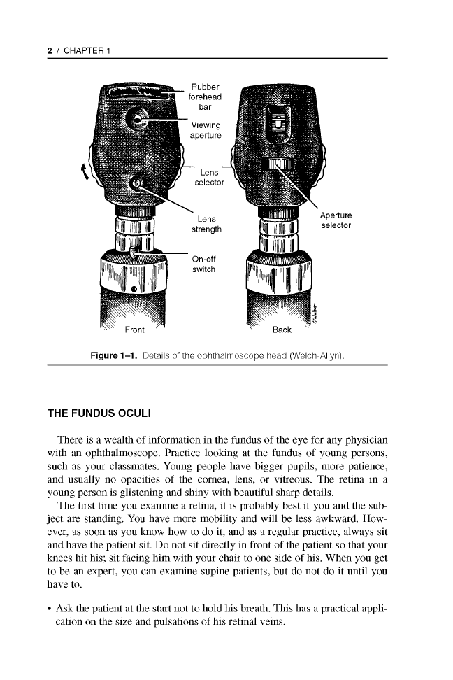
THE OPHTHALMOSCOPE
There are five mechanical details you need to know about the head of the
ophthalmoscope. The following remarks apply to the Welch-Allyn ophthal-
moscope (
Figure 1–1
).
Buy an ophthalmoscope with a halogen bulb and handle that takes D-size
batteries. A handle containing a rechargeable cell is almost as big as the
D-size battery model and just as good. The ophthalmoscope that takes AA
batteries is undesirable. The power does not last long enough, and the scope
is difficult to hold.
When you attach the head to the handle, have the on-off button in front,
as in
Figure 1–1
. To turn it on, push in the on-off button and turn the disc
that contains it. It turns only one way. As you rotate the lens selector
wheel, the numbers change in the lens strength window. This changes the
amount of magnification between your eye and the patient’s retina when
you are looking through the viewing aperture. If the lens selector wheel is
turned clockwise in the direction of the heavy arrow, increasingly stronger
plus lenses continue to appear in the viewing aperture and increasingly
higher black numbers continue to appear in the lens strength window. On
the back of the ophthalmoscope there is another adjustable wheel, the aper-
ture selector, which rotates in a horizontal plane. You will find that rotating
this wheel produces a green circle, a large white circle, a small white cir-
cle, or a grid. Turn it back to the large white circle and leave it there. (If
the white circles appear orange, get new batteries, and if still orange, get a
new bulb.)
1

OPHTHALMOSCOPE, FUNDUS OCULI, AND CENTRAL AND PERIPHERAL VISION / 3
• With the patient sitting with his back to the window, pull the blinds down
and ask the patient to remove his glasses. You do not need a blacked-out
room, but remove any incidental light.
There are two things working against you when you use the ophthalmo-
scope. First, the eye adds all the light it is exposed to. The sum of the ambi-
ent light and the ophthalmoscope light, plus the balance between sympa-
thetic and parasympathetic tone, plus the fact that the pupillary sphincter
muscle is stronger than the dilator, determines the pupil size. Second, trying
to look through a reflecting surface is difficult. On a sunny day you cannot
look into a lake to any depth because of the reflection off the surface. How-
ever, if you hold a hat close to the water and look into the lake in the shadow
of the hat, you can see into the water. Similarly, side light or ceiling light re-
flected on the patient’s cornea or surface of his glasses will hinder you.
When you examine the patient’s fundus, you will partially eliminate reflec-
tions by holding the ophthalmoscope so close to the patient that it is touch-
ing her forehead. If you rotate the ophthalmoscope 5 or 10 degrees while
holding it vertically, the light from the scope strikes the cornea at different
angles and you will find the best “reflection-free” angle.
Some doctors keep their glasses on when using the ophthalmoscope; most,
however, remove them.
1. Unless you have marked astigmatism, take your glasses off. With
your glasses off, the head of the ophthalmoscope can be closer to your
eye and you will see a larger area of the patient’s retina. Try it both
ways, beginning with your glasses off. The pinhole effect (see under
“Near Vision”) of looking through the viewing aperture (
Figure 1–1
)
may take care of your refractive error. If you are astigmatic, your
glasses contain a cylinder; hold your glasses at arm’s length, look
through one lens with one eye and slowly rotate your glasses to the
right and then to the left. If the object you are looking at tilts and elon-
gates at one point in the rotation, then your prescription includes a
cylinder; you are astigmatic, and you may have to wear your glasses
when using the ophthalmoscope.
2. Hold the ophthalmoscope in your right hand, turn it on, and look
through the viewing aperture. You must get the viewing aperture as
close as possible to your eye. The soft rubber bar (
Figure 1–1
) hori-
zontally placed across the top of the ophthalmoscope head is meant to
fit firmly against or just below your eyebrow. There should be some
contact between your skin and the rubber bar at all times when you are
looking at the fundus oculi. Keep your index finger on the lens selector
wheel. If you have the ophthalmoscope halfway down your nose, you

4 / CHAPTER 1
are, in effect, looking through a tube with your pupil at one end and the
viewing aperture at the other.
3. Use your right eye to examine the patient’s right eye and vice versa.
However, if you have one “weak” eye, use the other for examining both
of the patient’s eyes.
4. Hold the ophthalmoscope in your right hand when examining the
patient’s right eye and in your left hand when examining the pa-
tient’s left eye.
5. Keep the ophthalmoscope vertical, and keep both your eyes open.
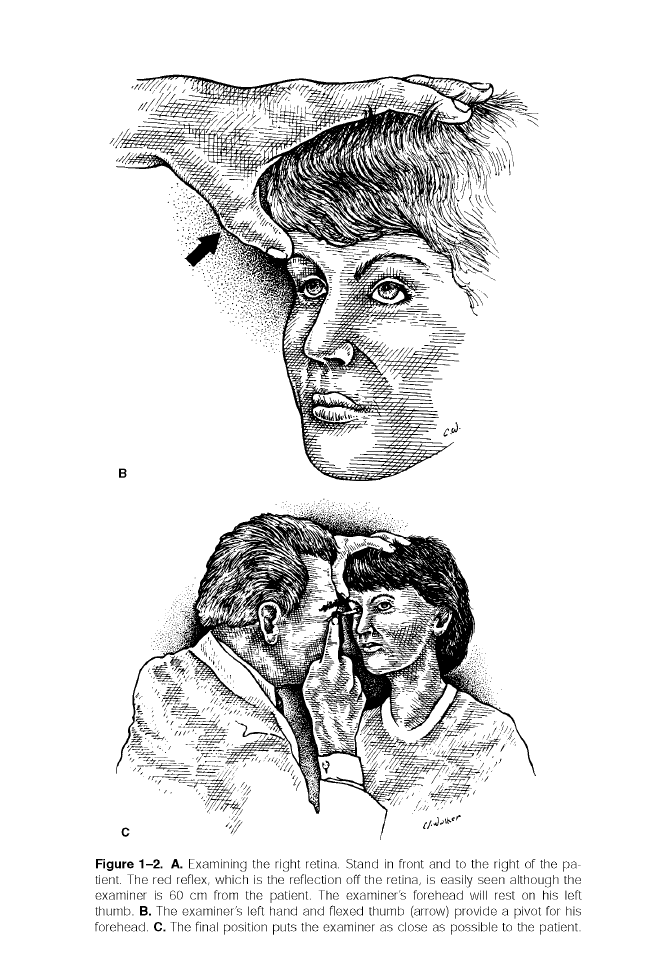
6. When examining the patient’s right eye, put your left hand on top
of the patient’s head (
Figure 1–2B
) and rest your forehead on your
flexed thumb and vice versa when examining the left eye. Ask the
patient to stare at something at eye level on the other side of the room
and to keep her eyes as still as she can. To examine the patient’s right
fundus, stand to the patient’s right (not directly in front of her) about 60
cm from her. With the ophthalmoscope head up against your eye, shine
the light in her right and left eyes by rotating the vertically held oph-
thalmoscope (
Figure 1–2A
). You will see two red-orange circles, the
red reflexes (much like the reflections of a car’s headlights in a cat’s
eyes at night); these are the patient’s retinas.
Follow down onto the right red circle by moving closer to the patient (
Fig-
ure 1–2C
). If you lose the red reflex, move back and start over. Adjust the
A

6 / CHAPTER 1
lens wheel if necessary. This will bring any fine artery at the edge of the disc
(the head of the optic nerve) into clear, sharp focus. Starting at 0, turn the
lens selector wheel one or two clicks in either direction. If the definition of
what you see is worse, turn the wheel in the opposite direction, all the time
keeping the ophthalmoscope up to your eye.
You need to identify the following and know normal from abnormal:
• Arteries
• Veins
• Optic nerve head or disc
• Physiological cup
• Posterior pole, macula, and fovea
The following comments are helpful when examining each other. Before
examining patients, read Chapter 3.
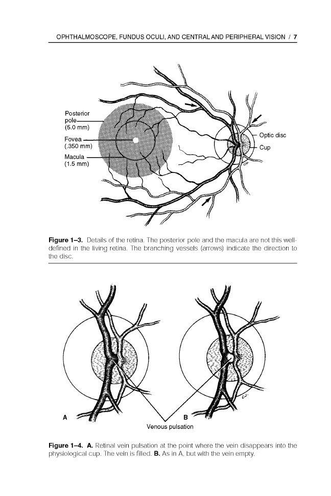
Retinal Arteries
Of the two kinds of vessels in the retina, the arteries are the smaller.
Orange-red, they reflect the light of the ophthalmoscope so that the center of
the artery shows a pale strip along its length. The thicker the wall of the
artery, the wider is the shiny white strip down its center. Arteries do not pul-
sate and are somewhat angular. They cross and more commonly are crossed
by retinal veins (
Figure 1–3
).
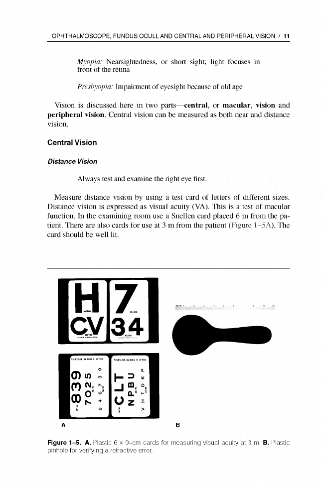
Retinal Veins
Retinal veins are larger than retinal arteries and are a dusky red. More sin-
uous and less angular than arteries, they pulsate. Retinal veins do not have as
distinctive a clear white strip of reflected light down the center. To see the
pulsations, look for a bend or change in direction of the vein. The pulsation
is not only an expansion of the caliber of the vein, but a shunting of the col-
umn of blood up and down within the length of the vessel (
Figure 1–4
).
Veins change direction at the disc or physiological cup edge. One can often
see a filling and emptying of the vessel at the bend.
Retinal veins pulsate spontaneously in 80% of people. If you cannot see
the pulsations, try the following:
• Watch the biggest vein you can see at the edge of the disc or where it dis-
appears over the edge of the cup.
• Put your finger on the patient’s eyelid and press gently against the eye. You
will see the vessels empty as the intraocular pressure rises.
• If you let go suddenly, the venous pulsations often become visible.

8 / CHAPTER 1
Alternatively, ask the patient to take in a big breath, hold it (which means
closing off the glottis), and then bear down (the Valsalva maneuver). As the
intracranial and intraocular venous pressure rises, you can see the retinal
veins distend. While still looking at the retinal vessels and after 30 s, ask the
patient to slowly breathe out. This should make retinal venous pulsations
visible. The pulsation indicates that intracranial pressure is below 200 mm of
water at that moment.
Follow the vessels as far as you can peripherally. You can help yourself as
follows:
When examining her left eye, ask the patient to turn her eyes
to her left while you move to her right; you are now looking
at the temporal periphery of her left retina.
This is not easy. Instead of looking through the patient’s round pupil, you
are now looking through a slit pupil and an oblique lens that induces astig-
matism. However, if you brace the ophthalmoscope against yourself and the
patient and ask her to look left, right, up, and down while you look and
move in the opposite directions—and persist at it—you will be able to see
the peripheral retina.
The Head of the Optic Nerve or Disc
The disc is located at the nasal side of the center of the retina. The V
formed by the bifurcations of veins and arteries is pointing toward the disc
(small arrows in
Figure 1–3
). The disc is pink and white and is much paler
than the orange-red retina. It is round or slightly oval, and the veins disap-
pear into it while the arteries arise from it. About 1.5 mm in diameter, the
disc usually has a distinct edge around it, but this edge may vanish and the
disc will blend into the retina without distinction for a portion of the periph-
ery. This blending into the retina is more evident on the nasal side. A cres-
cent of black pigment around the edge of the disc is common in myopic
(shortsighted) persons.
The Physiological Cup
Some part of the disc will appear to be deeper than the rest. This is the
cup.
The cup is less than one third of the disc’s diameter.
It is usually eccentric and is the point in the disc from which the arteries
arise and the veins enter. There may be pearly white fibers, the lamina
cribrosa, making a crosshatched appearance on the floor of the cup. It is im-

OPHTHALMOSCOPE, FUNDUS OCULI, AND CENTRAL AND PERIPHERAL VISION / 9
portant for you to learn the size of the physiological cup relative to the disc
and the depth of the cup in those with normal vision (see the section on
“Glaucoma” in Chapter 2).
The Posterior Pole
The central region of the retina is divided clinically and anatomically into
three areas. The clinical and anatomical terminologies are not the same. In
this manual clinical terminology is used. The anatomical equivalents are
given in parentheses.
We have to consider the following:
• Posterior pole (macula or area centralis)
• Macula (fovea)
• Fovea (foveola)
The posterior pole is about 5 mm in diameter. Its outer limits cannot be
clearly defined clinically. Histologically, it has more than one layer of gan-
glion cell nuclei.
The macula is at the center of the posterior pole and is a shallow depres-
sion about the same size as the disc. It is a darker red than the rest of the fun-
dus, opposite the center of the pupil, and you will not see it well without di-
lating the pupil. Never give your opinion on its appearance without first
dilating the pupil.
• You can see the macula by asking the patient to look into the ophthalmo-
scope light.
• Look at the retina. You will see several small groups of arteries coming off
the temporal side of the disc that rapidly curve toward each other in a ver-
tical direction. They end by surrounding the macula (
Figure 1–3
).
The fovea is the center of the macula. A depression seen as a yellow-white
reflecting spot, it lies two disc diameters to the temporal edge of the disc,
about 1 mm below its center.
Keep the following points in mind when using the ophthalmoscope:
• Examine the fundus of the eye with the patient sitting.
• Keep the patient’s back to the window, with the overhead lights out.
• Ask the patient not to hold his breath.
• Hold the ophthalmoscope vertically.
• It is easier for the patient if you decrease the intensity of the ophthalmo-
scope light when looking at the posterior pole.

10 / CHAPTER 1
• Keep your right eye opposite the patient’s right eye, and keep both your
eyes open.
• Do not breathe into the patient’s face.
If the patient wears glasses, ask him to take them off. Look at them. If
they are plus lenses, as you look through them and move them from side to
side, objects move in the opposite direction. The reverse is true for the my-
ope who wears minus lenses. For the myope you need a negative (red-num-
bered) lens in the ophthalmoscope to see retinal details clearly. Start with 0
showing at the lens strength window, and with your finger changing the lens
wheel in the opposite direction of the arrow (counterclockwise) (
Figure
1–1
), you will soon see a clearly defined retina.
You do not need to do anything to allow for a patient with astigmatism, al-
though many astigmatics are also myopic. For a hyperopic patient, reverse
the procedure described for a myope. Start with 0 at the lens strength win-
dow, but turn the lens selector in the direction of the arrow (clockwise).
When you become proficient at using the ophthalmoscope, you may leave
the patient’s glasses on and look through them; this is especially helpful in
high myopia. However, there are disadvantages: (a) both the inside and out-
side surfaces of his lens reflect incidental light and (b) his glasses prevent
you from getting close to his eye and thus reduce the area of his retina that
you can see.
Finally, do not spend too long on this part of the examination; you cannot
see all there is to see in each patient until you have looked in several hun-
dred eyes. Get to know each feature of the retina individually by looking in
every eye that you can, irrespective of the patient’s complaints.
VISION
The following terms should be familiar to you:
Amblyopia: Reduced visual acuity
Aphakia: Absence of the lens, such as following cataract re-
moval or dislocation of the lens out of the pupil area
Astigmatism: Impairment of eyesight usually caused by un-
equal curvature of the cornea
Hypermetropia: Same as hyperopia
Hyperopia: Farsightedness, or focusing of light behind the
retina

12 / CHAPTER 1
Ask the patient to put on her glasses, cover one eye, and
read the letters or numbers (or E chart for those who are illit-
erate) downward, that is, from the bigger to the smaller char-
acters.
The smallest line of readable symbols is the patient’s VA, which is ex-
pressed as a fraction. The numerator is the distance in feet or meters at
which the test is made, and the denominator indicates the distance at which
someone with normal vision would be able to read the line. If the patient can
read the 20-ft (or 6-m) line when the card is 20 ft (or 6 m) away, her vision is
20/20 (or 6/6) and she has normal macular function. Less than normal ranges
from 20/25 to 20/400.
If the patient can read all the letters except one in the 40-ft line, her vision
is recorded as 20/40-1. Similarly, if she can read all the letters except two in
the 60-ft line, her vision is 20/60-2. If she misses more than two letters in
any line, her visual acuity is that of the next line up.
If the patient cannot see the largest letter, hold your hand about 1 m in
front of the eye being tested and, with three or four fingers outstretched, ask
her, “How many fingers can you see?” If she answers correctly, record this
as CF (counting fingers) at 1 m.
Lesser vision than this should be tested and recorded as HM (hand move-
ments) only at 1 m, and even lesser vision should be tested by directing a
bright light into one eye from 0.3 m—recorded as LP (light perception) only.
A finding of no light perception indicates a sightless eye.
There are Snellen test cards to be used at 6 m, or with the card on the wall
above and behind the patient’s head, while the patient faces a mirror at 3 m.
Distance vision can be recorded from 6/60 to 6/6 (normal) and better than
normal as 6/4.
For the patient who has forgotten his glasses, see the remarks on using a
pinhole, under the heading “Near Vision.”
Test visual acuity in each eye with the patient’s other eye covered and
with both eyes open. Patients with cataracts may have VA 20/60 right and
left when the eyes are tested separately and 20/30 to 20/40 when tested with
both eyes open. Also, patients with latent nystagmus may have normal acuity
with both eyes open. When one eye is covered, the nystagmus occurs in both
eyes and the acuity decreases in the uncovered eye.
Remember:
• Visual acuity is a test of macular function.
• Visual acuity is expressed as a fraction.
• The numerator is the distance at which the test is made.
• The denominator is the distance that a person with normal vision can read

OPHTHALMOSCOPE, FUNDUS OCULI, AND CENTRAL AND PERIPHERAL VISION / 13
the smallest line readable by the patient.
• Measure the best corrected vision of each eye separately, right then left,
and both together.
Near Vision
The patient with cataracts may have quite good distance vision
and poor near vision, and the myope can read newsprint without his glasses
but has poor distance vision. Near vision can be tested using the print in a
telephone directory or a newspaper. Formal testing requires Jaeger’s test type
or Birmingham Optical Group test card or the American Medical Association
test card. The patient holds the card in his hand at a comfortable distance and
reads the smallest typed paragraph that he can. Glasses must be worn; test
each eye separately, then together.
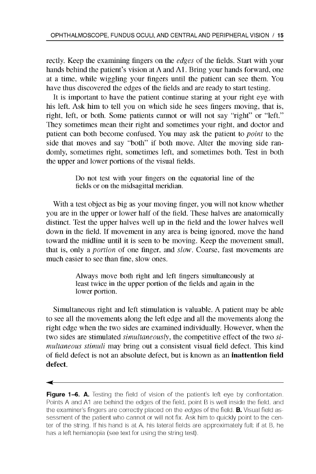
If the patient has forgotten his glasses, a pinhole will help (
Figure 1–5B
).
If the patient holds the pinhole up to his eye and reads the test card through
one of the holes, refractive errors are eliminated. The light coming through
the pinhole is axial and remains unrefracted by the patient’s cornea and lens
and is sharply focused on the macula.
Whether the person is myopic, hyperopic, or presbyopic, testing his vision
with the use of a pinhole will bring his vision up to normal. If it does not, his
loss of vision is not a result of refractive error.
Figure 1–5A
is a reproduction of two pocket cards used to test visual acu-
ity. They are lettered and numbered and are used at 3 m. Similar cards are
available at most surgical supply houses or medical college bookstores. Get
a set and put them in your ophthalmoscope case with the pinhole.
Peripheral Vision
Peripheral fields of vision reflect the function of the retina (the nonmacu-
lar part) and the visual pathways connected to the nonmacular retina. When
testing acuity, letters and parts of letters and numbers of different size that
subtend a portion of an arc are used. When testing peripheral fields, the
stimulus is movement.
To test visual fields by confrontation, seat the patient facing you with his
glasses off. Ask him to cover his right eye with his hand and to stare at your
right eye with his left eye. Hold your arms out to either side so that your fin-
gers are at the edges of his visual fields, as in
If the examiner
has his right hand at A in
, he will get no responses from the pa-
tient, as the test object (his moving finger) is behind the patient’s field and
the patient cannot see it, let alone say whether or not it is moving. If the ex-
aminer’s right hand is at B, then the test object is inside the field, not on the
edge, and a substantial crescent of blindness could be present in the patient’s
left temporal field and yet be missed because the test is being done incor-
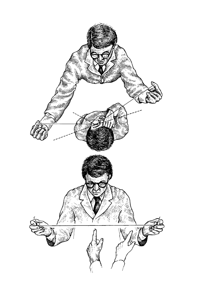
A
A
B
A1
A
B
B
A
1
2
4
3
B
C
D
E
F
G
H

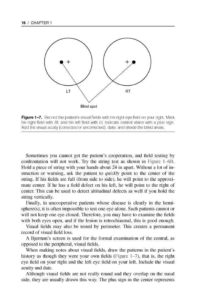
18 / CHAPTER 1
central vision. The shaded oval area, lateral to the center and partially astride
the equatorial line, is the normal blind spot. This is an absolute scotoma rep-
resenting the optic nerve head, or disc, which has no retinal function.
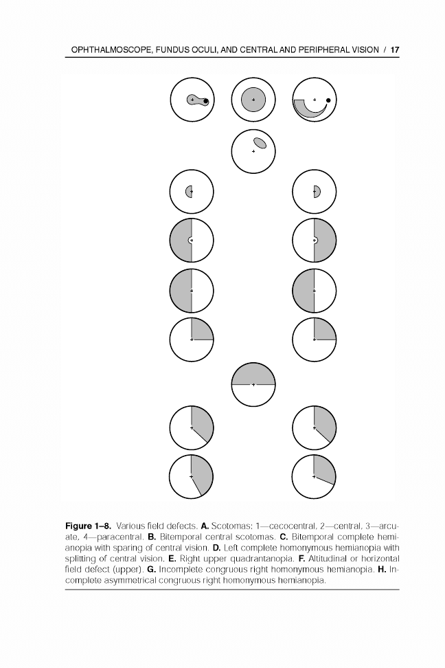
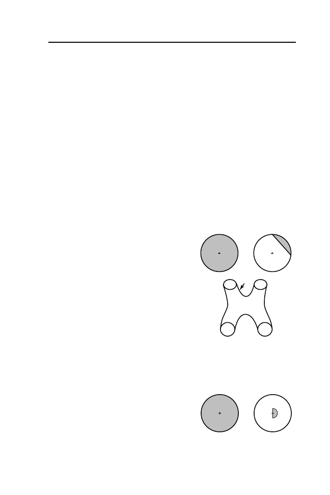
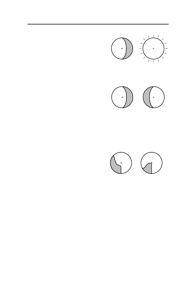
You need to be familiar with the types of field defects shown in
Figure 1–8
and their significance. (The blind areas are shaded.)
In testing visual fields by confrontation, remember:
• The patient must fix his gaze on your eye and keep this eye still.
• No one can detect color or definition in his peripheral fields of vision—the
stimulus is movement.
• Keep your testing fingers on the edge of his fields.
• Test in both the upper and lower quadrants of each eye.
• Always make the right half fields compete against the left half fields by of-
fering simultaneous right and left stimulation.

Loss of Vision
2
This chapter includes a list of definitions you need to know, a few anatom-
ical hints about the visual system, and a listing of some of the kinds of visual
problems you can expect to encounter.
Listed with each disease are some common or typical characteristics that
will help you with recognition. Some of these diseases do not “belong” in
the neurologist’s examining room, but you cannot preselect your patients.
Although the neurologist’s opinion is often not the final word on a problem
of visual loss, you must know how the visual system works and how dis-
eases of this system present themselves.
DEFINITIONS
Visual acuity: Literally, visual “sharpness” tested by evaluat-
ing the recognition of patterns of letters or numbers, graded
sizes, brightness discrimination, and color recognition
Macular vision: Same as central vision, the zone in the cen-
ter of the field of vision. It extends as a circle from fixation
about 15 degrees. Visual acuity is a measurement of the in-
tegrity of macular vision.
Peripheral field of vision: All of the area of space that can be
seen, exclusive of central vision, when the eye is stationary.
The extent of the peripheral field is (from fixation) about 60
degrees on the nasal side and upward, about 70 degrees infe-
riorly, and about 90 degrees on the temporal side. The nasal
fields of the two eyes overlap.
Scotoma: An area of decreased or absent vision surrounded
by an area of normal vision. A scotoma may be relative or
absolute. A scotoma is relative if a small test object cannot
be seen in it but a sufficiently large object can be seen. It is
absolute if nothing can be seen in it.
Central scotoma: A scotoma that includes the fixation point;
visual acuity is decreased. The bigger it is, the larger will be
the letter on the Snellen test type that may be “hidden” in the
19

20 / CHAPTER 2
scotoma and the worse will be the visual acuity (type 2 in
Figure 1–8A
).
Cecocentral scotoma: A horizontal oval scotoma including
fixation and extending to and including the blind spot (type
1 in
Figure 1–8A
)
Arcuate scotoma: As seen in type 3 in
Figure 1–8A,
corre-
sponds to and represents nerve fiber bundle loss
Blind spot, or physiological scotoma: The area of blindness
in the field of vision resulting from the head of the optic
nerve. It is oval, 5 degrees wide and 8 degrees high, and on
and mostly below the equator on the temporal side of fixa-
tion. Its nasal side is about 12 degrees from fixation. It can-
not be seen with both eyes open because of the visual field
overlap.
Find your own blind spot. Close your left eye. Stare at something at eye
level straight ahead of you with your right eye (do not move your eye!).
Hold a pencil in your right hand at arm’s length. Move the pencil in from the
temporal side, on the equator or just below it. As your pencil approaches fix-
ation, you will find you can “lose” the eraser on the end of the pencil in your
blind spot. You can slide the eraser around in the blind spot. This is how cen-
tral field defects are plotted. If you cannot find it, you are not fixing your
right eye on a distant object.
Hemianopia: Loss of half of the field of vision in both eyes
or either eye
Homonymous hemianopia: Loss of half of the field of vision
in each eye and on the same side of the midsagittal meridian
in each eye; that is, a left homonymous hemianopia means
the patient has lost vision in his temporal half field of the left
eye and nasal half field of the right eye. A bitemporal or bi-
nasal field defect may be hemianopic but it is not homony-
mous. Loss of vision in the upper or lower field respecting
the equator and ignoring the midline vertical meridian is
called an altitudinal defect (upper or lower) and may be con-
gruous or incongruous (discussed later in this chapter; see
also
Figures 1–8H
and
2–5
).
Bitemporal hemianopic central scotoma: The scotomas are
in the temporal halves of macular vision. The nasal sides are
intact and acuity is normal. The peripheral field is normal
(
Figure 1–8B
). (a) Hemianopia with splitting of the mac-
ula—the vision in half the peripheral field and half the mac-

LOSS OF VISION / 21
ula is gone, but visual acuity is normal (
Figure 1–8D
). (b)
Hemianopia with sparing of the macula—the vision in half
the peripheral field is gone, but all of macular or central vi-
sion is normal and, of course, acuity is normal
(Figures
1–8C
and
2–7
). (c) Hemianopia with macular loss—hemi-
anopia as above plus all of macular vision and abnormal vi-
sual acuity. This is not a hemianopia plus a central scotoma.
By definition, a scotoma is an area of blindness surrounded
by normal vision.
The visual cortex is all of Brodmann’s areas 17–19. Area 17 is the primary
visual cortex receiving the optic radiation from the lateral geniculate body.
Area 17 is the striate cortex, area 18 is the parastriate cortex, and area 19
is the peristriate cortex.
The striate cortex is striated by a light strip of myelinated fibers parallel to
the surface of the cortex and known as Gennari’s line. You can see it easily
with the naked eye in a fresh brain.
Seven important points about the visual pathways are:
1. Optic inversion: Everything you can see from the right eye is divided in
two ways. There is a vertical meridian dividing it into the right (or tem-
poral) field and the left (or nasal) field. Everything in the temporal field
is perceived by the nasal retina, and vice versa for the nasal field.
Also, there is an equatorial division. Everything below it is perceived by
the upper retina, and vice versa for things above it.
In addition, the nerve fibers in the visual pathways arising from both tem-
poral retinas do not cross the midline and they end in the ipsilateral occipital
cortices. All the fibers arising from both nasal retinas do cross the midline
and thus end in the contralateral occipital cortices.
On the medial surface of the occipital lobe you can see the calcarine sul-
cus, which is roughly horizontal and divides the occipital lobe into upper and
lower portions.
All the nerve fibers arising from cells in the retinas above the horizontal
division end up in the occipital cortex above the calcarine sulcus and below
it for the fibers originating below the horizontal.
The retina is well organized, like a pie divided into four more or less equal
pieces, plus the central plum for the posterior pole. In contrast, the visual
fields are not equal. The temporal field is 60–70% bigger than the nasal field,
yet each has about the same amount of retina. Each of the four retinal
“pieces” is anatomically distinct with its own connections. The occipital cor-
tex is clearly divided by the calcarine fissure in the horizontal plane. How-
ever, the arrangement of fibers in the visual pathways between the retina and

22 / CHAPTER 2
the occiput is not so methodical. For example, macular fibers in the optic
nerve immediately behind the eye are in the upper lateral part of the nerve.
Immediately in front of the chiasma the same fibers are in the center of the
nerve. The chiasma, optic tract, lateral geniculate body (LGB), and optic ra-
diation each have their unique fiber arrangements and proportions of fibers
or cells (LGB) devoted to peripheral or central vision. [There is an excellent
picture of this on page 916 of the 35th edition of Gray’s Anatomy (Warwich
and Williams, editors), Longman, 1973.]
As an example of proportion, the posterior pole of the retina is about 5 mm
in diameter and the macula is about 1.5 mm. The whole retina, when laid out
flat, is about 50–55 mm across; thus, over 90% of the retina is devoted to pe-
ripheral vision. By contrast, in the occipital cortex the amount of brain repre-
senting central vision is huge relative to the amount concerned with the pe-
ripheral fields. The occipital cortex representing central vision is the most
posterior part of the occipital pole, and the peripheral retina is represented
by the most anterior part of the occipital cortex.
In general, upper retinal fibers remain upper and lower ones remain lower
except in the optic tract and the LGB, where a 90-degree rotation occurs.
The rotation becomes undone in the optic radiation.
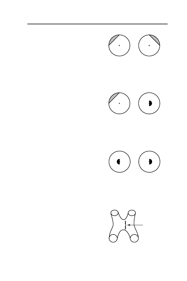
2. Lesions of the retina and optic nerve are characterized by unilateral,
central, altitudinal, ring, and cecocentral scotomas; diminished visual
acuity; altered color vision; and afferent pupillary defects. The opposite
eye remains normal.
3. Chiasmal lesions are characterized by bitemporal defects. They may be
in the peripheral field, the central field, or both (
Figure 1–8B
and
C
).
Progressive lesions posterior to the chiasma will eventually cause
homonymous field defects. The chiasma is about 1 cm above the pitu-
itary gland. This large space can accommodate relatively large
suprasellar tumors with no field defect. Although the chiasma is di-
rectly above the dorsum sella in most patients, it may be located more
anteriorly or posteriorly. This variability will account for atypical field
defects. There are unique fiber arrangements in the chiasma.
a. Ventral nasal optic nerve fibers (serving the superior temporal
field) cross in the anterior chiasma and loop into the distal opposite
optic nerve and then turn posteriorly to the contralateral optic tract
(see
b. Macular fibers make up a large part of the chiasma and are located
largely inferiorly.
c. Nasal macular fibers decussate in the posterior chiasma. (
are applicable to chiasmal lesions.)

LOSS OF VISION / 23
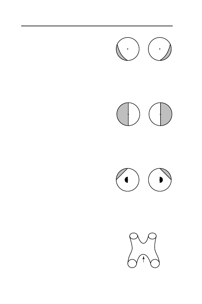
4. Optic tract lesions are characterized by their incongruity. They usually
begin as a quadrantic defect with early sparing of macular vision (
5. Optic radiation:
a. In temporal lobe lesions, field defects are principally superior and
quadrantic; if hemianopic, they are denser and earlier in the upper
quadrant.
b. In parietal lobe lesions, field defects may be inferior and quadrantic
but, more commonly, hemianopic. When early and progressive, they
will be denser and first evident in the lower quadrants (
6. Occipital lobe: Monocular temporal crescent, field, or scotomatous de-
fect, in anterior occipital lobe lesions (
a. Central, congruous scotomas, in lesions of the tip of the occipital
b. Hemianopic with macular sparing; bilateral lesions will leave the
patient with tubular vision, that is, all blind but for 10 degrees
around fixation, or all central and peripheral loss.
7. The more posterior the lesion, the more likely are the areas of field de-
fect in the two eyes to be congruous and macular vision spared. Macu-
lar fibers in tract, LGB, and anterior optic radiation are confined to a
relatively narrow area. Posteriorly, they form a large part of the radia-
tion and cortex.
8. Visual field defects are congruous when they are the same shape in
each eye and incongruous when they are not. Unless they are hemi-
anopic, they are seldom the same size. Optic radiation lesions are com-
monly congruous, and tract lesions are usually incongruous.
DIAGNOSIS
The location of the lesion is often obvious in visual system disease be-
cause of the pattern of the area of visual loss; for example, when visual acu-
ity in the right eye is reduced to 20/200 with a central scotoma but the left
eye is normal, the lesion must be between the right cornea and the optic chi-
asma. When the visual acuity is normal and there is a right upper homony-
mous quadrantanopia (
Figure 1–8E
), the lesion must be in the left temporal
lobe involving the optic radiations.
As a generalization, the findings of central (type 2 in
Figure 1–8A
), ring,
arcuate (type 3 in
Figure 1–8A
), or cecocentral (type 1 in
Figure 1–8A
) sco-
tomas, visual loss in one eye only, abnormal color vision, and afferent pupil
defect (see Chapter 5), or an altitudinal field loss (
Figure 1–8F
) are indica-
tive of disease in the retina or optic nerve.

24 / CHAPTER 2
Normal visual acuity with field defects on the same side of the vertical
meridian (ie, homonymous) are caused by lesions in the optic tract, LGB, op-
tic radiation, or occipital cortex. The more posterior the lesion, the more
likely the field defect in one eye will be congruous with the defect in the other
eye (
Figure 1–8G
). If the field defect is a complete homonymous hemianopia,
you cannot tell where the lesion is, except that it must be behind the chiasma.
More diagnoses are missed because of a failure to appreciate the arrange-
ment of the fibers in the optic chiasma than any other part of the visual sys-
tem.
Histories vary from helpful to misleading. The presbyopic patient com-
plains he cannot get far enough from his newspaper to read it. (The ability to
read the fine print in the average newspaper requires 20/30 vision.) The pa-
tient with pigmentary retinal degeneration will often forget to mention the
progressing night blindness, an extremely important symptom. Another pa-
tient with an inattention left hemianopia has no complaints and is being ex-
amined only at the insistence of, for example, his wife or the motor vehicle
licensing authorities. Typically, in the past months he has been driving with
the left side of his car over the dividing line into oncoming traffic.
The patient who says he covered one eye with his hand and realized he was
virtually blind in the other eye has told you nothing about the tempo of the dis-
ease, but has revealed that the lesion is between his cornea and the chiasma.
Also, some people are born with one eye myopic and the other eye normal
and they discover the fact incidentally. A man of 40 who wears no glasses
and has no visual complaints gets a foreign body in his eye (not in the line of
vision). When the foreign body is successfully removed, he discovers that
the vision in the previously injured eye is not as good as in the other eye. In
his mind the injury and the diminished visual acuity are cause and effect.
Examination shows reduced visual acuity but an otherwise normal eye. The
pinhole improves his visual acuity to the 20/20 line. Refraction by an oph-
thalmologist confirms that his best corrected vision is normal. This eye has
always been myopic and the trauma has simply brought it to his attention.
When confronted with an apparent recent loss of vision in one eye that
makes no sense, always get a full refraction before conducting more elabo-
rate investigations.
SOME DISEASES OF THE VISUAL SYSTEM
Retina and Optic Nerve Lesions
Retrobulbar Neuritis
Retrobulbar neuritis typically presents in young
adults, with onset over 24–48 hr, a large central scotoma, and a painful eye

LOSS OF VISION / 25
on palpation and movement. There may be a past history of episodes of mul-
tiple sclerosis or this illness may be the first manifestation of it. The fundus
is usually normal. Afferent pupillary defect is commonly present.
Optic Neuritis
The presentation of optic neuritis is like that of retrobulbar
neuritis, except that it may be painless. The terms retrobulbar neuritis and op-
tic neuritis are used interchangeably. Edema of the optic nerve head may be
present. (See the section on papillitis in Chapter 3.)
Central Serous Retinitis
Central serous retinitis has subacute onset (days)
of slight to moderate vision loss in one eye, usually in a male patient aged
20–40. The disease is caused by fluid exudate lifting the retina, usually at the
macula. The patient complains that objects look smaller with the affected eye.
Giant Cell Arteritis of the Central Retinal Artery
This disease occurs in
those age 60 or older, with a sudden onset of central blindness. Superficial
temporal arteries are typically tender, pulseless, and tortuous. There is almost
always an elevated erythrocyte sedimentation rate and a low-grade fever. The
history will often contain complaints of headache and stiff, aching, weak
shoulder and hip muscles. Funduscopy shows total retinal ischemia (see the
following section). Diagnosis, including temporal artery biopsy, is an emer-
gency.
Anterior Ischemic Optic Neuropathy
In patients over age 50, anterior is-
chemic optic neuropathy is a manifestation of giant cell arteritis, vasculitis,
diabetes mellitus, or Takayasu’s disease or is frequently idiopathic. There is a
sudden onset of visual loss, sometimes followed by increasing and progres-
sive visual failure over 5–7 days. Altitudinal (inferior) field defects are com-
mon. Funduscopy examination shows a swollen disc, hemorrhages near the
disc edge, and cotton-wool spots. A macular star is common with vascu-
lopathies. The other eye is commonly involved within weeks or months of the
first eye.
Retinitis Pigmentosa
The most common symptom of retinitis pigmentosa
(RP) is night blindness. The onset and severity of symptoms depend on the
pattern of inheritance: the autosomal recessive type presents earliest and is
the most severe, X-linked is intermediate, and autosomal dominant is fre-
quently mild. The fundus shows black “bone spicules” of pigment clustered
around vessels in the midperiphery. The disc is waxy pale, and there is attenu-
ation of both retinal vessels. Visual fields show ring-shaped arcuate or annular
scotomas.

26 / CHAPTER 2
Pseudoretinitis Pigmentosa
Pseudoretinitis pigmentosa refers to the fundus
findings of a number of disorders that mimic RP. Some pigmentary
retinopathies that are not RP are those following congenital and acquired
syphilis, childhood exanthemas, phenothiazine usage, and trauma.
Atypical RP includes sectoral RP (fundus changes in one sector of the
fundus bilaterally, which can mimic chiasmal lesions) and pericentral RP
(fundus changes central). Unilateral RP probably does not exist.
Diseases and syndromes associated with RP are Bassen-Kornzweig,
Bardet-Biedl, Kearns-Sayre, and Usher’s.
Glaucoma
Glaucoma exists when elevation of intraocular pressure is suf-
ficient to damage optic nerve fibers at the optic disc level. This is the second
leading cause of blindness in North America. There are two types: acute
angle-closure glaucoma (ACG) and chronic open-angle glaucoma (OAG).
ACG presents with severe pain in the eye and head, blurred vision, and
colored halos around lights, plus nausea and vomiting. Pain results from a
rapid rise in intraocular pressure. Attacks are precipitated by reduced illumi-
nation and may be relieved by sleep, bright light, or miotic agents, all of
which constrict the pupil. ACG usually presents unilaterally. The predispos-
ing factors are often bilateral, and the uninvolved eye is therefore at risk.
This is an emergency.
OAG is the most common form of glaucoma and is dangerous because the
onset is often gradual and asymptomatic. Central vision is preserved until
late, and the field loss is often unnoticed by the patient until the disease is
well advanced. It is usually a bilateral disease, although asymmetrical.
Visual field defects in glaucoma respect the horizontal division of the vi-
sual fields. The arcuate nerve fibers arching above and below the macula
from the temporal region are most susceptible to glaucomatous damage.
Therefore, the characteristic field defects are paracentral and arcuate nasal
scotomas (type 3 in
Figure 1–8A
). The central area of the visual field is the
most resistant, so visual acuity may be normal even in advanced glaucoma.
Optic disc changes are an increase in the diameter and depth of the physi-
ological cup and increased pallor.
Central Retinal Vein Occlusion
Central retinal vein occlusion (CRVO) pre-
sents with painless, always unilateral, loss of vision. The patient is commonly
a young adult. The extent of visual loss is variable, depending on the degree
of venous occlusion, the amount of macular edema, and the presence or ab-
sence of complications such as retinal neovascularization and neovascular
glaucoma.
The fundus shows dilated, tortuous veins; retinal hemorrhages usually in
the peripheral retina; and retinal edema. Cotton-wool exudates are usually

LOSS OF VISION / 27
seen only around the disc. The disc edge is indistinct, although the physio-
logical cup remains visible.
Visual fields reveal a relative central scotoma and acuity is usually 20/100,
improving to 20/60 with time if there are no complications.
Branch retinal vein occlusion is most common in the superotemporal
branch. Fundus changes are confined to the distribution of the branch, and
field loss is segmental.
Hypertension often coexists with CRVO, and associated retinal artery dis-
ease is a common finding. A picture similar to that of CRVO may be seen in
hyperviscosity syndromes, leukemia, and myeloma.
Retinal Artery Occlusion
Retinal artery occlusion (RAO) is accompanied
by sudden, painless, unilateral loss of vision. It is commonly discovered by
the patient on awakening in the morning and occurs in the stroke-prone age
group. If the central retinal artery is involved, vision will be completely lost
(no light perception). If a cilioretinal artery is present, an island of central vi-
sion will be preserved. Visual loss may be confined to a segment of the field
if only a branch of the retinal artery is occluded.
The fundus reveals a gray, opaque, edematous retina with a cherry-red
fovea. This is normal choroid contrasted against gray retina. Splinter hemor-
rhages are rarely seen. Branch arteries may show segmented columns of
blood (boxcars). There may be a history of transient monocular visual loss
lasting minutes, with altitudinal progression as vision fails, that is, the win-
dow blind effect.
Causes of RAO include emboli, thrombosis, giant cell arteritis, and collagen-
vascular diseases.
Retinal Detachment
Retinal detachment (RD) presents with a history of re-
current flashes or floaters in the same area of the visual field. The patient typi-
cally complains of a cloud or curtain obscuring part of the field. It usually starts
and is most dense peripherally and extends toward central vision. Visual acuity
is normal unless or until the macula detaches. An inferior detachment with a su-
perior field defect can be present for a long time before the patient is aware of
it. The fundus shows an elevated retina that is gray and wrinkled and undulates.
The elevation will be appreciated as you rack in progressively more plus
lenses (black numbers) on the ophthalmoscope to keep the elevated retina in
focus.
The most common predisposing factor to RD is degenerative retinal
change (as seen in high myopia). Other causes include trauma; a simple
“black eye” history may be significant. Malignant melanoma of the choroid
can cause a secondary RD, and the retinal neovascularization of diabetes
mellitus may result in a tractional RD.

28 / CHAPTER 2
Macular Degeneration
Macular degeneration begins at any age but is most
common over 60 years. Slow loss of visual acuity occurs bilaterally and is
worse in bright light and better in the dark; recovery of vision is slow after
exposure to a bright flash of light. The macula may appear normal initially.
There are central and paracentral scotomas. Fluorescein retinal angiography
and the Amsler grid are helpful in diagnosis.
Optic Neuropathy
Optic neuropathy can occur:
1. Without other diseases (eg, Leber’s). The typical patient is a young
adult male with loss of central vision in one eye, followed by the other
eye in days or weeks, with a positive family history. Characteristic fun-
dus changes in the acute stage are followed by optic atrophy. A central
scotoma will become cecocentral with upper nasal breakout.
2. With other central nervous system (CNS) diseases, with one or more of
the following: congenital deafness, ataxia, spastic quadriparesis, mental
deterioration, polyneuropathy, Friedreich’s ataxia, Marie’s cerebellar
ataxia, and Charcot-Marie-Tooth disease
3. With inborn lysosomal disorders: (a) mucopolysaccharidoses or (b)
lipidoses
Optic Atrophy
Optic atrophy occurs as a consequence of the foregoing and
other diseases, such as methyl alcohol or chloroquine ingestion, isoniazid tox-
icity, and any compressive, ischemic, or toxic disorder of the retinal ganglion
cell or fiber from the retina to the LGB. This includes papilledema, which, if
severe enough or chronic enough, can result in optic atrophy. Diagnosis of the
cause is often impossible. Clinical diagnosis of atrophy is dependent on the
color and structure of the disc.
The lesion responsible for optic atrophy may be anywhere from the retina
to the LGB inclusive.
Chiasmal Lesions
The most common visual field defect is bitemporal, ei-
ther central or peripheral or both.
Be careful and persistent with patients with visual loss. Lesions in the chi-
asmal region can be deceptive and extremely chronic. The chronicity and
slow progression seem to make the symptoms more acceptable and less de-
manding. Every neurologist and neurosurgeon has had some bad experience
with patients thought to have multiple sclerosis, amblyopia from childhood,
low-tension glaucoma, retinitis pigmentosa sine pigmento, or atypical macu-
lar degeneration as an explanation for their blindness who eventually turn
out to have a chiasmal lesion as the true cause. It does not help the patient to
LOSS OF VISION / 29
make the diagnosis after the optic atrophy is marked and the acuity is down
to 20/200.
Another reminder about chiasmal lesions is the place of exploratory in-
tracranial surgery. Ordinarily, there is no such operation as an exploratory in-
tracranial procedure. However, if an eye and field examination point to a chi-
asmal lesion, even if the skull x-ray, carotid angiography, computerized
tomography (CT) scan, and nuclear magnetic resonance (NMR) studies are
all normal, then the next “investigative” step is exploration of the chiasmal
area. The neurosurgeon may make the diagnosis of chiasmal arachnoiditis
that cannot be treated, but he may find the otherwise undiagnosable 4-mm
meningioma, pituitary tumor, or craniopharyngioma.
Finally, lesions of the visual pathways behind the optic chiasma never in-
terfere with color vision (ie, without an accompanying loss of light percep-
tion). There are reported examples of defective color appreciation from a
parietal cortical lesion, but this is color agnosia, not loss of color vision.
Anterior Chiasmal Syndromes
1. The ipsilateral eye is blind and
there is an upper temporal field
defect in the contralateral eye.
The lesion is shown at a in
2. The ipsilateral eye is blind and
there is a central temporal sco-
toma in the contralateral eye
(The same lesion as in
A
a
Figure 2–1A
B
Figure 2–1B
30 / CHAPTER 2
3. The ipsilateral eye is normal and
the contralateral eye is as in
or B (The same lesion
as in
4. The ipsilateral eye has a central
scotoma and there is a paracen-
tral scotoma in the temporal field
of the contralateral eye (The
same lesion as in
This can progress to the condi-
tion shown in
5. The ipsilateral eye is blind and
there is a paracentral scotoma in
the temporal field and full pe-
ripheral field of the contralateral
eye (The same lesion as in
D
Figure 2–1D
C
Figure 2–1C
E
Figure 2–1E
LOSS OF VISION / 31
Chiasmal Body
Bitemporal periph-
or central temporal scotomas oc-
cur as in
sions occur as in
. The
lesion is usually below the chiasma
(eg, a pituitary tumor).
A
Figure 2–2A
B
Figure 2–2B
C
Figure 2–2C
D
Lesion area
related to fields
2.2A to 2.2C
Figure 2–2D
32 / CHAPTER 2
Suprasellar lesions (eg, cranio-
pharyngioma, aneurysm, meningioma,
chordoma, or third ventricle disten-
tion) can start with a central or periph-
eral temporal defect that initially may
be most marked inferiorly as in
Any of these lesions (suprasellar or
infrasellar) may eventually produce
the field defect shown in
Posterior Chiasma
Posterior chi-
asma presents usually with a bitempo-
ral central scotoma with peripheral de-
fects as well, as in
. A
central scotoma usually occurs first.
The lesion is shown at a in
. When big enough to involve the
tract at b, a homonymous, hemianopic
defect is added.
F
Figure 2–2F
A
Figure 2–3A
Figure 2–3B
B
a
b
E
Figure 2–2E
LOSS OF VISION / 33
Lateral Chiasma
Lateral chiasmal
lesions present with nasal field de-
fects. Very rare, they can be unilateral
as in
or bilateral as in
up to the midline. Unilateral nasal de-
fects have been reported as a result
of infarction of the optic nerve,
aneurysm, pituitary tumor, and ectatic
carotid artery. Bilateral nasal defects
have been reported resulting from chi-
asmal arachnoiditis and secondary to
obstructive hydrocephalus and pres-
sure from above from the third ventri-
cle. Glaucoma is probably the most
common cause of nasal field defects.
Optic Tract Lesions
Optic tract lesions between the chi-
asma and the LGB produce incongru-
ous field defects as in
Most lesions behind the chiasma,
whether tract, LGB, radiation, or cor-
tex, present as homonymous field de-
fects with normal visual acuity. The
lesions cause loss of vision in some or
all of the two homonymous half
fields. When the hemianopia is com-
plete, the site of the lesions cannot be
located by visual field examination
alone.
Blind areas resulting from tract le-
sions are homonymous and extremely
incongruous in shape and size.
A
Figure 2–4A
B
Figure 2–4B
Figure 2–5
34 / CHAPTER 2
1. When a tract lesion produces a complete hemianopia, the eye contralat-
eral to the lesions will show an afferent pupil defect (see Chapter 5).
This may be difficult to elicit.
2. When a tract lesion presents with some degree of hemianopic visual
loss plus decreased visual acuity and color vision, the lesion has in-
volved the chiasma or the optic nerve(s). This area is compact, and dis-
tances are small.
3. Some lesions originate in or near the sella and then expand laterally to
involve the tract. The patient may present with the confusing combina-
tion of a chiasmal (bitemporal) defect plus an optic tract (incongruous
hemianopic) defect.
4. Many patients with lesions anterior to the LGB (ie, tract, chiasmal, and
nerve) are aware of a dimness or loss of vision. Conversely, most pa-
tients with lesions posterior to the LGB are unaware of their visual de-
fect.
Optic Radiation
Within the Temporal Lobe
A ho-
monymous defect, superior and quad-
rantic, is shown in
Defects can be congruous or incon-
gruous. When incongruous, the larger
defect is usually in the eye on the
same side as the lesion. It is also
denser in this eye. The blind area may
cross the horizontal division of the vi-
sual fields.
When the defect is incongruous, it
is never as incongruous as with optic
tract lesions.
Within the Parietal Lobe
A hemi-
anopic homonymous defect may ini-
tially be only, or denser, in the lower
fields, as in
This is congruous with sparing of
the central vision and normal acuity.
Peripheral defects result from me-
dially placed lesions.
There is an abnormal optokinetic
response (see Chapter 6).
Figure 2–6
Figure 2–7
LOSS OF VISION / 35
Occipital Lobe and Visual
Cortex Lesions
The more posterior the homony-
mous defect, the more likely it is to be
completely congruous.
Tip of occipital pole lesions can
produce central homonymous sco-
tomas with precise congruity.
shows a field defect recorded by
Sir Gordon Holmes. The tip of the pa-
tient’s right occipital lobe was injured
with the resulting left incomplete
homonymous hemianopic central sco-
tomas.
Anterior Visual Cortex
or Posterior Optic Radiation
There are unpaired nasal fibers that
correspond to the extreme temporal
peripheral field and the most anterior
aspect of the area striata. In patients
with posterior optic radiation lesions,
there may be a unilateral temporal
crescent defect as in
may be above or below the horizontal
division and often is the forerunner of
a hemianopic defect. The defect may
be a temporal crescent scotoma not
touching the edge of the field (not
shown). Thus, a postchiasmal lesion
may produce a purely monocular field
abnormality.
The sparing of a thin temporal cres-
cent of vision when the rest of the
temporal field is hemianopic also oc-
curs as in
Figure 2–8
Figure 2–9
Figure 2–10

36 / CHAPTER 2
Cortical Blindness
The term cortical blindness is used synonymously with bilateral homony-
mous hemianopia. The definition includes
• Loss of all vision, including perception of light
• Loss of reflex blinking on being threatened
• Normal pupil response to light and convergence
• Full eye movements
It is common for patients with this disorder not to complain of the vision
loss or to deny the blindness when confronted with the fact (Anton’s syn-
drome). Remember that the condition commonly occurs from occlusion of
the upper end of the basilar and the two posterior cerebral arteries. As the
latter supply the inferior and medial surface of the temporal lobes as well as
the primary visual cortex, an acute confusional amnesic syndrome may ac-
company the acute loss of vision. The latter may account for the denial of
the blindness; that is, it is an agnosia.

The Abnormal Retina
3
PAPILLEDEMA
Terms synonymous with papilledema which are in common use are
choked disc, swollen disc, and swelling of the nerve head.
Definition
Papilledema is a swelling (laterally) and an elevation (anteriorly) of the
disc. It is caused by increased intracranial pressure and is measured in
diopters. (A diopter is a unit of refraction; that is, a lens with a power of 1
diopter has its principal focus at a distance of 1 m.) Only the elevation and
swelling of the disc are papilledema.
Restrict the term papilledema to nerve head swelling caused by increased
intracranial pressure. Other causes of swelling and elevation of the nerve
head should be named from the causative process, for example, optic neuri-
tis, anterior ischemic optic neuropathy, or hemorrhagic retinopathy.
The characteristics of early papilledema include the following:
1. Disc hyperemia results from dilatation of capillaries on the surface of
the disc.
2. The retina immediately surrounding the disc becomes dull, loses its lin-
ear light reflexes, and is a dusky red. The disc margins blur.
3. Venous pulsations—which disappear late in fully developed papilledema—
are not a useful early sign. The level of increased intracranial pressure
varies, and when it drops below 200 mm of water, the veins then pulsate.
Also, not all people with normal intracranial pressure have pulsatile retinal
veins.
4. Venous dilatation and tortuousity is not in itself a good early sign.
5. Nerve fiber hemorrhages—thin radial streaks on the disc or near its
edge—are a highly reliable confirming sign.
6. Nerve head elevation is the sine qua non of papilledema (see below).
In fully developed papilledema the following occurs:
1. Engorged, tortuous, dusky retinal veins are present, the disc is elevated
and margins of the disc are unidentifiable, and there is marked capillary
37

38 / CHAPTER 3
dilatation over the disc. In addition, there are many flame-shaped and
splinter hemorrhages in the retina and (if the pressure increase has been
rapid) globular subhyaloid hemorrhages. In advanced papilledema you
will also see cotton-wool spots, macular exudates forming an incom-
plete stellate pattern around the macula, and retinal wrinkling (Paton’s
lines).
2. In chronic papilledema the disc elevation remains, the hemorrhages and
exudates resolve, and nerve fiber loss may be seen. The end state is op-
tic atrophy.
How to See It
• When examining patients, start with a plus 8 (black number); you see an
orange blur.
• Turn the lens selector wheel counterclockwise using progressively weaker
plus lenses until the surface of the disc is sharply defined. Note the magni-
fication (eg,
+
4).
• Look at the patient’s fundus again beside the disc and keep decreasing the
plus lens until retinal details come into focus (eg,
+
1).
The difference in these two readings is the amount of papilledema in
diopters, that is, 3. (We cannot use a lens of less than 1 diopter. A difference
between the nerve head and surrounding retina of less than 1 diopter is too
subjective to be meaningful.)
About 3 diopters are equal to 1 mm of change. The target to be seen
clearly is usually the smallest vessel you can see. You do not need a patient
with papilledema to learn this; one of your classmates has a normal physio-
logical cup. You can see the bottom of it with great clarity using a (for exam-
ple)
−
2 (red number) in your ophthalmoscope. He has a fine artery in the
retina to one side of the disc. You will see this vessel with great clarity by
using a
+
1 (black number), that is, 3 diopters, or three clicks on the scope;
the retinal vessel is about 1 mm closer to you than the bottom of the cup.
Remember, the higher the plus lens you use, the shorter the focal length
and the more anterior is the part of the patient’s eye you can clearly see, for
example, with a
+
20 lens in the window of the ophthalmoscope you will fo-
cus on his cornea. The more negative the lens, the longer the length and the
farther back in the patient’s eye is the point of focus; for example,
−
50 to
−
10 enables you to focus on the retina of the myopic patient with the long
oval eye.
Do not look at the fundus with a zero lens in the ophthalmo-
scope and plan to increase the plus lenses as you concentrate

THE ABNORMAL RETINA / 39
on the elevated disc, thinking it will become clear while the
surrounding retinal details blur. Your own accommodation
will not allow it, and you will miss the papilledema.
Remember:
1. Start plus (eg,
+
8); aim the light at the disc.
2. Diminish the lens strength (by a counterclockwise turn on the lens se-
lector wheel).
3. Stop when the disc is clearly seen.
4. Note the lens number.
5. Aim beside the disc at the retina.
6. Diminish the lens strength.
7. Stop when retinal details are clearly seen.
8. Note the lens number (the difference is the amount of papilledema).
The normal eye is shown in cross-section in
Figure 3–1A,
and the disc
area is shown in more detail in
Figure 3–1B
. The disc is 1.5 mm in diameter
and, seen with the ophthalmoscope, looks like those in
Figures 1–3
and
1–4
.
The disc edges are of different elevations at various points on the circum-
ference of the nerve head. The physiological cup is a true depression in the
nerve head. There is no retina overlying the nerve head, hence the normal
blind spot.
The optic nerve is surrounded by a sheath space continuous with the sub-
arachnoid space surrounding the brain (
Figure 3–1B
). This space, which
contains spinal fluid, may be filled with blood, inflammatory products, or
spinal fluid under increased pressure in various disease states.
Figure 3–2A
and
B
shows cross-sections of a papilledematous eye. The
nerve is swollen, bulging forward, and expanded. Notice in
Figure 3–2B
how the retina is pushed laterally by the bulging nerve head.
Examine both eyes, because the amount of papilledema is frequently dif-
ferent in the two eyes. Look in both eyes and judge them independently.
Visual Acuity
Uncomplicated papilledema of moderate duration and degree does not in-
terfere with visual acuity. Clearly, acute papilledema with a hemorrhage into
the macular area will diminish visual acuity.
Severe and long-standing papilledema can produce optic atrophy and
blindness. In the chronically compressed optic nerve both glial cells and
small vessels proliferate, and if the swelling is severe, necrosis of the nerve
may occur.
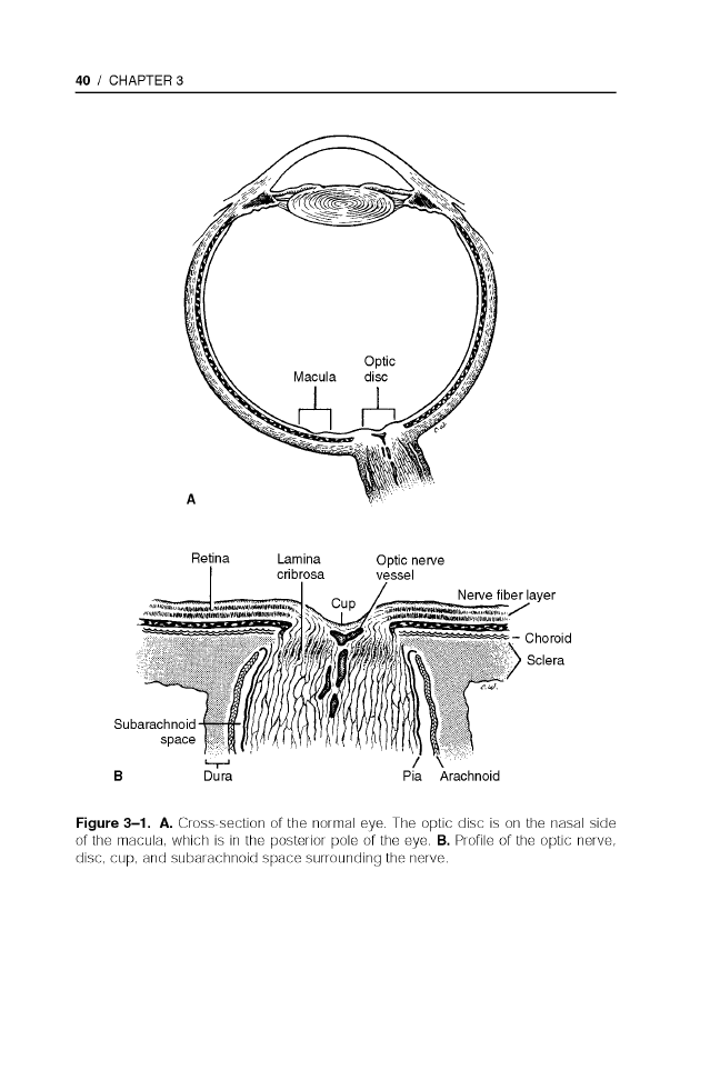
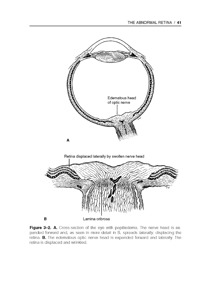

42 / CHAPTER 3
Retinal Wrinkling
In the presence of papilledema (especially chronic), one can see wrinkling
of the retina. You will see concentric folds or ripples parallel and close to the
edge of the disc and most marked on the temporal side of the disc. As long
as the swollen nerve head pushes the retina laterally, these folds will persist.
OTHER DISEASES
Papillitis, optic neuritis, retrobulbar neuritis, and neuroretinitis, by their
word endings, suggest inflammation of the nerve head (papillitis) or nerve
trunk (neuritis). They are usually part of a demyelinating, vascular, or inflam-
matory process. Some of them produce swelling of the disc, and therefore can
be mistaken for papilledema caused by increased intracranial pressure.
Papillitis, or Optic Neuritis
Papillitis, or optic neuritis, causes swelling of the disc of about 2 diopters.
Loss of central vision is the prominent symptom. There will also be evidence
of a vascular, inflammatory, or demyelinating disease of the optic nerve
head. There may be pain in the eye at rest or on eye movement only.
Retrobulbar Neuritis
A normal fundus with no swelling of the disc is usually seen with retrobul-
bar neuritis. The symptoms are loss of central vision and pain in the eye,
which is worse on eye movement and palpation. The demyelinating process
is well behind the eye, hence the term retrobulbar.
Neuroretinitis
Similar to the situation with papillitis, the process of neuroretinitis has ex-
tended farther into the retina.
These diseases (eg, optic neuritis) have several distinctive features:
1. Loss of vision—This may be a central or paracentral scotoma or a huge
island of blindness involving the whole or almost the whole field of vi-
sion (in contrast, in early papilledema an enlarged blind spot is usually
the only vision change).
2. Pain on eye movement (or on palpation of the eye and sometimes at
rest)—This is presumably a result of traction and swelling of pain-
sensitive structures at the apex of the orbit. The pain is more common
with retrobulbar neuritis than with papillitis and may be present or ab-
sent with either. The pain usually precedes the blindness by 1–2 days
(in contrast, papilledema is painless).

THE ABNORMAL RETINA / 43
Hypertension
There are several systems for grading the fundus changes in hypertension
(eg, grade I or grade II). Do not do this: describe what you see. Hypertension
produces a number of changes and signs in the fundus:
1. Diffuse and focal or segmental constriction of the retinal arteries—The
older the patient, the less significant is the arterial narrowing. The earli-
est narrowing occurs in the retinal periphery. Tortuousity of the arteries
is most evident at the disc edge, and this change is indistinguishable
from arteriosclerotic changes unrelated to hypertension.
2. Disc edema—It looks the same as disc edema resulting from any other
cause.
3. Arteriovenous “nicking”––This may produce relative venous obstruc-
tion and retinal hemorrhages distal to the arteriovenous crossings.
4. Retinal hemorrhages—Widely distributed over the retina (in contrast, a
radial arrangement is seen around the disc in papilledema), petechial
and larger and often round, as well as flame-shaped. Look distal to the
arteriovenous crossings.
5. Exudates (spots, streaks, and clusters of white to gray material in the
retina)—These may be arranged radially around the macula in a star
configuration.
Remember:
1. Increased intracranial pressure may produce papilledema.
2. Start with a
+
8 to
+
10 lens, diminishing the magnification until the disc
is in focus. Note the magnification and then diminish the magnification
further until the retina is in focus.
3. The more acute the onset of the papilledema, the more hemorrhages are
present; these are arranged radially around the disc.
4. Vision is initially normal, with an enlarged blind spot.
5. Papillitis and retrobulbar neuritis may cause nerve head swelling; char-
acteristically, these cause central blindness and eye pain.
6. Elevated blood pressure may cause disc edema:
a. With diffuse and focal arterial changes
b. With venous nicking and venous obstruction at the arteriovenous
crossings
c. With a variety of hemorrhages diffusely seen in the retina often from
veins distal to the arteriovenous crossings
d. With exudates

Eye Movements,
4
Diplopia, and Cranial
Nerves 3, 4, and 6
DIPLOPIA
When a patient says he has double vision, his eyes are no longer in align-
ment. He has a disease of the third, fourth, or sixth cranial nerve or a disease
of one of the six muscles that move the eye, or of the myoneural junctions.
You can make yourself have double vision. Do this and you will under-
stand better what the patient is talking about. Keep both your eyes open. Put
your left index finger horizontally on your left upper eyelid. Do not cover
the pupil. Gently press the left eye down. Hold your pen horizontally at
arm’s length in your right hand. Look at the pen as you move it toward the
floor. You see two pens, one above the other. The farther downward you
move the pen, the greater is the separation between the two images. They are
parallel, one above the other. If you close your right eye, the top pen (the
true image, the macular image, the sharp one) disappears. If you close your
left eye, the bottom pen (the false image, the fuzzy one) disappears.
Double vision results from light reflected off an object and onto the mac-
ula of one eye (the nonparalyzed one) and onto the retina, near the macula,
of the other eye. The true image is the macular one, and the false image,
which is always less distinct, is the nonmacular one. The greater the non-
alignment of the paralyzed eye, the greater is the separation between the two
images. When a patient says she is seeing double and her right eye is turned
in toward her nose, she has a weak right lateral rectus. If her right eye is
turned down and out and she has a drooping right upper eyelid and an en-
larged pupil, the patient has a third nerve lesion on the right. Usually, how-
ever, the eyes seem to move in a normal way. Therefore, a system of exam-
ining eye movements is necessary.
Definitions
Abduction: Movement of the eye in the horizontal plane
away from the midline
45

46 / CHAPTER 4
Adduction: Same as for abduction but toward the midline
Cover test: Have the patient fix on the Snellen chart at his
line of best vision. Cover one eye. If the other eye moves to
take up fixation, a tropia exists. The movement will be seen
in the uncovered eye irrespective of which eye is covered.
On the other hand, if a covered eye moves to fixate when
uncovered, a phoria exists. The other uncovered eye does
not move.
Depression: Same as for elevation but downward
Elevation: Movement of the eye in the vertical plane upward
Esophoria: A deviation of the eye toward the other eye
when the stimulus of vision has been removed
Esotropia: A manifest deviation of the visual axis of one eye
toward the other; also known as convergent or internal stra-
bismus, or cross-eye
Exotropia: A manifest deviation of the visual axis of one eye
away from that of the other; also known as divergent or ex-
ternal strabismus, or walleye
Heterophoria (latent strabismus): Failure of the eyes to re-
main parallel after the stimulus for binocular single vision
has been removed (by covering one eye, as in the cover
test). A phoria may be inadvertently demonstrated in the
course of retinal examination when the head and ophthalmo-
scope of the examiner interfere with the patient’s fixation
and the eye being examined turns as the examiner with-
draws.
Hypertropia: A deviation of the visual axis upward
Hypotropia: A deviation of the visual axis downward
Rotation (as you face the patient): Inward, or internal—Ro-
tation of the eye so that the 12-o’clock point on the cornea
turns toward the nose, that is, clockwise rotation of the right
eye, counterclockwise rotation of the left eye. Outward, or
external—Rotation so that the 12-o’clock point turns away
from the nose, that is, counterclockwise rotation of the right
eye, clockwise rotation of the left eye
Squint: In North America this word sometimes means to
screw up the facial muscles around the eyes as though look-

EYE MOVEMENTS, DIPLOPIA, AND CRANIAL NERVES 3, 4, AND 6 / 47
ing into a bright light. In medicine, it means to have an ex-
ternal eye muscle palsy or a permanent strabismus.
Strabismus: A deviation of the eye that the patient cannot
overcome
The other heterophorias are hypophoria, in which the visual axis of one
eye sinks below the other; hyperphoria, in which the visual axis of one eye
rises above the other; and cyclophoria.
See the section on “Paralytic Versus Concomitant (Nonparalytic) Strabis-
mus.”
How to Discover the Muscles Responsible for Double Vision
• Test the function of the third, fourth, and sixth cranial nerves together.
• Have the patient seated, facing you.
• Put your left hand on top of the patient’s head so it will not move when he
is moving his eyes from side to side.
We test saccadic movements (command) by asking the patient to look
right, left, up, and down and pursuit movements by having him follow an
object. Test both. Some diseases affect one type of movement but not the
other.
1. Hold a pen 0.5 m in front of the patient’s nose. If your pen is any
closer, you are making his eyes converge at the same time the other
movements are being examined.
2. When examining lateral eye movements, hold the pen vertically;
when testing up-and-down movements, hold the pen horizontally. If
you test lateral eye movements by holding the pen horizontally, the pa-
tient will have to separate the two images by the length of the pen in or-
der to see two pens. If you hold the test pen vertically, he will need
only to separate the two images by the width of the pen in order to see
two.
3. Move your pen from side to side, asking the patient to follow it with
his eyes. The area of lateral binocular vision is limited by the bridge of
the nose and how far the eyes are sunk into the head. From
Figure 4–1
it is clear that double vision can occur only when an object is between
C and B. There cannot be binocular double vision lateral to C or B.
4. Test the vertical movements once with the patient’s eyes turned to
the right halfway between looking straight ahead and an extreme
right lateral gaze, and once with the eyes to the left, halfway be-
tween looking straight ahead and an extreme left lateral gaze.
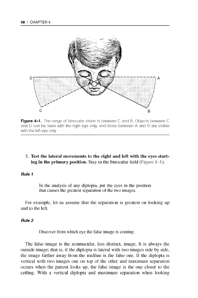

EYE MOVEMENTS, DIPLOPIA, AND CRANIAL NERVES 3, 4, AND 6 / 49
down, the false image is closer to the floor. This is true whether the ocular
palsy is divergent or convergent.
The next step is to cover either eye. Ask the patient which
image disappears. Give him several chances while you
slowly cover and uncover one of his eyes and he holds his
gaze so that the two images are maximally separated. If he
tells you the near or inside image always disappears when
you cover his right eye, then the outside image must vanish
when you cover his left eye. The false image is coming from
the left eye, and the left eye is thus the paretic one.
Rule 3
Determine what muscle moves the eye in this particular di-
rection and what is the nerve supply of that muscle.
Maximal separation is up and to the left, and the false image is from the
left eye. The elevator of the left eye when the eye is abducted is the superior
rectus, the nerve supply of which is the third cranial nerve. Therefore, the
cause of diplopia is a partial left third nerve palsy.
FUNCTIONS OF THE EXTERNAL OCULAR MUSCLES
Six muscles move each eye, and none of them has a single function. Each
has a major and a minor function. The six muscles are the four recti (medial,
lateral, superior, and inferior) and the two obliques (inferior and superior).
Rectus means “straight.”
Lateral Rectus
Primary Function
The lateral rectus muscle is supplied by the sixth cranial
nerve (the abducens).
1. Its primary function is abduction, the most common extraocular paresis
you will encounter.
2. Head posture is important. With paresis of the left lateral rectus, the
chin is turned toward the left shoulder as the patient faces you. The
eyes are thus out of the area of control of the left lateral rectus.
3. Images are side by side; the outside image, farther from the patient’s nose
(the paralytic, or false, image), disappears when the left eye is covered.
Conversely, the inside image disappears when the right eye is covered.

50 / CHAPTER 4
4. The diplopia will be greatest at a distance and lesser for near vision if
weakness of either lateral rectus muscle is the cause of the diplopia.
Secondary Function
The lateral rectus muscle has a slight but definite in-
hibition of vertical eye movements, both up and down.
In a left lateral rectus palsy, when the patient looks to the left there are two
images, the most lateral from the left eye, the most medial from the right
eye. When she looks to the left and up, the most lateral image is from the left
eye but there is now vertical separation of the two images and the top one is
from the right eye.
If she looks down and to the left, the image from the left eye is most lat-
eral but the bottom one is from the right eye. The logical conclusion from
this mess is (a) a left lateral rectus palsy (correct) and (b) a right superior and
inferior oblique muscle palsy (wrong).
When the left lateral rectus is paralyzed, the inhibition of elevation and
depression is lost as well as the abductor function. Therefore, the eye has
increased mobility upward and downward when it moves to the left as far as
the weak lateral rectus will take it. This overly mobile up-and-down move-
ment of the left eye may be misinterpreted as reduced mobility, up and
down, of the adducted right eye.
A comparable secondary function with vertical separation exists for pare-
sis of the medial rectus (see below).
To summarize, the lateral rectus is a primary abductor and secondarily in-
hibits elevation and depression of the abducted eye.
Medial Rectus
The medial rectus muscle is supplied by the third cranial nerve (the oculo-
motor).
1. Its primary function is adduction.
2. Its secondary function is to inhibit elevation and depression of the ad-
ducted eye as the lateral rectus does of the abducted eye.
3. When the right medial rectus is paretic, the patient will sit facing you
with his chin toward his left shoulder and his right eye abducted.
4. With the images side by side, the greatest separation occurs when the
patient looks to the left. The outside image will vanish when you cover
the right eye.
Superior Rectus
The superior rectus (one of two eye elevator muscles) cannot do its princi-
pal job of elevation with the eye in the primary position because the long

EYE MOVEMENTS, DIPLOPIA, AND CRANIAL NERVES 3, 4, AND 6 / 51
axis of the muscle is not parallel to the anteroposterior axis of the eye (
Fig-
ure 4–2A
).
1. The superior rectus is supplied by the third cranial nerve. Fibers arising
from the right oculomotor nucleus supply the left superior rectus and
vice versa. It is a pure elevator of the eye, only when the eye is ab-
ducted as in
Figure 4–2B
.
2. When the right eye is adducted as in
Figure 4–2C
, the superior rectus
acts as an inward rotator—its secondary function.
3. In all other positions it has mixed functions, elevation and rotation.
4. When the superior rectus is paretic, the patient’s chin is up, with the
head extended and tilted toward the shoulder of the opposite side.
5. When the two images are positioned one above the other (you may
have to hold up the patient’s ptotic eyelid with your finger), maximum
separation (for right superior rectus weakness) is up and to the right.
The top image is from the right eye.
Inferior Rectus
The inferior rectus is one of the two eye depressor muscles.
1. It is supplied by the third cranial nerve.
2. The primary function is as a depressor when the eye is abducted.
3. Its secondary function is as an outward rotator when the eye is ad-
ducted (opposite to the superior rectus).
4. When the right inferior rectus is paretic, the patient’s chin is down, the
image separation is greatest when the gaze is down and to the right, and
the false image is closer to the floor.
Superior Oblique
The superior oblique is one of the two eye depressor muscles.
1. It is supplied by the fourth cranial nerve (the trochlear). An anatomical
reminder: The fourth cranial nerve is the only cranial nerve coming off
the dorsum of the brain stem. Also, the nerves decussate before they
emerge, so the left trochlear nerve supplies the right superior oblique
muscle and vice versa. The oblique muscle rises in the apex of the orbit
and its tendon runs through the pulley (trochlear) and then runs pos-
terolaterally to insert in the posterior part of the eye behind the equator
(
Figure 4–3A
and
B
).
2. Its primary function is depression of the adducted eye.
3. Its secondary function is inward rotation of the abducted eye.
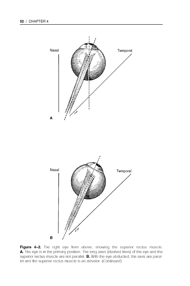
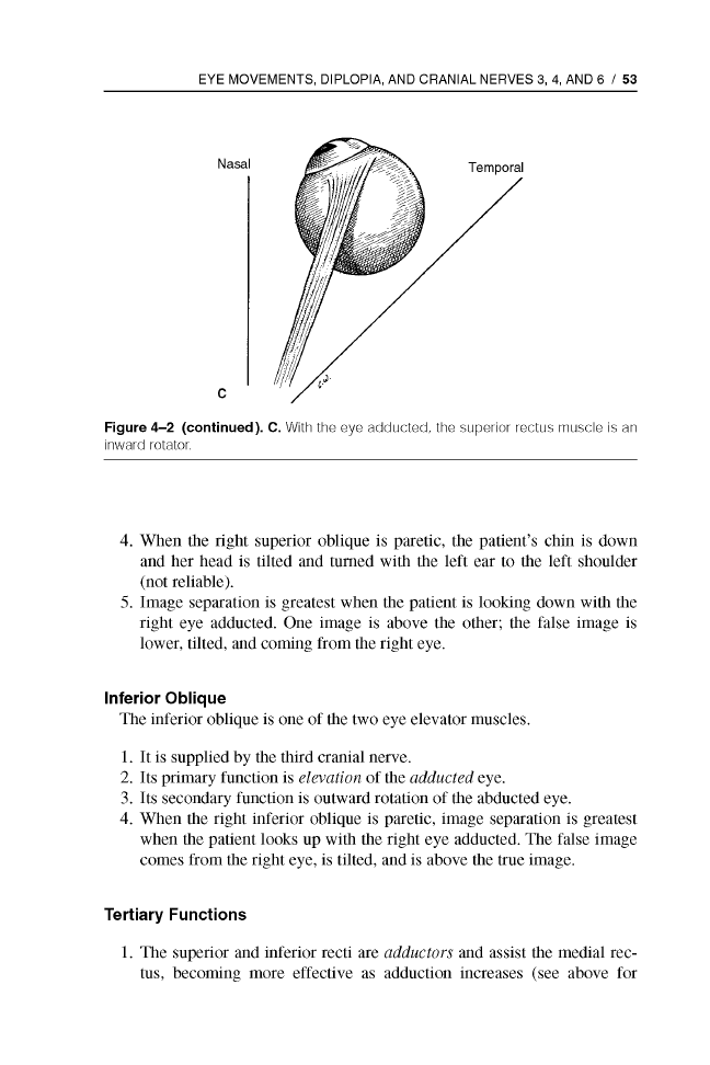
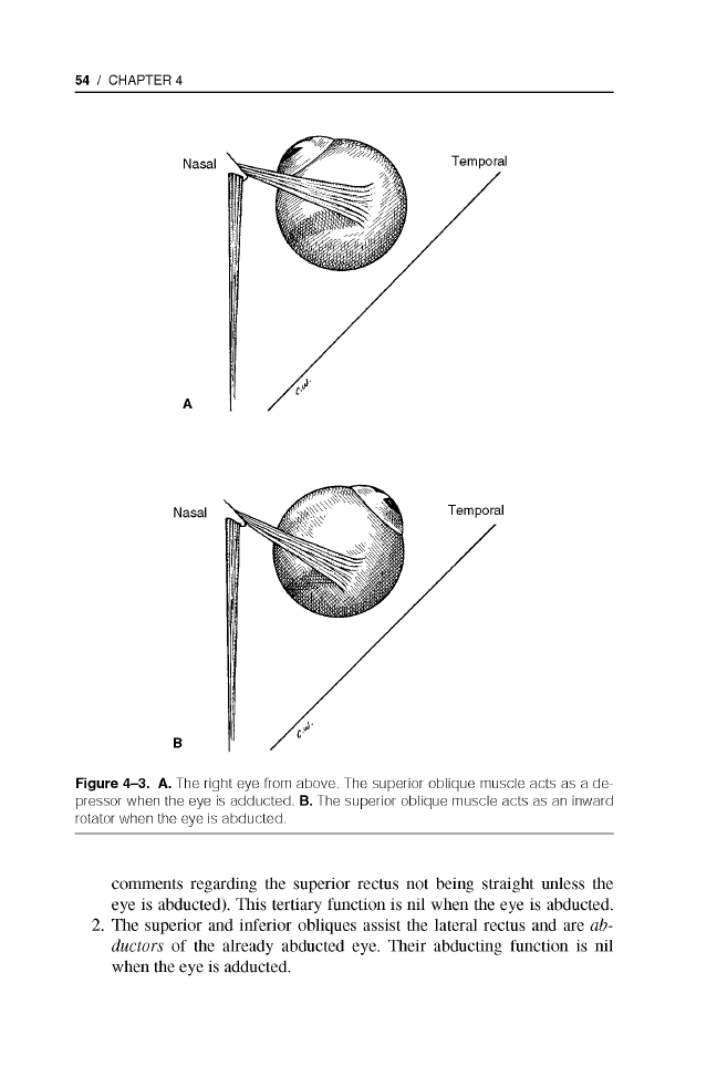

EYE MOVEMENTS, DIPLOPIA, AND CRANIAL NERVES 3, 4, AND 6 / 55
Summary
• When the eye is abducted, the superior rectus elevates, the inferior rectus
depresses, and the obliques rotate.
• When the eye is adducted, the superior oblique depresses and the inferior
oblique elevates, while the superior and inferior recti rotate.
• The two elevators are the superior rectus and the inferior oblique.
• The two depressors are the inferior rectus and the superior oblique.
• The superior rectus and superior oblique are inward rotators (“SIN” may
be used as a memory jogger).
• The inferior rectus and inferior oblique are outward rotators.
• The superior and inferior recti add to adduction; the two obliques add to
abduction.
• The lateral and medial recti inhibit elevation and depression.
• When an extraocular muscle is paralyzed, have the patient fix with the
paretic eye alone and the normal eye alone. The deviation (of the normal
eye) is always greater when the paretic eye is fixating and less (of the
paretic eye) when the normal eye is fixating.
WHEN TO EXAMINE FOR ROTATION
If both elevator muscles and both depressor muscles are working, you can-
not see rotation, as the rotatory effects of the superior rectus and superior
oblique (inward) and the inferior rectus and inferior oblique (outward) bal-
ance each other.
In a third nerve palsy the unopposed lateral rectus muscle abducts the eye.
The medial rectus is paretic and cannot adduct. The superior oblique will not
depress the abducted eye. The only movement left to test on the superior
oblique is its rotary function, as both outward rotators (inferior oblique and
inferior rectus) are paretic and cannot oppose the inward rotation of the su-
perior oblique. Do the following:
• Pick out a scleral conjuctival vessel on the temporal side and another on
the nasal side of the globe and watch them.
• Ask the patient to look down and out; the movement will not be marked.
• If the fourth cranial nerve and superior oblique muscle are working, the
temporal vessel moves up and the nasal vessel moves down (ie, inward ro-
tation).

56 / CHAPTER 4
PARALYTIC STRABISMUS VERSUS CONCOMITANT
(NONPARALYTIC) STRABISMUS
Concomitant Strabismus
A concomitant strabismus is a deviation of the eye in which the angle of
deviation does not vary with the direction of gaze or the fixating eye.
In paralytic strabismus the deviation of the eye becomes greater as the
eye is moved in the direction of function of the paretic muscle.
There are a number of possible causes of a concomitant strabismus. These
include defects of innervation, refraction, and accommodation plus a genetic
predisposition as well as a large group in which the cause is unknown. Com-
mon causes are imbalance of the near reflex (accommodative strabismus)
and unilateral reduced vision in an infant or child.
By about age 7 years the accommodative and fusion reflexes are set. Mus-
cle imbalance before this age results in concomitant strabismus. Muscle im-
balance after this age is usually paralytic in type. It is not always easy to
classify strabismus as concomitant or paralytic.
Concomitant strabismus does not usually produce the complaint of double
vision. When a patient has a latent strabismus (phoria) and, because of fa-
tigue or a debilitating illness, presents with a manifest strabismus (tropia), he
may complain of the sudden onset of double vision. If the distance between
the two images is the same in every direction of gaze, this is not paralytic.
In concomitant strabismus monocular eye movements are full in all direc-
tions, the angle of deviation is the same in all directions of gaze, and the pri-
mary and secondary deviations are identical irrespective of which eye fix-
ates.
Paralytic Strabismus
In a paralytic strabismus the secondary squint position or secondary devia-
tion of the normal eye is greater than the primary squint position of the
paretic eye.
Example: With a left lateral rectus palsy, the left eye deviates from the
midline while the right eye fixes straight ahead. The left eye might be ad-
ducted 5 degrees. If the right eye is then shielded, the left eye fixes in the
straight-ahead position and the right eye adducts 10 degrees. This is the sec-
ondary deviation.
The more recent the paralytic squint, the greater is the angle of deviation
in the various gaze positions. A single extraocular muscle palsy is easiest to
recognize on the day it occurs. As time goes by, some degree of concomi-
tance occurs. This will involve (a) overreaction of the yoke muscle (ie, the
right lateral rectus and the left medial rectus are yoke muscles; each extraoc-
ular muscle has a yoke, or partner, muscle of the other eye); (b) contracture

EYE MOVEMENTS, DIPLOPIA, AND CRANIAL NERVES 3, 4, AND 6 / 57
of the unopposed ipsilateral agonist of the paretic muscle; and (c) underfunc-
tion of the contralateral yoke muscle related to the agonist in (b).
MISCELLANEOUS
Abnormal Head Posture
In the analysis of any patient with double vision, the way he holds his
head may be helpful. When a head turn is present, the patient turns his head
into the field of action of the paretic muscle; that is, if the left lateral rectus
muscle is paretic, he turns his chin to his left shoulder. As he faces you, he
adducts the left eye and abducts the right eye. He then sees only one doc-
tor—and that is enough for anyone, especially if he is sick! However, the pa-
tient may do the opposite—in our example, he would turn his chin to his
right shoulder and look at you from the nasal corner of his right eye, with the
left eye as abducted as the weak lateral rectus will take it.
The separation of the two images is so great and the false one is on such a
peripheral aspect of the retina that it is suppressed.
Eye Patch
The patient almost always covers or closes the paretic eye. She may be a
myasthenic with multiple weak extraocular muscles for whom the side of the
patch is of no lateralizing help. However, if a single muscle or two muscles
of the same eye are at fault, the closed eye is usually the paretic eye.
Ask the patient to take off the patch, to keep both eyes open, and to put
her head in the position where there is no double vision. If her chin comes
up and her head goes back into extension, she has weak eye elevators. Side-
to-side movement has already been discussed. The head is tilted with the ear
to the shoulder most commonly with oblique muscle palsies. With a superior
oblique palsy the head is turned and tilted away from the weak side.
Monocular Diplopia
Monocular diplopia can result from opacities in the vitreous, lens, or
cornea or from a dislocated lens (Marfan’s syndrome) as well as astigmatism
and early cataract. There are well-documented cases of occipital cortex injury
resulting in monocular diplopia and quadruple vision with both eyes open.
CAUSES AND SITES OF LESIONS OF THE SIXTH CRANIAL NERVE
1. Within the pons, causes include
a. Multiple sclerosis
b. Infarct

58 / CHAPTER 4
c. Mass (eg, glioma or tuberculoma)
d. Wernicke’s syndrome
Look for ipsilateral
• Seventh nerve lesion
• Gaze palsy
• Horner’s syndrome
• Facial hypesthesia
• Internuclear ophthalmoplegia
• Ataxia
Look for contralateral
• Hemiparesis
• Nystagmus (possibly in more than one direction)
• Hemisensory pain and temperature loss in the trunk or limbs
2. At the base of the brain, causes include
a. Acoustic neuroma
b. Meningitis
c. Subarachnoid hemorrhage
d. Compression of an aneurysm or anomalous vessel
e. Basal meningioma
f. Nasopharyngeal carcinoma
g. Chordoma
h. Wegener’s granulomatosis
Look for signs of involvement of the fifth, seventh, and eighth cranial
nerves.
3. In relation to the petrous portion of the temporal bone, causes include
a. Infection, fracture, or tumor of the petrous bone
Look for pain, paresthesia, or hyperesthesia of the face (same side) and
deafness.
4. In the cavernous sinus/superior orbital fissure, causes include
a. Aneurysm (carotid)
b. Carotid—cavernous fistula (trauma)
c. Tumor—pituitary, meningioma, or nasopharyngeal carcinoma

EYE MOVEMENTS, DIPLOPIA, AND CRANIAL NERVES 3, 4, AND 6 / 59
d. Tolosa-Hunt syndrome
e. Cavernous sinus thrombosis
f. Mucormycosis (in diabetics)
g. Herpes zoster
Look for signs of involvement of the third and fourth cranial nerves as
well as first and second divisions of the fifth.
5. In the orbit, causes include
a. Tumor
b. Trauma
6. Common and not so common causes, but with an uncertain site, include
a. Postviral infection (especially in children)
b. Diabetes
c. Hypertension
d. Postlumbar puncture or myelogram
e. Increased intracranial pressure
f. Migraine
g. Arteritis or angiitis of sarcoidosis
h. Fisher variant of Guillain-Barré syndrome
7. Other causes of abduction defect include
a. Thyroid ophthalmopathy
b. Myasthenia gravis
c. Orbital pseudotumor
d. Trauma
e. Duane’s syndrome
f. Convergence spasm
CAUSES AND SITES OF LESIONS OF THE OCULOMOTOR NERVE
Remember that the right superior rectus is supplied by part of the nucleus
of the left oculomotor nerve and vice versa. There is only one midline nu-
clear structure that supplies both the right and left levator palpebrae superi-
oris muscles.
1. Within the midbrain, causes include
a. Complete unilateral nuclear lesion, although it probably does not
exist
b. Partial unilateral (nuclear or fascicular) infarction or hemorrhage,
demyelination, or tumor

60 / CHAPTER 4
Look for contralateral
• Hemiparkinsonian signs
• Hemihypesthesia
• Hemiparesis in the face, arm, and leg
• Ataxia and possibly tremor
2. Interpeduncular causes include
a. Aneurysm (in the posterior communicating artery or basilar artery)
b. Meningitis
c. Infarction (in diabetics the pupil is usually normal and the infarction
is often painful)
3. At the edge of the tentorium (herniation of the uncus from an expanding
intracranial mass), causes include
a. Contralateral upper motor neuron signs
b. Deteriorating vital signs
c. Progressing drowsiness
4. Within the cavernous sinus, causes are the same as those for the ab-
ducens nerve (see above).
5. Within the orbit, causes are the same as those for the abducens nerve.
Additionally, thyroid ophthalmopathy with tethered medial rectus may
be a cause, that is, defective abduction is seen, and with inferior rectus
restriction defective elevation is seen.
6. Common and not so common causes, but with an uncertain site, include
a. Migraine
b. Diabetes (common)
c. Hypertension
d. Arteritis
e. Collagen-vascular disease
CAUSES AND SITES OF LESIONS OF THE TROCHLEAR NERVE
The causes and sites of lesions of the trochlear nerve are the same as those
of the oculomotor nerve. The most common cause is trauma and the next is
diabetes. The latter probably produces an ischemic infarction of the nerve
trunk.

Ptosis and the Pupils:
5
Myasthenia Gravis and
Other Diseases of the Eye
and Eyelid Muscles
PTOSIS
Ptosis is often the greatest abnormality in familial, chronic progressive ex-
ternal ophthalmoplegia (CPEO).
In other diseases ptosis is often the presenting (and only) symptom. If it is
bilateral, long-standing, and familial, the diagnosis is probably CPEO (see
below).
If it is unilateral, recent, partial, and the only abnormality, the problem is
more difficult to diagnose.
The space between the lids (palpebral fissure) varies. Increased sympa-
thetic function increases the space and so does adduction of the eye in many
people. With the eyes in the primary position, the space is about 7–12 mm in
the vertical midline. The cornea is about 10.5 mm in vertical diameter; the
upper lid usually covers the top 0.5–1 mm, and the bottom lid touches the
lower limbus.
Buy an inexpensive transparent plastic ruler marked in millimeters and
start to measure the fissure every time you have the chance.
Blepharospasm or Ptosis?
Blepharospasm is eye closure resulting from a contraction of the orbicu-
laris oculi muscle. Ptosis is partial or complete eye closure caused by pare-
sis or paralysis of the levator palpebrae muscle or the superior tarsal muscle.
Blepharospasm may be a result of some painful or irritating ocular dis-
ease, or it may be voluntary to abolish the false image of diplopia. It may
also be a dystonia, that is, an involuntary movement because of organic dis-
ease, in this case always bilateral. When unilateral, how is blepharospasm
distinguished from ptosis? In blepharospasm the eyebrow is pulled down
below the superior orbital margin. In ptosis it is on the margin or above. If
above, the forehead is wrinkled as the patient uses his frontalis to compen-
sate for the drooping lid.
61

62 / CHAPTER 5
Is Ptosis Part of a Third Nerve Palsy or of a Horner’s Syndrome?
If there is a big pupil on the side of the ptosis, with or without the appro-
priate extraocular muscle palsies, the diagnosis is a third nerve lesion. If
there is a small pupil on the same side, it is a Horner’s syndrome (see below
under “Pupils,” with regard to the response of the pupil to decreased illumi-
nation in Horner’s syndrome). A third nerve lesion can cause complete pto-
sis; a Horner’s syndrome cannot. On upward gaze, the ptosis of a third nerve
palsy does not change; however, if it is part of a Horner’s syndrome, it will
diminish.
If the pupils are equal, look at the lower lid. The inferior tarsal muscle
(which pulls the lower lid down) is paretic in a Horner’s syndrome and the
lower lid is higher than the normal lower lid. It covers more of the limbus at
6 o’clock.
Bilateral ptosis without the other signs of either third nerve or sympathetic
lesions has been seen following basilar artery infarction and ischemia. The
ptosis was an isolated ocular finding. The pupils and eye movements were
normal, and the patients were alert.
PUPILS
Pupillary size, equality between right and left, and pupil responses to vari-
ous stimuli in the conscious and comatose patient are prime physical signs.
Size
Pupil size depends on the light, the near reaction, and sympathetic and
parasympathetic tone. Newborns and the elderly have smaller pupils than do
youths. Schizophrenic patients and frightened people have big pupils. Blue-
eyed people have bigger pupils than brown-eyed people, and myopic people
also have larger pupils. Use a transparent plastic ruler to measure pupil size.
The pupils in young people often constrict and dilate in a repetitive rhyth-
mic way. This is hippus and is normal. Its absence, however, is also normal.
Most pupils in the examining room are 2–5 mm in diameter.
Equality
Most people have pupils of equal size. About 15–20% of patients have un-
equal pupils (anisocoria) that cannot be explained, but the difference is usu-
ally not more than 1 mm. If the difference remains constant in both bright
and dim illumination, it is not significant. If the difference in size changes
with the illumination, then disease is present. The small pupil of a Horner’s
syndrome will not dilate in dim light as much as the pupil of the other nor-

PTOSIS AND THE PUPILS / 63
mal eye. The larger pupil of a partial third nerve palsy will not constrict as
much as the normal pupil when the patient is in a bright light. Therefore,
measure pupil sizes in average room light, dimness, and bright light. (Prac-
tice this first with classmates or patients with equal pupils.)
Two things that do not cause unequal pupils are
1. Unilateral or bilateral diminished or absent visual acuity or peripheral
visual field defects
2. Differences in refractive errors between the two eyes
Pupils are displaced slightly to the nasal side of the center of the irises and
are round. Eccentric pupils are not necessarily abnormal, and irregularities
of shape are usually caused by trauma.
How to Examine the Pupils
Light Reaction
Examine the patient while she is sitting, with moderate
background illumination. Inspect and compare the pupils while your flash-
light is held vertically, pointing up, below the eye, and just in front of the pa-
tient’s cheek.
Then ask the patient to look at the far wall and direct a bright light at first
one and then the other eye. Be careful not to illuminate the two eyes at the
same time.
Both pupils should react to the light in either eye with equal speed and to
the same degree. This tests the second cranial nerve and other parts of the vi-
sual pathways (afferent) and the pupillary fibers of the third cranial nerve
(efferent).
Direct and Consensual Light Reaction
A direct pupil response to light
means that the right pupil constricts when the right eye is exposed to a bright
light. A consensual response means that the right pupil constricts when the
left eye is exposed to a bright light, and vice versa.
If the right eye has a normal direct response and an abnormal consensual
response, the lesion is in the afferent pathway of the left eye. If the right eye
has an abnormal direct response and a normal consensual response, the le-
sion is in the afferent pathway of the right eye. If the right eye has neither a
direct nor a consensual response, the lesion is in the efferent pathway on the
right (third nerve palsy). A lesion at the apex of the right orbit involving both
efferent and afferent limbs of the reflex may also abolish direct and consen-
sual responses for the right eye.

64 / CHAPTER 5
Near Reaction
If the light reflex works, so will the near reflex. If one or
both pupils do not react to light, ask the patient to look at the far wall and
then at the tip of your pencil, which should be 5–10 cm in front of his nose.
If his visual acuity is seriously compromised, ask the patient to look at the
tip of his own finger held in front of his nose.
Look for two things: bilateral constriction of the pupils as the patient
changes his gaze from far to near and convergence of the eyes. Accommoda-
tion also occurs. This increases curvature of the lens and is a result of con-
traction of the ciliary muscle and loosening of the suspensory ligament.
If the right eye has a sluggish direct pupillary response to light, relative to
the direct left response, but has a brisk near response, the lesion is in the
right afferent limb of the light reflex.
Afferent Pupil Defect, Swinging Flashlight Test, and Marcus Gunn Pupil
Take, for example, the case of a patient with retrobulbar neuritis of the right
optic nerve. His vision is 20/80 right, 20/20 left; peripheral fields are full;
and the retina, optic nerve head, and media are all normal. Examine his pupil
responses in dim illumination. Shine a strong light into his left eye only. Both
pupils constrict as expected. Shield the left eye from light and shine the light
into the right eye only. The right pupil seems to constrict momentarily and
then dilates widely, as does the left. Swing the light back to the left eye only
and again both constrict.
The high-intensity light-induced impulses from the left eye have their nor-
mal influence on the two pretectal and Edinger-Westphal nuclei. The efferent
systems (third nerves) are normal, so both pupils constrict. However, when
the light is directed into the right eye, the pupil changes from the constric-
tion of a normal consensual light reaction to the relative dilatation of a low-
intensity direct reaction. This low-intensity response is caused by diminished
conduction through the diseased afferent system of the right eye. The left
pupil also dilates as the consensual response in this eye is determined by the
afferent system on the right.
Causes and Types of Abnormal Pupils
Argyll-Robertson Pupils
Argyll-Robertson pupils are usually bilateral and
result from tertiary syphilis of the nervous system, diabetes, or the late signs
of bilateral tonic pupils (see the section on “Adie’s Syndrome”). The pupils
are small, irregular, and unequal. They do not react to light, they do react to
near vision, they respond poorly to mydriatics, and they do not dilate in the
dark. However, they can be made to constrict even more by the use of mi-
otics. The poor light response and good near response may be relative; that
is, the light response need not be absent, but is much less evident than the
near response. Visual acuity is not impaired.

PTOSIS AND THE PUPILS / 65
Horner’s Syndrome
Horner’s syndrome, or oculosympathetic palsy (unilat-
eral), presents with abnormalities of the eyelids as described in the section
on “Ptosis.” The pupil is small and round with good response to light and
near. The difference between normal and abnormal pupil size in the dark is
even greater; that is, the pupil on the side of the lesion dilates later and less.
A lesion of the sympathetic fibers can be in the brain stem, cervical cord,
apex of the lung, carotid sheath, or orbit.
Additional signs are apparent enophthalmos and a warm, dry, nonsweat-
ing, ipsilateral face. A number of pharmacological agents can be used to aid
the diagnosis and localize the lesion as central, preganglionic, or postgan-
glionic.
Horner’s syndrome, with pain in the eye and ipsilateral face and forehead,
has been reported in dissection of the carotid artery and with cluster
headaches with a normal carotid artery.
Oculomotor Nerve Lesion (Unilateral)
Other features of the unilateral ocu-
lomotor nerve lesion are as described above. The pupil is mid-dilated. There
is no response to light or near, and the difference in pupil size is greater in
the light (in contrast with Horner’s syndrome). Mydriatics and miotics are
both effective. Diabetic oculomotor nerve palsies usually have normal
pupils, and lesions can be painful.
Adie’s Syndrome
Adie’s syndrome, or tonic pupil (which may be unilateral
or asymmetrically bilateral), is also known as “the big, slow pupil.” The con-
dition presents as an enlarged pupil that either does not react to light or
eventually constricts after being exposed to very bright light for 15–20 min.
It eventually constricts for near after a similarly long effort. Redilatation is
just as protracted. The difference in the pupil sizes is best seen in the light.
The pupils respond to mydriatics and miotics and demonstrate denerva-
tion supersensitivity. This means that the tonic pupil will constrict from
2.5% solution of Mecholyl or 0.125% pilocarpine. Normal pupils do not re-
spond to these weak solutions.
Accommodation is just as slow as the light response and may be the pre-
senting complaint. When the deep tendon reflexes are diminished or absent,
the syndrome is called Holmes-Adie. The cause is unknown.
Benign Anisocoria
Usually a young adult with benign anisocoria reports a
difference in pupil size. The longer it has been present, the less likely it is to
be important. Ask for some old photographs of the patient, and examine
them for pupil inequalities. The response to light and near in both eyes is
normal, the difference in pupil size is no greater in dimness or light, mydriat-
ics and miotics have a normal response, and there is no diagnosis.

66 / CHAPTER 5
The Factitious Big Pupil
Sometimes proprietary eyedrops have impuri-
ties in them with atropine-like properties. Patients using ointments with
atropine-like properties may inadvertently introduce them into the eye.
There is occasionally a deliberate atropine abuser. He or she is usually work-
ing or studying in a hospital and has some access to medications and is under
pressure or stress of some kind. The patient presents with the biggest pupil you
have ever seen. There is not a flicker of response to light or near. The difference
is greatest in bright light, and neither mydriatics nor miotics change the pupil.
Keep the patient talking (about anything), get the whole story, change the en-
vironment (if a medical student or nurse, keep him off the wards for the next 3
days if possible), and reexamine the eye daily for 3 consecutive days. If this big
pupil is caused by medication, each day its response to light and near will be a
little better and your chances of getting the full history improve. Pilocarpine will
not constrict an atropinized pupil; it will, however, constrict an Adie’s pupil.
Midbrain Lesions
With midbrain lesions the pupils are large, particularly if
the lesion involves the parasympathetic fibers of the third nerve. If the mid-
brain is totally interrupted, including the sympathetic fibers, the pupils are
big, but less so.
Carotid Artery Occlusion (Unilateral)
An enlarged pupil ipsilateral to the
occlusion has been reported in atheroma and Takayasu’s disease. The pupil
reacts poorly to light (direct and indirect) and near. The explanation is proba-
bly ischemic atrophy of the iris, rather than nerve disease.
Pontine Miosis (Bilateral)
The classic sign of pontine infarction or hemor-
rhage is small (1- to 1.5-mm) pupils. They will constrict to light if a bright
enough stimulus is used and if examined through a magnifying glass.
ACCOMMODATION AND CONVERGENCE
For near vision the eyes converge (ie, turn toward the midline), the pupils
constrict, and the lenses thicken. In order to complete an examination, the
patient must cooperate. When the simultaneous contractions of the two me-
dial recti produce convergence, they have a different central connection than
when either is being used for conjugate lateral gaze in conjunction with its
appropriate yoke muscle, that is, the contralateral lateral rectus.
Failing accommodation is most commonly related to aging, as the lens be-
comes less resilient. The parasympathetic nerve fibers subserving accommo-
dation, as well as those subserving the pupillary near response, will be inter-
rupted in a complete peripheral third nerve palsy.

PTOSIS AND THE PUPILS / 67
The diabetic, out of control or of recent onset, can have a sudden increase
in the refractive power of the lens; that is, a sudden improvement in near vi-
sion is as much a symptom as the reverse. Anticholinergic drugs commonly
produce a complaint of blurred vision from diminished accommodation if
taken in large enough doses.
Near Response
1. If the pupils react to light, they will react to near.
2. If the pupils do not react to light, it is important to know whether the
near response is also abnormal.
3. If there is a defect in adduction, it is important to know whether or not
convergence is present. (See the section on “Internuclear Ophthalmo-
plegia” in Chapter 7.) You can verify the patient’s efforts to converge
by the attendant miosis, which means she is trying.
4. In any bilateral paresis of external ocular muscles, convergence assess-
ment is part of the examination that may help you localize and diag-
nose the lesion.
5. The sudden onset of diplopia, with eyes divergent, but with full monoc-
ular eye movements, is a paresis of convergence.
6. The periaqueduct syndrome, discussed in the section on “Parinaud’s
Syndrome” in Chapter 7, includes paresis of convergence.
7. Convergence excess looks like a unilateral or bilateral lateral rectus
palsy. The patient is staring at the tip of his nose. This is often a hysteri-
cal disease. When you ask the patient to look laterally, which he says
he cannot do, his pupils constrict, proving that he is overconverging.
However, it can be organic following head injury and can be part of the
periaqueduct syndrome.
8. Miscellaneous: The toxin of Clostridium botulinum can produce large,
nonreacting pupils, paralysis of accommodation, ptosis, and extraocular
muscle palsies. The patient is wide awake, with progressing respiratory
distress. He may have vomited but is usually constipated. Diphtheria
can produce paralysis of accommodation and also affects bulbar origi-
nating nerves and cardiac rhythm.
OTHER DISEASES WITH WEAKNESS OF THE EYE
AND EYELID MUSCLES
Myasthenia Gravis
Myasthenia gravis is a common cause of ocular complaints, often diag-
nosed late. It may present as weakness of one muscle or one eye or any com-

68 / CHAPTER 5
bination of muscles. The essence of the disease is excessive fatigability, that
is, the patient cannot sustain upward gaze or sustain the upper lids in a fixed
open position. Of all the possible combinations of myasthenic muscle weak-
ness (eg, ocular, pharyngeal, or limb), bilateral fluctuating undulating pto-
sis is probably the most common.
Look at the patient’s eyes and keep looking. She may blink three times
with blinks of the same duration and the same interval between blinks, but
she does not open as wide after each blink. Then, in the fourth blink, she
keeps her eyes closed for 10–20 s. She is not sleeping and you are not that
boring—when she finally opens her eyes after the long blink, her ptosis is
less pronounced and you can see more of her eyes. She then has three or four
of her usual blinks with her ptosis increasing and then has another long
blink. You are looking at a patient with myasthenia gravis.
When the eye opening muscles are weak and the eye closing muscles are
also weak, the diagnosis is almost always either myasthenia gravis or one of
the CPEOs (see the following section). Therefore, when the patient has pto-
sis (you know the eye openers are weak) test the eye closers. The diagnosis
of myasthenia gravis starts when you first consider it. There are immunolog-
ical, neurophysiological, pharmacological, and therapeutic ways to help
make the diagnosis, but it usually all starts when a young woman or an old
man says, “My eyelids are drooping” or “I see double when I’m watching
the late news on TV.”
Chronic Progressive External Ophthalmoplegia
CPEO is actually a number of diseases. They have in common a progres-
sive, restricted range of eye movements and ptosis. Some, however, have
ptosis only. The patient may know nothing about this. Most of his family
members look the same, the diseases are very chronic, and they usually do
not cause diplopia. Often, an associated symptom unrelated to the abnormal
ocular mobility brings the patient to the doctor. Included in this group of dis-
eases are:
1. Oculopharyngeal dystrophy—Common in French Canadians, is autoso-
mal dominant; ptosis is more marked than gaze limitations, and trouble
swallowing is often the presenting complaint.
2. Myotonic dystrophy—Presents with action and percussion myotonia;
weak neck (sternomastoid), temporal and masseter, and limb muscles;
marked ptosis; and cataracts
3. Thyroid ocular myopathy—Patients commonly complain of diplopia,
thyroid status may be normal, proptosis may be absent, and lid lag on
downward gaze and inability to elevate the globe are the most common

PTOSIS AND THE PUPILS / 69
signs. This myopathy commonly involves the medial rectus muscle,
producing a secondary limited abduction that mimics a lateral rectus
palsy.
The syndromes are
• Stephens—A positive family history of CPEO plus peripheral neuropathy
and cerebellar ataxia
• Kearns-Sayre—Childhood onset of CPEO, retinal pigment degeneration,
heart block plus progressive encephalopathy and sometimes cerebellar
signs, short stature, and a negative family history
• Bassen-Kornzweig—Abetalipoproteinemia
• Chronic multiple sclerosis
• Chronic myasthenia gravis
• Myotubular myopathy

Nystagmus
6
Nystagmus is a rhythmic oscillation of the eye or eyes.
Nystagmus is usually a sign of disease in the posterior fossa or peripheral
vestibular apparatus. However, the most rostral structure, when diseased
enough that it can cause nystagmus, is the thalamus; the most caudal is the
neural axis at the level of the craniocervical junction. The nature of the nys-
tagmus may reveal the site of the lesion causing it; that is, nystagmus of the
abducting eye only is always a result of intrinsic brain stem disease and ver-
tical nystagmus is never vestibular in origin. In many cases other physical
signs are used to determine the site of the nystagmus-producing lesion. In
some patients the precise site of the lesion is unknown.
Alcohol and other drugs are a common cause of nystagmus and are the most
common cause of transient coexisting horizontal and vertical nystagmus.
Nystagmus is classified into several types with considerable overlap: these
include acquired or congenital; jerk or pendular; present with the eyes in the
primary position or gaze-evoked; present or modified when the patient is up-
right, supine, prone, or with his head tilted; and present in the light or the dark.
TYPES OF NYSTAGMUS
Jerk
Jerk nystagmus is the most common type. When the patient is asked to
follow the examiner’s finger to the right, his eyes are seen to drift off the
point of fixation (the finger) and back to the midline. They then snap back to
the finger. The nystagmus is named from the direction of the fast (ie, correct-
ing) movement, that is, right beating jerk nystagmus on right lateral gaze. In
jerk nystagmus the two movements are never of the same velocity.
Pendular
In pendular nystagmus the movement of the eyes in one direction is at the
same speed as the movement in the other direction. For this reason pendular
nystagmus is not named right, left, and so on, but is named from the plane of
the movement, that is, horizontal, vertical, or rotary. Pendular nystagmus can
be acquired or congenital.
71

72 / CHAPTER 6
Gaze-Evoked
With the eyes in the primary position, no nystagmus is seen.
• Ask the patient to follow your finger in all the principal directions of gaze
with the finger 60 cm from him and both eyes open.
• Have him do it again with one and then the other eye closed.
• Repeat the above with the finger close enough to the patient to make him
converge while he looks in all the principal directions.
Nystagmus elicited from any of these procedures will be described as, for
example, “bilateral left beating nystagmus on left lateral gaze,” or whatever
eye position is necessary to evoke it.
Primary Eye Position
When the patient looks straight ahead, nystagmus may be present. It can
be jerk or pendular. When the former, it is often so fine that you need to
watch a retinal vessel with your ophthalmoscope in order to see it. It may be
vertical (fast component) or lateral. It may become coarser or vanish when
the patient looks to either side.
Congenital
Pendular
Congenital nystagmus is most commonly pendular. The history
is of vital importance. The patient may “do something” (ie, consult a doctor)
about his jumping eyes for the first time at age 16. Diminished visual acuity
is a common but not causal accompanying feature. Nystagmus is usually
horizontal, is always in both eyes, and diminishes with convergence. It will
change from pendular in the primary position to jerk on lateral gaze. These
patients often have a head tremor that only appears or worsens when they try
to read. The patient will usually have a head and eye position where the nys-
tagmus is least or absent, known as the null position.
There is often rhythm to congenital pendular nystagmus. The patient will
have five or six oscillations of the eyes, followed by a pause of 1–2 s, during
which the movements almost vanish, and then the oscillations resume. (See
the section on “Optokinetic Nystagmus,” below, and its effect on congenital
nystagmus, also described below.)
Probably the most common cause of nystagmus is congenitally poor vi-
sion in both eyes from any cause.
Latent
Latent nystagmus is also congenital. When the patient has both eyes
open and the eyes in the primary position, there is no nystagmus. When ei-
ther eye is covered, both eyes develop marked jerk nystagmus, fast phase to

NYSTAGMUS / 73
the side of the uncovered eye. Visual acuity is decreased when one eye is
covered. Many patients with this type of nystagmus have a strabismus.
LESION SITES
In general, when the patient looks in the direction of the fast movement,
the amplitude of the nystagmus becomes greater. The fast component of nys-
tagmus is dependent on the integrity of the opposite cerebral hemisphere,
basal ganglia, and diencephalon; that is, an acute destructive right vestibular
lesion will produce conjugate ocular deviation to the right and nystagmus to
the left. If the patient has an additional right middle cerebral artery territory
infarct, there will be no nystagmus.
Brain Stem, Cerebellar Nuclei, and Their Connections
1. Jerk (the most common type of nystagmus resulting from acquired dis-
ease)
a. May be in the abducting eye only, and the intensity increases when
looking in the direction of the fast phase. It is often easily exhausted
and can be three or four coarse beats in the abducting eye only and
then stop.
b. May be equal in the two eyes
c. May be associated with limited adduction of the other eye as part of
an internuclear ophthalmoplegia (see Chapter 7)
d. Downbeat—May be difficult to see in the primary eye position as
described in the section on primary eye position nystagmus, above.
Will become coarser on conjugate downward gaze and much
coarser (while remaining downbeating) on lateral conjugate gaze to
either side. Usually vanishes on conjugate upward gaze. Usually in-
creased when posture changes from upright to supine. Responsible
lesions are usually cerebellar herniation or degeneration or Arnold-
Chiari malformation.
e. Upbeat (two types)—One type is present and easily seen in the pri-
mary position and will increase with upward gaze and decrease with
downward gaze; the lesion is in the anterior vermis of the cerebel-
lum. The other type of upbeat nystagmus is difficult to see in the pri-
mary position and is paradoxical in that it increases with downward
gaze. The responsible lesion may be in the tegmental gray matter at
the pontine-medullary or pontine-midbrain junction. Both upbeating
and downbeating nystagmus may be modified by head tilting or may
appear only on head tilting.

74 / CHAPTER 6
2. Rotary only
3. Mixed rotary and jerk
4. Acquired pendular—Present in many directions (ie, vertical or hori-
zontal) and may be different in the two eyes. It is often accompanied by
signs of disease of nearby structures, for example, slurred speech,
ataxia of the limbs and gait, diplopia, and tremor.
Vestibular (Peripheral or End Organ) Lesions
Nystagmus resulting from vestibular lesions occurs in one direction only,
always away from the lesion. It is usually mixed, lateral, and rotary. It is
never vertical or rotary only, is reduced by visual fixation, and is always ac-
companied by marked vertigo and usually by tinnitus and hearing loss.
Diencephalon
This is a seesaw, pendular nystagmus with no fast component. There are
conjugate rotational eye movements plus a bizarre vertical movement. The
eye that rotates inward rises as the eye that rotates outward drops. The verti-
cal seesaw movements may be elicited in downward gaze only. It can be
congenital. The site of the lesion responsible for this type of nystagmus is
not firmly established.
ASSOCIATED SIGNS AND SYMPTOMS
1. With nystagmus of brain stem or cerebellar origin,
a. Ataxia, dysarthria, diplopia, internuclear ophthalmoplegia (see
Chapter 7), other cranial nerve lesions, long tract motor and sensory
signs
b. Vertigo, absent or relatively mild and improved by lying still with
the eyes closed
2. With nystagmus of vestibular origin,
a. Vertigo and vomiting are intense, onset of both is abrupt, and the pa-
tient usually cannot be induced to move or get up; the patient feels
that the environment is rotating away from the side of the lesion.
b. Other signs of eighth cranial nerve dysfunction are present (see
Chapter 9).
c. There are no signs of brain stem or cerebellar disease but the eyes
are forced to the side of the lesion.
d. There is usually a history of tinnitus or reduced hearing.
e. If the patient can be induced to stand and walk, he staggers to the
side of the lesion.

NYSTAGMUS / 75
MISCELLANEOUS
End-Point Nystagmus
Normal, seen at the extremes of lateral gaze, end-point nystagmus is of the
small, irregular jerk type, best seen in the abducting eye. It is more marked
and common in association with even minor alcohol intake.
Convergence Nystagmus
Convergence will usually dampen congenital nystagmus and peripheral
vestibular nystagmus. Nystagmus seen only during convergence can be part
of the dorsal midbrain syndrome.
Rebound Nystagmus
With this condition, the patient looks, for example, to the left and has left
beating jerk nystagmus. It stops and when he moves his eyes back to the pri-
mary position, nystagmus reappears as right beating nystagmus in the pri-
mary position. After a few seconds it stops again. This signifies intrinsic
brain stem/cerebellar disease. It is important to watch the patient’s eyes as
they come back to the primary position, as this may be the only position in
which you see any nystagmus.
Periodic Alternating Nystagmus
This is an uncommon, startling type of nystagmus. The patient will have a
spontaneous constant jerk nystagmus in one direction; it will stop after some
seconds and, after a short interval of no nystagmus, will resume in the oppo-
site direction. This alternating goes on constantly. It is significant in a num-
ber of acquired and congenital posterior fossa disorders. It can occur in oth-
erwise normal people.
Convergence-Retraction Nystagmus
On attempting to look upward, rapid repetitive convergent movements
with retraction of the eyes occur. The lesion is in the midbrain (see the sec-
tion on “Parinaud’s Syndrome” in Chapter 7).
Voluntary Nystagmus
Voluntary nystagmus is not a true nystagmus. It is a very rapid, usually
side-to-side, oscillation or rotation with the eyes in the primary position and
with voluntary overconvergence. Like the ability to voluntarily move one’s
ears, a few of us can do it, but most of us cannot. The person with voluntary
nystagmus can keep it up for only a few seconds at a time. This is not a man-
ifestation of disease. When present in a medical student, it can win small wa-

76 / CHAPTER 6
gers from other medical students, as in “Did you know I can have nystagmus
any time I want to?”
Optokinetic Nystagmus
Optokinetic nystagmus (OKN) is also referred to as “railroad” nystagmus.
If you have ridden on a train and watched the utility poles go by, you have
had OKN in the direction (ie, fast phase) that the train is moving.
• Clinically, patients are tested for this by having them watch a revolving
drum that has alternating black and white vertical stripes.
• At the bedside you may use a cloth 1.0-m measuring tape; however, if you
want even better contrast, buy a roll of black electrician’s tape and stick
15-mm pieces onto your 1.0-m cloth tape, 15 mm apart. Draw it through
your fingers from left to right, asking the patient to look at a number on the
tape as it appears and follow it until it disappears and then look at another
number and follow it.
Repeat the process from right to left. Watch the patient’s eyes. For a few
seconds after the tape has stopped moving or the drum has stopped rotating,
spontaneous nystagmus is seen.
Most people have OKN. The slow phase is in the following direction, and
the rapid phase is the direction the tape is coming from. It can be evoked in
the horizontal or vertical plane.
Who Does Not Have OKN?
1. The drowsy or uncooperative patient, who looks “through” the tape. He
has no OKN to the right, left, up, or down.
2. The patient with a defect of fixation. Fixation requires
a. A functioning macula and normal visual acuity
b. Contours or contrast in the object being seen; that is, you cannot fix
your vision on a perfectly cloudless blue sky
c. The object seen must arouse the attention of the subject
3. The patient with a gaze palsy (see Chapter 7). OKN is absent in the di-
rection of the gaze palsy.
4. The patient with a homonymous field defect—absolute or inattention—
may have absent OKN to the side of the defect. This is most consistent
when the lesion is in the middle or posterior part of the optic
radiation, that is, the parietal lobe. If OKN is equal to the right and left
and a homonymous defect is present, the lesion is probably in the oc-
cipital lobe.

NYSTAGMUS / 77
5. The patient with congenital nystagmus (see the section earlier in this
chapter). The OKN response in most of these patients is the opposite of
what one would expect and is known as “inverted.” The OKN response
can be used to differentiate acquired from congenital nystagmus; that
is, if the patient has vestibular nystagmus to her right and a tape is pre-
sented moving to her left, the OKN will be added to the vestibular nys-
tagmus. If her nystagmus stops or reverses its direction after she looks
at the tape, her nystagmus is congenital. This is a reliable way to differ-
entiate congenital from acquired nystagmus and may save a patient
from expensive, possibly dangerous, and needless, investigations.

Conjugate Gaze Palsies
7
and Forced
Conjugate Deviation
Conjugate gaze palsies or forced conjugate gaze may result from disease
of the cerebrum, diencephalon, brain stem, cerebellum, or peripheral
labyrinth apparatus.
Conjugate gaze palsies do not cause double vision. Patients usually do not
know there is anything amiss with the way their eyes move in tandem. A de-
creased level of consciousness frequently accompanies a gaze palsy or
forced conjugate gaze of acute onset.
When a patient has a conjugate gaze palsy to the left, he cannot look to the
left of the midline with either eye but he does not have paresis of the left lat-
eral rectus muscle or of the right medial rectus.
There is more than one way to demonstrate a gaze palsy, and the method
is important; that is, if the patient cannot look to the left when asked, can
he pursue an object moving to his left, or can he fix on a stationary object
in front of him and keep looking at it as his chin is rotated to his right
shoulder? Will his eyes move conjugately with stimulation of the labyrinth
with hot or cold water? (See the section on eighth cranial nerve examina-
tion in Chapter 9.) Do his eyes respond normally to doll’s eye testing (see
below)? Therefore, a gaze palsy may be voluntary, pursuit, or reflex with
different anatomical implications (supratentorial, brain stem, or periph-
eral).
Forced conjugate deviations are usually caused by acute lesions in seri-
ously ill and often stuporous patients. All the centers concerned with conju-
gate gaze have opposing centers that have the opposite function. If the right
cerebral hemisphere center concerned with conjugate gaze to the left is sud-
denly destroyed by a hemorrhage, the eyes will be forced to the right tem-
porarily by the now unopposed left hemisphere center. Forced conjugate
gaze is the result of either destruction or irritation of a nervous system gaze
center or of a labyrinth lesion.
79

80 / CHAPTER 7
SUPRATENTORIAL GAZE “CENTERS”
Anterior, or Frontal, or Volitional, Saccadic Center
The caudal part of the middle frontal gyrus (part of Brodmann’s area 8) is
concerned with voluntary eye movements and is independent of visual stim-
uli. If the lesion is
1. Irritative, that is, epileptic seizure, the eyes and head will turn to the
opposite side. This phenomenon has been reproduced experimentally,
although the direction of the turn is not to the opposite side 100% of
the time.
2. Destruction of the frontal cortex or its connections through the internal
capsule, the eyes are forced, by the opposite healthy cortex, to the side
of the lesion and away from the side of the paretic arm and leg. As the
lesion becomes chronic, the eyes will again be in the primary position
and will move to the side of the lesion, but for a time will not cross the
midline to the side of the paretic arm and leg. In short, the stroke pa-
tient will not look toward his paretic arm and leg, whereas the convuls-
ing patient must look toward his twitching arm and leg (usually).
To determine that the lesion is in the hemisphere and not the brain stem,
• Rapidly turn the patient’s head to the side of the normal arm and leg and
you will see her eyes move conjugately to the side of the paresis (this is
the doll’s eye test; see the section on “The Doll’s Eye Test and Caloric
Testing”).
• Irrigate the external auditory canal on the side of the abnormal arm and leg
with cold water and the eyes will deviate to that side (see the section on
examination of the eighth cranial nerve in Chapter 9).
• If the patient is alert enough to cooperate, have her fix her gaze at your fin-
ger (or her own) about 0.5 m in front of her. Ask her to keep staring at the
finger as you rotate her head away from her paretic arm and leg. If the eyes
stay fixed on the stationary finger, they will conjugately move to the side
of the hemiparesis.
If the cause of the gaze palsy is in the brain stem, these three procedures
will not produce conjugate gaze movement.
It is often difficult to tell whether a patient has a conjugate gaze palsy to
one side of the midline, or a hemianopic field defect on the same side (he
may have both; see the following section on the occipital eye center).The
difficulty may be compounded because the patient is obtunded or dysphasic
or both.

CONJUGATE GAZE PALSIES AND FORCED CONJUGATE DEVIATION / 81
The presence of a field defect can be inferred by the following method:
• Bring your hand rapidly up to the patient’s eyes from the side of his head
in a threatening gesture.
• If he consistently blinks when the hand comes from one side but not from
the other, he is probably hemianopic on the latter.
• If he blinks on both sides, he has a gaze palsy only and full visual fields.
Patients with conjugate gaze palsy from vascular disease of the anterior gaze
“center” or its connections usually recover full eye movements. The recovery of
the gaze palsy is usually much better than recovery of the paretic arm and leg.
Posterior, or Occipital, or Following, Smooth Pursuit Center
This center (approximately Brodmann’s area 18) is less well defined than
the anterior center, and lesions here are less common.
Destruction of the occipital eye center causes deviation of the eyes toward
the lesion, and stimulation causes deviation away from the side of the lesion.
This center is concerned with following, largely involuntary, movements
of the eyes. When diseased, the eyes will move in response to frontal,
labyrinth, and doll’s eye stimuli, but will not follow a moving object. The
loss of conjugate gaze is almost always accompanied by a hemianopic field
defect ipsilateral to the gaze palsy plus defective optokinetic nystagmus.
Summary
A lesion of the frontal gaze center prevents the patient from moving his eyes,
on command, to the side opposite the lesion. He can follow an object to this
side, however, provided that he will fix on it. A lesion of the occipital gaze cen-
ter prevents the patient from following an object to the side opposite the lesion.
“WRONG WAY” CONJUGATE DEVIATION
Acute destructive lesions (hemorrhage) in the medial aspect of the thala-
mus on one side can result in a hemiparesis of the contralateral half of the
body and the eyes conjugately forced to the side of the paresis.
CEREBELLAR GAZE PALSY
Cerebellar gaze palsy is seen with an acute unilateral cerebellar lesion.
The eyes are deviated in the opposite direction. Ocular responses to
labyrinth stimulation are normal.

82 / CHAPTER 7
DOWNWARD GAZE
Forced
With forced downward gaze the patient seems to be looking at his nose.
He is usually comatose, there is some convergence as well as the fixed
downward gaze, and his pupils are miotic. The most common cause is thala-
mic hemorrhage with extension into the midbrain. In acute Parinaud’s syn-
drome, the eyes may be in a fixed downward position (see the section on
“Paralysis,” below). This physical sign has also been found, however, in pa-
tients with raised intracranial pressure and has been reversed when this was
corrected. Doll’s eye movement (in this case vertical, moving the patient’s
chin to his chest) results in no change in eye position.
Paralysis of Downward Gaze
Paralysis of downward gaze is an uncommon finding. It may exist as a
congenital and familial finding. When an acquired sign, the lesion is likely to
be destruction of the posterior thalamus or lesions of mesencephalic peri-
aqueduct gray matter, ventral to the aqueduct. Loss of downward gaze may
be part of Parinaud’s syndrome and progressive supranuclear palsy. Diffi-
culty with reading is often an early symptom.
UPWARD GAZE
Paralysis of Upward Gaze
Many people over 65 years of age and most patients with parkinsonism
have defective, symptomless loss of conjugate upward gaze. This is evi-
dent on command and following. However, if they fix on a stationary ob-
ject at eye level and the chin is depressed to the chest, the eyes will move
upward.
Bilateral lesions of the pretectum will produce an upward gaze palsy.
BELL’S PHENOMENON
Bell’s phenomenon is the normal turning up and out of the eye when a
person closes his eyes. This is obvious in a patient with a seventh nerve
palsy, as the eye on the paralyzed side cannot close and the movement of the
globe is visible. It has some application in the assessment of patients with
defective upward gaze.

CONJUGATE GAZE PALSIES AND FORCED CONJUGATE DEVIATION / 83
To elicit the phenomenon, have the patient sit facing you.
Tell him to close his eyes as tightly as he can and resist your
attempt to force them open. You can overcome his orbicu-
laris oculi and you will see the globe turned up and out.
This is normal, and almost all people have it. The pathway for the reflex
between the orbicularis oculi and the extraocular muscles is at brain stem
level. Therefore, if a patient with defective upward gaze to command or on
following has a normal Bell’s phenomenon, the lesion is above the brain
stem. Disease of the temporoparietal region can produce an abnormal Bell’s
phenomenon. The eyes will deviate laterally away from the side of the cere-
bral lesion.
PARINAUD’S SYNDROME
Parinaud’s syndrome (also known as dorsal midbrain syndrome, or sylvian
aqueduct syndrome) results from a lesion (glioma, hydrocephalus, germi-
noma, pinealoma, or vascular disease) of the supranuclear tectal or pretectal
areas.
Signs consist of paresis of upward gaze and retraction nystagmus or con-
vergence nystagmus on attempted upward gaze. There may be lid retraction
or ptosis.
The pupils are enlarged and react poorly to light, but have normal near re-
sponse. Convergence may be absent or in spasm. Downward gaze may also
be defective, and vertical diplopia is common. Most patients will have pa-
pilledema. Upward optokinetic nystagmus is absent. The three most com-
mon components of the syndrome are
• Defective conjugate upward gaze
• Defective convergence
• Large pupils that respond more briskly to accommodation than to light
PONTINE RETICULAR FORMATION LESION
When unilateral, a permanent ipsilateral conjugate horizontal gaze palsy
results. There is forced conjugate deviation to the opposite side only when
the lesion is acute. When the lesion is rostral to the sixth nerve nucleus,
labyrinth stimulation (cold) on the ipsilateral side will result in the eyes turn-
ing to that side. When the lesion is at the level of the sixth nerve nucleus,

84 / CHAPTER 7
there is no response to labyrinth stimulation. Vertical movements and con-
vergence are normal.
INTERNUCLEAR OPHTHALMOPLEGIA
Internuclear ophthalmoplegia (INO) results from a lesion in the medial
longitudinal fasciculus (MLF) and may be demyelination, tumor, vascular
disease, Wernicke’s disease, systemic lupus, and others. Lesions at the ante-
rior end of the MLF will have defective convergence. Posterior lesions have
better medial rectus function on convergence than on gaze.
A unilateral INO will cause weakness of the medial rectus of the adduct-
ing ipsilateral eye varying from complete to partial as well as nystagmus of
the abducting contralateral eye. The nystagmus of the abducting eye may be
very coarse, slow, and easily exhausted. A bilateral INO reveals defective
adduction to the right and left and nystagmus of the abducting eye on both
directions of gaze. There may be nystagmus of the adducting eye as well.
However, it is always of different and usually lesser amplitude and fre-
quency than the abducting eye nystagmus.
THE “ONE-AND-A-HALF” SYNDROME
The “one-and-a-half” syndrome was first described by C. Miller Fisher
(“Some Neuroophthalmological Observations.” J Neurol Neurosurg Psychi-
atry 1967;30:383). It is a lesion (eg, vascular or demyelinating) of the pon-
tine reticular formation and the ipsilateral medial longitudinal fasciculus.
Clinically, the ipsilateral eye has no movement in or out. The contralateral
eye can abduct only but cannot cross the midline toward the nose.
(The pontine reticular formation lesion on the right, for example, will
abolish conjugate gaze to the right for both eyes, the MLF lesion will abol-
ish adduction for the right eye, and the only remaining lateral movement is
abduction of the left eye.) Vertical movements are normal, as are the pupils.
SKEW DEVIATION
With skew deviation one eye is elevated and the other is depressed. The
patient typically complains of diplopia. He may present with the elevated
eye turned in. Skew deviation may be transient and may be the result of basi-
lar artery ischemia, migraine, tumor, and trauma. Lesions may be in various
parts of the brain stem and the cerebellum and the sign is not of precise lo-
calizing value.

CONJUGATE GAZE PALSIES AND FORCED CONJUGATE DEVIATION / 85
THE DOLL’S EYE TEST AND CALORIC TESTING
In a comatose patient it is of great practical significance to be able to as-
sess the integrity of the brain stem. The doll’s eye test can help and is highly
reliable. This test can be used in the conscious or unconscious patient.
The patient lies on his back without a pillow. Stand at the
head of the bed. Hold the patient’s eyes open with your
thumbs or tape. Rapidly rotate the head to one side and hold
it there. If the brain stem reflexes are intact, the eyes will
move in the direction opposite the head rotation.
You can try this to the left and right, in flexion, and extension. If you are
testing a conscious patient, ask him to fix on an object.
Caloric testing gives useful information when examining the unconscious
patient.
Irrigate the external auditory canal with 20 mL of ice water
(before doing this, see the section on “How to Test Vestibu-
lar Function” in Chapter 9). The eyes will turn toward that
ear (or toward the opposite ear, if you use warm water).
If these simple tests show the appropriate eye movement, then the lesion
causing the coma is not in the brain stem. When the brain stem is damaged,
the eyes do not move.
Two cautions: If there is a possibility that sedative drug overdose or
freezing may be contributing to the coma, both the doll’s eye and caloric
tests may lead you to think the brain stem reflexes are gone. They may not
be. You cannot make a valid decision in these two conditions until you have
examined reflex eye movements daily for several days.
PARALYSIS OF CONVERGENCE
In paralysis of convergence the patient complains of double vision with
greater separation of the images when the object is closer to her than when
it is farther away. This is the opposite of a lateral rectus palsy, in which the
separation is greater when the object is farther away.
Further, the separation is the same in all directions of gaze, and testing of
individual eye muscles, particularly the medial rectus, reveals a complete
and full range of movement.
Inability to converge can be organic or functional. If you ask the patient to
converge by looking at the tip of her own finger 30 cm from the tip of her

86 / CHAPTER 7
nose, and there is no convergence but her pupils constrict, she has an organic
convergence palsy. If the pupils do not constrict, you cannot say whether or
not she has an organic lesion.
SUPRANUCLEAR CONVERGENCE PALSY
There is probably a cortical “center” concerned with convergence, and it is
probably posterior somewhere in the occipital lobe. Head injuries to this re-
gion have been reported in association with convergence defects.

Cranial Nerves 1, 5, and 7
8
FIRST CRANIAL NERVE (OLFACTORY NERVE)
Doctors get into trouble more from carelessness than from ignorance. The
examinations of taste and smell are neglected more often than are any other
parts of the nervous system.
How to Examine Smell
The test object is important. Do not use substances (eg, ammonia) that irri-
tate the nasal mucosa. Granulated coffee, oil of peppermint, or oil of cam-
phor will do.
Ask the patient to close his eyes. Move the test substance to-
ward him, under his nose, and then away from him. Ask him
in advance to tell you when he smells something.
The objective is to see whether the patient will know when the test sub-
stance is under his nose. Can he tell the difference between this substance
and nothing? The name of the substance is of no importance. Few people
can identify ground coffee, peppermint, and camphor by smelling them, al-
though almost every person can tell one from the other. Furthermore, the en-
vironment seems more important than the test substance; that is, almost
everyone examined in a hospital says that peppermint is offensive and smells
like some objectionable medicine. When smelled in a candy store, however,
it is considered pleasant.
If anosmia exists, is the cause intranasal or intracranial? The common
cold, allergic rhinitis, and hay fever can all abolish the sense of smell tem-
porarily. Reexamine the patient if this may account for the anosmia.
Bilateral anosmia can be diagnosed easily and with certainty. However,
because air diffuses throughout the nasal passages so quickly, a finding of
unilateral anosmia is always suspect.
The most common cause of permanent anosmia is head injury. The lesion
involves the 20 or so “nerve” fibers that leave the nasal mucosa and enter the
skull through the cribriform plate of the ethmoid bone and join the olfactory
bulb. These “nerves” can be sheared off with or without a fracture of the
cribriform plate during a frontal or occipital injury. Because these fibers are
87

88 / CHAPTER 8
unmyelinated central processes of olfactory sensory cells, they are not
nerves and cannot repair themselves. The anosmia is permanent. The patient
complains that she has lost her taste (most of what we call taste is really
smell) and she is at risk because she cannot smell smoke, gas, or a skunk.
One of the great pleasures in life (the ability to smell food, drink, and per-
fume) is gone forever to such patients.
Anterior fossa tumors, usually the meningioma, metastases to the skull,
and nasopharyngeal carcinoma account for most of the other causes of anos-
mia. Look for frontal lobe signs, vision loss, and papilledema. The olfactory
groove meningioma may be the explanation for the patient with chronic pa-
pilledema and no other obvious abnormalities. The diagnosis is obscure until
the sense of smell is tested.
Dementia, old age, and central nervous system sarcoidosis can also cause
anosmia.
When examining patients for the late effects of head injury, always test
their sense of smell, irrespective of their complaints.
A chronic, distorted, unpleasant smell is a fairly common complaint and
has no explanation. A short, strong, repeated, unpleasant smell familiar to the
patient but which he cannot name is a common manifestation of a temporal
lobe seizure.
The pathways subserving the sense of smell, paleobiologically one of our
most important protective sensory systems, have no apparent thalamic con-
nections. Central anosmia does not exist, although anosmia is marked in
demented patients.
The patient with fictitious anosmia can sometimes be identified with am-
monia. It irritates the nose, causes tearing, and stops respiration. The irrita-
tion is transmitted via the fifth cranial nerve. Test results for the other func-
tions of the fifth cranial nerve are normal. The patient says he cannot tell the
difference between ammonia and tap water when they are consecutively
placed under his nose. This is not consistent with organic disease.
FIFTH CRANIAL NERVE
The fifth cranial (trigeminal) nerve is a mixed nerve. It contains motor and
sensory fibers. Most of the diseases of this nerve and its connections are dis-
turbances of sensation. Motor lesions are relatively rare.
Peripheral Anatomy
The first division, or ophthalmic branch, leaves the cranial vault through
the superior orbital fissure and surfaces onto the face through the superior
orbital foramen. Read again the sections on lesions of the third, fourth, and
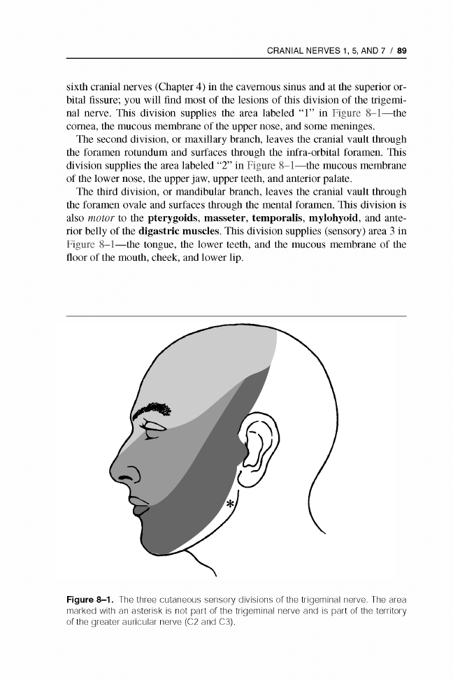
1
2
3

90 / CHAPTER 8
There is no overlap between the areas supplied by the three peripheral di-
visions of the trigeminal nerve, unlike the situation with the spinal nerves.
All three divisions supply sensation to the dura.
Sensory Testing
All sensory deficits are subjective. There are reflexes, conduction veloci-
ties, and evoked responses that can assess sensory function with variable
sensitivity and objectivity. However, most of our information about the pa-
tient’s sensory systems is obtained at the bedside or in the office with a pin, a
tuning fork, a wisp of cotton wool, and a few other crude instruments. Only
the patient can judge the quality of what he feels. You must stay out of the
decision; that is, if you are testing his response to pinprick on the right and
left sides of the face, you may obtain one answer by saying to him, “Is this
[touching the pin to the right] the same as this [touching the left]?,” and a
completely different answer by saying (under the same circumstances), “Is
this . . . different from this . . . ?” You will have planted the word different
or same in the patient’s mind. Most patients are helpful and agreeable and
may honestly think that one side is less sensitive than the other because of
the way the question was asked.
It is essential that you have the patient’s cooperation and confidence, and
he must not be tired or so ill that his answers are meaningless.
Keep your questions and instructions simple and short. If the patient is a
child or an adult of low intelligence, you will usually get a short, definitive
answer. From others, usually the very anxious, you may get an overinter-
preted answer, such as “Well . . . , maybe not exactly the same” or “Would
you do that again?” In these cases, the patient is beginning to alert you that if
this diagnosis is heavily dependent on sensory signs only, it is probably go-
ing to be wrong.
Complex sensory abnormalities deserve an examination session devoted to
the sensory system only or should be verified by a repeat examination the
next day.
Pain Perception
Do not hurt the patient. Use a sterilized common pin.
Each patient should be examined with a new pin that is discarded at the end
of the examination.
Compare the perception of pain between the right and left
sides in the first, second, and third divisions of the nerve;
that is, touch the patient’s forehead lightly several times on
the right side with the pin. Do the same on the left, asking
the patient whether they feel the same. Then compare the
right cheek with the left cheek and the right chin with the
left chin.
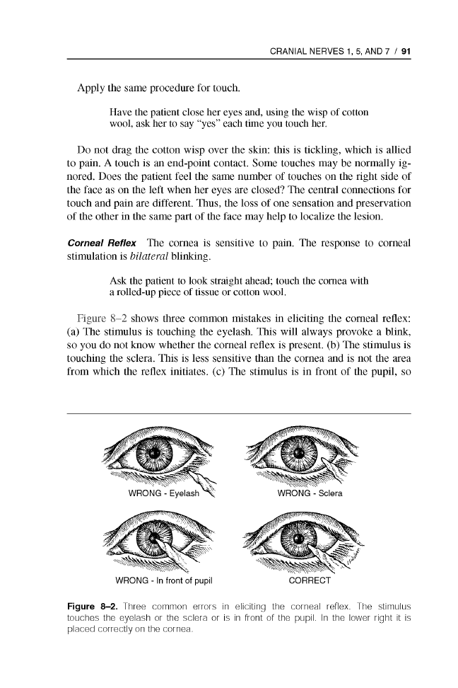
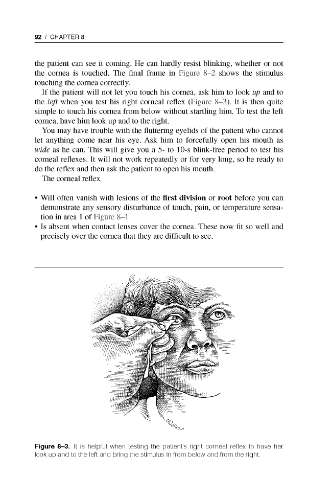

CRANIAL NERVES 1, 5, AND 7 / 93
• Is often absent after cataract surgery
• Is often absent in elderly people, for unknown reasons
• Has a supranuclear connection to the opposite thalamus. In acute destruc-
tive lesions (hemorrhage) into the thalamus, the contralateral corneal reflex
will be absent and then present but delayed for days or a week or so after
the ictus. Superficial hemisphere lesions do not do this.
The Spinal Nucleus and the Tract of the Trigeminal Nerve
The spinal nucleus and tract of the trigeminal nerve
• Extends from the midpons at the level of entry of the trigeminal root to the
upper cervical spinal segments
• Blends into and is continuous with the substantia gelatinosa of the cervical
cord
• Subserves the functions of pain and temperature sensation, touch being a
function of the main sensory nucleus, which is at the rostral end of the
spinal nucleus
Fibers from the ophthalmic division of the nerve are most ventral in the
tract and those from the mandibular division are most dorsal, with the maxil-
lary fibers in the middle.
The spinal trigeminal nucleus is medial to the tract and divided into three
areas corresponding to the areas shown in
Figure 8–4
at A, B, and C. These
areas do not have the same cutaneous relationships as the three peripheral
branches. Area A cuts through all three peripheral divisions and has its cellu-
lar components in the most rostral part of the nucleus, area B is intermediate,
and area C is related to the most caudal end of the nucleus. (For an excellent
account of the anatomy of the nucleus, see A. Brodal, Neurological Anatomy
in Relation to Clinical Medicine, 3rd ed. Oxford University Press, New
York, 1981, p. 528.)
When pain loss involves parts of all three peripheral divisions of the
trigeminal nerve, consider the possibility of a nuclear location of the lesion.
Temperature Sensation
Hot and cold sensations are not commonly examined on the face. How-
ever, it may be a useful thing to do. When a patient complains of pain or
numbness in the face and the conventional pain-touch sensory examination
as outlined above reveals no abnormalities, there may be some decreased
awareness of temperature differences over the face. This can precede the loss
of pain. Temperature sensation should not be examined as a routine measure
(see the section on “Temperature” in Chapter 14).
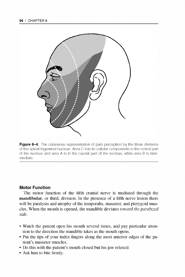
B
C
A
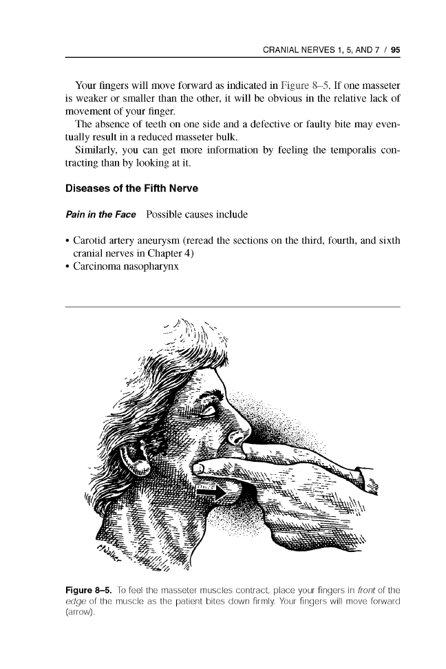

96 / CHAPTER 8
• Raeder’s paratrigeminal neuralgia
• Metastases to the base of the skull
• Herpes zoster
• Cluster headaches
• Angina pectoris
• Tic douloureux, or short, sharp, excruciating pain that travels in the anatomi-
cal distribution of one or sometimes two cutaneous divisions of the nerve.
The mandibular division is most common. Pain lasts for seconds, with
20–200 stabs per day, precipitated by talking, eating, drinking, washing the
face, or exposure to cold air. There is often a precise trigger point; touching it
invariably initiates pain. It is most common in middle and late life, although it
also occurs in younger people as a manifestation of multiple sclerosis.
Numbness (Analgesia, Hyperesthesia, or Paresthesia) in the Face
Causes
include
• Lesions in the cerebellar pontine angle, for example, acoustic neuroma—
as it grows upward, it will elevate and stretch the root of the trigeminal
nerve before the latter perforates the tentorium. There will be signs of in-
volvement of the seventh (late) and eighth (early) cranial nerves.
• Multiple sclerosis, brain stem infarct, glioma, syringobulbia, and some
medications, for example, streptomycin, pyridoxine, isoniazid, and some
kinds of penicillin
• Fractures or metastatic and primary tumors of the bones of the face or the
base of the skull
• Trigeminal neuropathy of unknown cause, usually in women. It is progres-
sive, bilateral, and will eventually involve the entire face.
• Bilateral, circumoral numbness does not mean the patient necessarily has
a bilateral lesion. Focal sensory seizures, the somatic paresthesias of a mi-
graine, or a transient ischemic attack can produce numbness around the
mouth, on both sides of the midline. The secondary representation of all
the mouth and lips (and tongue) is in both the right and left hemispheres.
(The trigeminothalamic tracts subserving touch and pressure and probably
pain and temperature are both crossed and uncrossed.)
Weakness of Jaw Movements
Myotonic dystrophy will produce bilateral
weakness and wasting of both temporalis and masseters although jaw open-
ing muscles seem strong.
Myasthenia gravis will produce weakness without wasting of both jaw
openers and closers.
Amyotrophic lateral sclerosis and poliomyelitis also cause weakness and
wasting of both jaw openers and closers.

CRANIAL NERVES 1, 5, AND 7 / 97
Unilateral—fracture or neoplasm or inflammation of the foramen ovale; the
jaw is pulled over to the paretic side by the pterygoids of the normal side (PPP).
In a unilateral upper motor neuron lesion, jaw opening and closing is nor-
mal. Both hemispheres innervate both right and left muscles that open and
close the jaw.
• Bilateral upper motor neuron lesions produce an abnormality of chewing
that is part of pseudobulbar palsy (described in Chapter 16).
SEVENTH CRANIAL NERVE
The seventh cranial (facial) nerve is a great nerve—with it we can laugh,
frown, cry, taste, and spit!
Anatomy
The seventh nerve is an important mixed nerve with motor, sensory, and
autonomic divisions.
Motor Function
The motor fibers supply the muscles of facial expression, from frontalis to
platysma, the stapedius muscle in the middle ear, the stylohyoid, and the
posterior belly of the digastric. A lesion of the nerve trunk or its nucleus pro-
duces a lower motor neuron palsy and is described as a seventh nerve lesion.
It affects all the muscles partially or completely supplied by the nerve. By
contrast, facial weakness caused by a contralateral upper motor neuron le-
sion presents a distinctly different clinical picture.
The terms seventh nerve lesion and facial weakness are not interchange-
able. The first means an ipsilateral lesion of the lower motor neuron and
paralysis of all the muscles of one side of the face. The latter means a con-
tralateral lesion of the upper motor neuron and variable weakness of some of
the muscles of one side of the face.
Remember:
• The frontalis muscle is supplied by the ipsilateral and contralateral hemi-
spheres. In facial weakness (the upper motor neuron lesion) the forehead
on the side of the weakness appears to wrinkle in a normal way. If you ask
the patient to wrinkle upward against resistance, you can feel a slight
weakness on the abnormal side relative to the normal. In a seventh nerve
lesion the ipsilateral forehead is flat and will not wrinkle at all.
• The orbicularis oculi also have bilateral suprasegmental innervation. With
the upper motor neuron lesion causing facial weakness, the eye will close

98 / CHAPTER 8
but is weaker than the normal side and the difference is easy to detect with
your fingers. With the seventh nerve lesion the eye will not close at all and
is open wider on the paretic side.
How to Examine the Seventh Cranial Nerve
1. Look at the patient when she is at rest and while she is talking,
smiling, and blinking.
2. Ask her to wrinkle her forehead quickly two or three times (the
Groucho Marx maneuver) (
Figure 8–6A
). Watch the rate and ex-
tent of the movement of the two eyebrows. When the patient is re-
laxed, put your finger on her eyebrows and ask her to wrinkle her
forehead against this gentle resistance.
3. Ask her to close her eyes gently, then tightly (
Figure 8–6B
). Try to
open them while the patient resists you. If she can wrinkle her fore-
head and can close her eyes, this is not a seventh nerve lesion.
4. Now compare the lower right side of the face with the left side and
look for relative weakness on one side, as in facial weakness result-
ing from an upper motor neuron lesion. Ask the patient to close her
eyes as tightly as she can. Did the patient
• Tuck in the eyelashes between the edges of the lids equally well right
and left? They will not tuck in as far on the side of a facial weakness.
• Deepen the nasolabial groove equally well right and left? It will not
deepen as much on the side of a facial weakness.
• Retract the corners of her mouth away from the midline equally far
right and left? The corner of the mouth on the side of a facial weak-
ness will not move as far (
Figure 8–6B
).
5. Ask the patient to show her teeth. (The occasional patient with den-
tures will hand them to you at this point; everyone else with dentures will
tell you their teeth are not their own.) Ask the patient to clench her teeth
together and retract the corners of her mouth. Again, notice whether the
two corners go equally far and equally fast. Sometimes the only manifes-
tation of a facial weakness is the momentary delay by one corner of the
mouth, which begins to move 1 s later than on the normal side.
6. Ask the patient to whistle. Ask her to press her lips together and
blow up her cheeks; tap on one and then the other inflated cheek.
Air will escape between the lips when you tap the weak side (
Figure
8–6C
).
7. Ask the patient to open her mouth as wide as she can. The opening
should be symmetrical, with the same number of teeth showing on the
two sides of the midline. The weak side of the face covers more teeth
than the normal side.

CRANIAL NERVES 1, 5, AND 7 / 99
To see the patient’s platysma, you have to act as a model (
Figure 8–6D
).
Do the following, by way of example, then ask her to repeat your actions.
8. Clench your teeth and pull the corners of your mouth forcefully
downward with a grimace, revealing your platysma.
At rest, the patient with a seventh nerve lesion has, on the paretic side,
• An eye open wider than on the normal side; it does not blink; increased
tears are stimulated by the dry and irritated cornea and flow over the
paretic cheek.
• A flat, creaseless forehead
• A flat, sometimes drooping cheek
• The corner of the mouth lower than on the normal side
• A “flappy,” loose cheek as he talks
• The midsagittal line of the mouth pulled over to the normal side
At rest, the patient with a facial weakness may look perfectly normal or
show a minor flattening and asymmetry.
Remember that normal elderly people may have an asymmetrical lower
face at rest. This is because of an asymmetrical loss of teeth, a lifetime habit
of talking out of the corner of the mouth, or simple passage of time. (Look
carefully at the next 12 people over 65 years of age that you meet. Most of
them have some asymmetry about the mouth and difference in the depth of
the two nasolabial folds.)
The patient with a seventh nerve lesion is equally unable to perform vol-
untary, reflex, or emotional movements of half of the face.
In contrast, the patient with a facial weakness has paresis and slowness,
mostly of the lower half of the face, for voluntary movements but smiling is
normal and symmetrical. If you can make him laugh, the paretic side of the
face seems to move as well or more than the normal side. Why? Either emo-
tional facial movements have bilateral supranuclear connections (which is
probably correct) or the upper motor neuron for voluntary movements is
completely different from the supranuclear fibers concerned with smiling,
frowning, laughing, and crying. A patient with a hemifacial defect for emo-
tional movements only, but a normal face for voluntary movements and a
solitary lesion in the opposite thalamus, has been described (See N Engl J
Med 1998; 338(21):1515).
Blinking
Watch the patient blink. If he blinks less often than you do, he may have
parkinsonism or may be abusing some sedative. The eyes blink at exactly the
same moment and the eye is completely covered with each blink. If he
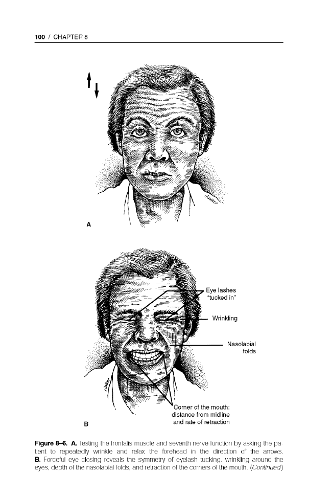
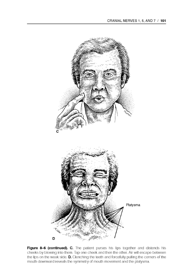

102 / CHAPTER 8
blinks less often on the right side or does not cover the entire right eye with
each blink, he has a partial right seventh nerve lesion, new or old. This is
never the result of an upper motor neuron facial weakness or a fifth cranial
nerve lesion. Asynchronous blinking is most often seen in the patient with a
partially recovered Bell’s palsy.
Sensory and Autonomic
The intermediate nerve or sensory root of cranial nerve 7 emerges from
the brain stem between the facial motor root and the vestibular (eighth) nerve.
If it had its own cranial nerve number, it would be 7
1
/
2
. Its functions are
• Taste—From the anterior two thirds of the tongue, cells are in the genicu-
late ganglion, and central termination is on the rostral part of the solitary
nucleus. The peripheral pathway is via the chorda tympani and lingual
nerves.
• Saliva—The cells are in the dorsolateral reticular formation, called the su-
perior salivatory nucleus. Fibers that are preganglionic and parasympa-
thetic travel in the intermediate nerve, then in the chorda tympani and lin-
gual nerves to the submandibular ganglion. Postganglionic fibers go to
the submandibular and sublingual salivary glands.
• Tears—Have the same reticular formation cells of origin as described
above for saliva. Parasympathetic preganglionic fibers leave the intermedi-
ate nerve to enter the greater superficial petrosal nerve to the pterygopala-
tine ganglion. Postganglionic fibers go to the lacrimal gland, and secretory
and vasomotor fibers proceed to the mucous membrane of the nose and
mouth.
• Pain fibers—Are from the external auditory canal and behind the ear. The
fibers are part of the intermediate nerve, and the nucleus is part of the
spinal trigeminal tract. Most patients with a seventh nerve palsy complain
of numbness over the cheek but have no demonstrable sensory loss to
touch, pain, or temperature examination on the cheek or anywhere else.
Taste, Tears, and Saliva
Taste from the anterior two thirds of the tongue is
an afferent function of the seventh nerve. Taste from the posterior tongue
and palate is via the glossopharyngeal nerve, which is more important than
the seventh nerve in this function.
From the anterior two thirds of the tongue the pathway is complex as fol-
lows: (a) the fibers are first in the lingual nerve, which is a branch of the
mandibular (the third division of the trigeminal); (b) they are then in the
chorda tympani nerve (a branch of cranial nerve 7) to their cell station,
which is the geniculate ganglion; and (c) from here are in the intermediate
nerve, which is the sensory root of the seventh cranial nerve.
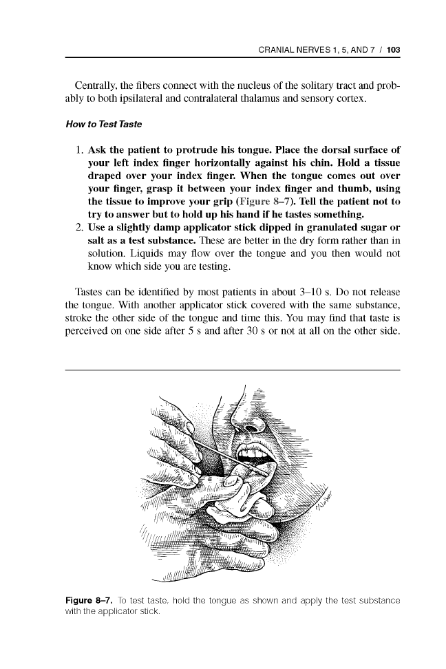

104 / CHAPTER 8
The slow side is the abnormal one. As soon as you let go of the tongue the
examination is over. The sugar is then tasted from many areas inside the
mouth. The test can be repeated after 5 min and after the patient has rinsed
his mouth.
Remember:
• There is not much taste sense on the tip of the tongue.
• Elderly people lose their sense of taste and the number of taste buds.
• Some people cannot perceive sugar on either side of the tongue. Allow
such a patient to rinse out his mouth and try a different substance.
• In general, taste is perceived better on the more posterior aspects of the
tongue, palate, and pharynx rather than on the anterior.
• There are only four taste substances: sweet, sour (acid), bitter (quinine),
and salty.
• A lesion of the seventh nerve at the stylomastoid foramen where it emerges
from the skull (commonly caused by trauma or disease of the parotid
gland) produces a seventh nerve lesion as described above, with no taste
loss and no hyperacusis.
• A lesion distal to the geniculate ganglion (Bell’s palsy being the most com-
mon) produces the paresis as above, loss of taste in the anterior two thirds
of the tongue, hyperacusis (the branch to the stapedius), and a decrease of
salivary secretion on the same side. (Review the anatomy of the intermedi-
ate nerve mentioned briefly at the start of this chapter.)
• A lesion proximal to the geniculate ganglion (a cerebellopontile angle tu-
mor being the most common) produces all of the above plus a dry eye on
the same side.
Diseases of the Seventh Nerve
Diseases of this nerve include
• Bell’s palsy—Onset may be at any age with pain behind the ear and down
the side of the neck. The cause is unknown. The lesion is in the facial
canal; the patient complains of a numb cheek and hyperacusis on the same
side, as well as facial paralysis. Examination reveals weakness of all the
muscles supplied by the seventh nerve and absent taste on the ipsilateral
anterior two thirds of the tongue. It can be bilateral, it is rarely recurrent,
and the prognosis is generally good.
• Guillain-Barré syndrome (also called infectious polyneuritis)—Often bilat-
eral, it may start with a seventh nerve lesion(s) or facial involvement may
occur after the arms and legs are involved.
• Sarcoidosis, vasculitis, parotid gland tumor, leprosy, infectious mononu-
cleosis, and Lyme disease

CRANIAL NERVES 1, 5, AND 7 / 105
• Acute or chronic otitis media and mastoiditis
• The cerebellar-pontine angle tumor—Seventh nerve palsy signs are both
rare and late and generally follow the deafness and trigeminal nerve signs
and symptoms. The ipsilateral eye will not tear.
• Geniculate ganglion herpes—Herpes zoster in the external ear canal, con-
cha, and mouth and behind the ear; pain in the ear; paralysis of the face as
in Bell’s palsy with the same taste loss. Herpes, pain, and facial weakness
may all appear at once or consecutively over several days. Taste recovery
is unlikely.
• Within the pons—Vascular disease, poliomyelitis, neoplasm demyelinating
lesions, and syringobulbia
Bilateral Seventh Nerve Palsy
• Bell’s palsy
• Guillain-Barré syndrome
• Encephalitis
• Prepontine, intrapontine tumor
• Meningitis, cryptococcal or tuberculous, and as part of acquired immune
deficiency
• Sarcoid, systemic lupus erythematosus
Diseases Causing Facial Weakness
Any lesions that occur in the internal capsule or any other part of the up-
per motor neuron above the seventh nerve nucleus (eg, vascular lesion, neo-
plasm, abscess, or trauma) can produce an upper motor neuron facial weak-
ness.
Parkinsonism
Parkinsonism can be exclusively unilateral or at least much
more marked in one arm and leg, but if the face is involved it is always on
both sides. The patient can wrinkle his forehead, close his eyes, show his
teeth, whistle, and so on, so clearly the face is not paretic. However, the
emotional responses revealed by the face are absent. A hemifacial defect for
emotional expression can occur from a lesion in the contralateral thalamus
(see page 99).

Cranial Nerves 8–12
9
EIGHTH NERVE
The eighth (acoustic) nerve is really two nerves—the cochlear (meaning
“snail shell”), which is concerned with hearing, and the vestibular (meaning
“cavity at a canal entrance”), which is concerned with movement, position,
and balance.
Cochlear Nerve
Lesions of this special sensory nerve cause deafness. Nerve cells are in the
spiral ganglion of the cochlea. Their peripheral connections are to the audi-
tory cells of the organ of Corti, and their central connections are to the
cochlear nucleus. The next-order neuron is the lateral lemniscus. When im-
pulses concerned with sound have reached the cochlear nucleus, their further
passage is bilateral (ie, a unilateral lesion above the level of the nucleus can-
not cause deafness).
Deafness
Does the patient have some hearing loss, and if so, is it nerve
deafness (or sensorineural deafness) or conductive deafness? Conductive
deafness is caused by a disease of the external canal, middle ear, or ossicles.
The key features of nerve deafness are
• Loss of perception of high pitched sounds
• Loss of hearing of bone conducted sound
How to Examine Hearing
You can learn a lot from the patient’s history. If
the patient can hear on the telephone using either ear, his hearing is reason-
ably good.
A formal test is awareness of whispered speech at 6 ft. It is often difficult
to find a quiet room in which to test the patient. Follow this method:
• Have the patient stand, facing at right angles to you, at a distance of 6 ft.
• Ask the patient to put his finger in the ear farther from you. Whisper a se-
ries of numbers, asking him to repeat each number after you.
107

108 / CHAPTER 9
A person with normal hearing can hear and repeat nine out of 10 whis-
pered numbers at this distance. Then turn the patient so that he faces the op-
posite way, ask him to plug the far ear, and repeat the procedure. Your whis-
pered voice can vary from very faint to almost conversational speech. After
you have done this a few times, you will get the sense of a constant-volume
whisper and the test becomes quite reliable. If his hearing is normal, record
it as “Whispered voice @ 6
′
, Rt and Lt.” If you had to move up for the pa-
tient to hear the numbers when testing the left ear, record it as, for example,
“Whispered voice @ 6
′
Rt and 4
′
Lt.”
If the patient is deaf in one or both ears, do the following to establish
whether the deafness is caused by a defect in the conducting system of the
ear or in the nerve leading from the ear.
• Place the handle of a vibrating 256 tuning fork on the patient’s mastoid.
• Ask him to tell you when the vibrations can no longer be heard.
• At that point, put the vibrating tines of the tuning fork close to the patient’s
ear.
If the sound reappears, one can say that air conduction is better than bone
conduction. These are the findings in most people and in early nerve deaf-
ness. If your hearing is normal, put the handle of the 256 tuning fork on the
patient’s mastoid and when he says he can no longer hear it, put the handle
on your own mastoid. If you hear it through your mastoid longer than the pa-
tient does, he has nerve deafness. If nerve deafness is complete, there is no
appreciation of sound by either bone conduction or air conduction. This is
the Rinne test. It is reliable only with bilateral hearing loss. The opposite
situation—when bone conduction is longer than air conduction—is found in
conductive or middle ear deafness.
There is another way to tell nerve deafness from conductive deafness:
Put the handle of a vibrating 256 tuning fork on the center of
the patient’s head, over the vertex. Ask him if he hears it bet-
ter in one ear or the other or in the center of his head.
If the patient has a conductive hearing loss in one ear, he will hear the tun-
ing fork in this ear. You can simulate this for yourself, provided that you
have normal hearing. When the vibrating tuning fork is on the vertex of your
head, put your finger in one ear. The sound will immediately become loudest
in this ear. You have thus produced a degree of conductive deafness in your-
self. This is the Weber test. On the other hand, if the patient has nerve deaf-
ness in one ear, he will hear the tuning fork in the opposite normal ear. We-
ber’s test is no help in bilateral deafness and it will not work if there is both
nerve and conductive deafness in the same ear.

CRANIAL NERVES 8–12 / 109
Remember, nerve deafness results in decreased bone conduction and loss
of high-tone appreciation; conduction deafness results in decreased air con-
duction and loss of low-tone appreciation. Total hearing loss always means
nerve deafness.
Vestibular Nerve
Test the vestibular nerve and vestibular apparatus (the semicircular canals,
utricle, and saccule) by observing the movements and position of the eyes.
This also assesses the function of the brain stem from the upper medulla to
the midbrain.
The patient with an acute unilateral vestibular lesion has vertigo and vom-
iting and staggers to the side of the lesion. The vertigo is spinning of the en-
vironment (object vertigo) to the opposite side. Her eyes are forced to the
side of the lesion, and she has nystagmus to the opposite side, that is, the fast
component. A chronic or slowly progressive lesion of the semicircular canals
or the vestibular nerve will likely have none of these signs and symptoms.
How to Test Vestibular Function
The otologists and clinical neurophysiolo-
gists have sophisticated ways of testing the function of both divisions of the
eighth cranial nerve. At the bedside the principal testing method is observa-
tion of eye movements in response to hot and cold water in the external ear
canal, that is, caloric testing. Cold water inhibits the function of the semicir-
cular canals, whereas warm water enhances it. If a temporary imbalance can
be created between the two sides, the system can be tested. Cold-water stim-
ulation of the external ear canal simulates acute destructive labyrinthine dis-
ease, that is, eyes to the irrigated side, nystagmus away from the irrigated
side. A mnemonic in Joel S. Glaser’s Neurophthalmology (Harper & Row,
Hagerstown, Maryland, 1978) will help you remember the nystagmus direc-
tion:
COWS: Cold–Opposite–Warm–Same
• Sit the patient with her head in extension, occiput supported, looking at
the ceiling, or have her lie on her back with her head on a pillow 30 de-
grees from the horizontal. Her horizontal semicircular canal is now verti-
cal.
• Look in the external ear with an otoscope. Remove any wax in the external
canal. If the tympanic membrane is perforated or red and bulging, stop
now.
• Put a large kidney basin under the ear, with the concave side against the
neck. Put a folded towel under the basin, and slowly and gently inject 20
mL of cold (20
°
C) tap water over 20–30 s.

110 / CHAPTER 9
• Time the onset, duration, and direction of nystagmus. Wait 10 min and do
the same to the other ear (check first for wax, perforation, and so on).
The interval and duration should be approximately the same, right and
left, as should the degree of discomfort, nausea, spinning sensation, or vom-
iting.
• Repeat the irrigations after an interval using warm (42
°
C) water in first one
and then the other ear.
If a patient has just recovered from an episode that you suspect was acute
labyrinthitis or has had chronic recurrent episodes of vertigo, ataxia, vomit-
ing, or dizziness, try warm water first. Cold-water irrigation in such patients
is often less effective.
Vestibular Function in the Unconscious Patient
Reread the section on
“The Doll’s Eye Test and Caloric Testing” in Chapter 7.
Use water as cold as you can find. This is a stronger stimulus than the
hottest tolerable hot water. The purpose of the examination has changed.
This is an attempt to localize the lesion responsible for the coma.
The full ocular deviation to one side and nystagmus to the other test the
integrity of the vestibular nerve, nuclei, medial longitudinal fasciculus, ocu-
lomotor nuclei, brain stem reticular mechanisms, and hemispheres.
• When the cause of the coma is a pontine lesion, the response is absent. Be
careful! If the patient’s coma is because he is frozen or drug-overloaded,
this false absent response is reversible. If in doubt, repeat the examination
in 12 and 24 hr.
• If the response is normal (remember in the unconscious patient there is no
nystagmus, and normal means eye deviation only), the cause of the coma
is not in the brain stem.
• If the response is only abduction of one eye, the cause of the coma is in the
midbrain. The oculomotor nuclear constellation is damaged, and adduction
cannot occur.
Diseases of the Eighth Cranial Nerve
When the patient has vertigo, tinnitus, and deafness, the diagnostic possi-
bilities are many. As always, start with a careful history. Vertigo can be pos-
tural in the arteriosclerotic elderly patient and in others who are on tricyclic
antidepressant drugs. It can also be part of the symptom complex of a tem-
poral lobe seizure or can be perfectly benign in the long-legged male youth
who seems to be growing like a weed and says that every time he stands up
quickly the room spins and his vision fades, and he has actually fallen twice

CRANIAL NERVES 8–12 / 111
in the past 6 months.
Deafness must be differentiated as to nerve, conduction, or mixed, and the
lesion may result from an acoustic neuroma, presbyacusis, otosclerosis, Me-
niere’s disease, chronic ear infection, industrial abuse, trauma, or drugs (eg,
kanamycin).
Tinnitus
Tinnitus is described as a ringing, whistling, buzzing, clicking, or
roaring in the ears. It may be present in one or both ears and can sometimes
even be heard by persons other than the patient.
Tinnitus is a common complaint and is a symptom rarely relieved. It is
thought to be peripheral (nerve or more distal) in origin, but there may be
central causes.
Tinnitus can be present early with an acoustic neuroma. By the time the
patient is deaf and seeks help, months or years may have passed, and he is
no longer aware of the tinnitus. Some patients with Meniere’s disease seem
to be able to forget their violent attacks of vertigo, tinnitus, and vomiting and
will go to the doctor years later because of progressing deafness.
Aspirin causes and aggravates tinnitus, but some patients never make the
connection. The sound may be rushing or pulsating and is always louder at
night in the quiet of the bedroom. You may hear the patient’s noise with your
stethoscope. If the patient is elderly and invasive investigations would be
hazardous, avoid such procedures.
Occasionally, a patient with increased intracranial pressure complains of a
pulsating sound in her head that other members of the family can also hear.
Some vessel in the head is being compressed. The sound vanishes when the
pressure is reduced.
When a patient sleeps on her side, she may compress the occipital branch
of the posterior auricular artery between the skull and the pillow. This can
produce a pulsating, swishing sound. This is harmless.
NINTH NERVE
The ninth (glossopharyngeal) nerve is both sensory and motor. Its only
motor function is to the stylopharyngeus muscle. This cannot be tested clini-
cally. The nerve supplies sensation to the tympanic cavity, the tonsils, the
posterior aspect of the soft palate (as does the maxillary branch of the
trigeminal nerve), the posterior third of the tongue, and the pharynx. It car-
ries taste fibers from the posterior third of the tongue and supplies secretory
fibers to the parotid gland.
How to Examine the Glossopharyngeal Nerve
Sensation on the pharynx

112 / CHAPTER 9
can be tested with an applicator stick that has no cotton on it.
• Compare the response of the right and left halves of the pharynx by touch-
ing them gently with the end of the stick.
• You can ask the patient whether touching the pharynx feels the same right
and left, and you may provoke a gag reflex on one side, but not the other.
The motor side of this reflex is a prompt contraction of the pharynx with
or without gagging. In many people pharyngeal contraction and gagging
cannot be elicited from either side. Abnormalities of palatal and pharyngeal
sensation are soft physical signs, and there is not a high diagnostic yield in
this part of the examination. You cannot test the taste on the posterior part of
the tongue.
Diseases of the ninth and tenth cranial nerves are considered together after
the section on the tenth nerve.
TENTH NERVE
The tenth (vagus) nerve is also a mixed nerve. It has afferent fibers from
the dura of the posterior fossa, the external ear, pharynx, larynx, trachea,
esophagus, and thoracic and abdominal viscera. It sends parasympathetic
fibers to the thoracic and abdominal viscera and motor fibers to the muscles
of the palate, pharynx, and larynx. Review the anatomy of the vagus, with at-
tention to the superior laryngeal nerve and the two recurrent laryngeal nerves.
The neurological examination of the vagus concerns mostly the muscles of
the palate, the constrictors and sphincter of the pharynx, and the muscles of
the larynx.
Palate
In the palate there may be no symptoms from unilateral paresis. However,
on examination the arch on the paretic side is lower.
Ask the patient to quickly say “ah.”
The midline of the palate will go over to the normal side, the normal arch
will lift and curve, and the paretic side will remain lower and straighten
(
Figure 9–1A
and
B
). The uvula is not important. It is soft and floppy and
sometimes hangs in the midline or to the right or left. Ignore it.
Bilateral palatal paralysis causes nasal regurgitation and a nasal voice, and
it is difficult for the patient to pronounce b and g. You can hear air coming
out of his nose as he talks.
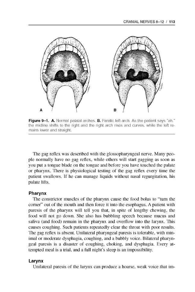

114 / CHAPTER 9
proves with time as the opposite normal cord crosses the midline. Even if the
patient, his family, and you think his voice is normal, he may still have a
unilateral vocal cord paresis. There is often trouble with swallowing liquids
if the lesion is peripheral and involves the recurrent laryngeal nerve only.
Both the abductor and adductor muscles of the larynx and the lower sphinc-
ter of the pharynx are paretic.
If the lesion is more proximal, palatal and pharyngeal paresis will also be
present.
Bilateral paresis with a distal lesion (thyroidectomy) presents as a stridu-
lous (see stridor), weak voice with shortness of breath. The intact superior
laryngeal nerve (a more proximal branch of the vagus nerve) keeps the cords
adducted and the airway dangerously narrow.
With more proximal lesions causing bilateral paresis, there is usually pare-
sis of the palate and pharynx as well. The vocal cords are in midposition, the
voice is weak without stridor, there is no dyspnea and no force to coughing,
and dysphagia is present.
How to Look at the Vocal Cords
1. Use a round dental mirror, 15–20 mm in diameter, on an 8-in handle.
Warm it under hot tap water and dry it.
2. Ask the patient to open his mouth, sit still, and breathe through his
mouth. Hold his tongue down with a tongue blade held in your left
hand. This hand holds the penlight on top of the tongue blade as shown
in
Figure 9–2
.
3. Put the warm (not hot) mirror far enough back in the pharynx until you
can see down the larynx and can see the vocal cords. Ask the patient to
say “eh, eh” and “ah, ah” several times.
Compare the amount and speed of movement of the two cords. If touching
his palate or pharynx with the mirror makes him gag, a little lidocaine spray
will temporarily abolish the afferent side of the gag reflex. If you use a cold
mirror, water vapor will condense on it and you will see nothing.
Things to Remember About Cranial Nerves 9 and 10
• Can the patient swallow liquids and solids?
• Is there nasal regurgitation or coughing when he swallows?
• Is the voice different in any way?
• Can the patient cough forcefully?
• An absent gag reflex (bilateral) is often normal; a unilaterally absent gag
reflex is not normal.
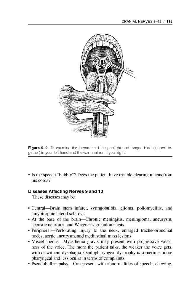

116 / CHAPTER 9
and swallowing, but the lesion is not in the ninth or tenth cranial nerve
• Both the ninth and tenth cranial nerves convey some sensation from the ex-
ternal ear, posterior palate, and other areas. Glossopharyngeal neuralgia con-
sists of attacks of severe, lancinating pain of short duration and is commonly
induced by swallowing. The pain is in the throat and ear, has the same quali-
ties as tic douloureux, and may be symptomatic of nerve trunk compression
(neuroma or an aberrant vessel). Each attack of pain can be accompanied by
a transient change in heart rate and rhythm with loss of consciousness (the
carotid branch of the glossopharyngeal nerve innervates the carotid sinus).
• The jugular foramen syndrome—Lesion of the ninth, tenth, and eleventh
cranial nerves. If the result of intracranial disease, there is usually brain
stem compression and long tract signs. If caused by extracranial disease,
the cervical sympathetic and twelfth cranial nerves are frequently in-
volved. The responsible lesions are usually primary or metastatic tumor of
the skull base, glomus jugulare tumor, meningioma, or epidermoid tumors.
• Polyneuritis cranialis—This disease is of unknown cause. It starts in el-
derly patients as multiple lower cranial nerve palsies. They are usually
thought to have nasopharyngeal carcinoma or metastatic carcinoma at the
base of the skull, but they have neither. It sometimes improves sponta-
neously.
• Diphtheria and botulism toxins—Both can cause bulbar palsy
ELEVENTH NERVE
The eleventh (spinal accessory) nerve is a motor nerve that originates in-
tracranially in the medulla and also in the cervical spinal cord. It is both a
cranial nerve and a spinal nerve. It innervates the sternomastoid muscle and
the upper portion of the trapezius muscle.
How to Test the Spinal Accessory Nerve
If you have the patient face you,
put your hand on the right side of his head as in
Figure 9–3A
, and ask him to
turn to the right against the resistance of your hands, you will not learn any-
thing. You cannot see or feel the left sternomastoid effectively.
However, if you ask him to turn his head to the right as far
as he can, unresisted, and then you attempt to bring the head
back to the facing position against his resistance, you will
see, as in
Figure 9–3B
, the size of the left sternomastoid and
feel the strength of it.
To test the trapezius,
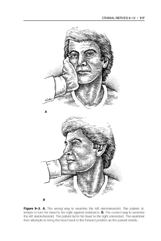

118 / CHAPTER 9
• Stand behind the patient. Look at the patient’s neck, back, and shoulders.
Do they appear to be symmetrical, and are the muscles the same size and
bulk on the two sides?
• Hold the upper edge of the muscle between your thumb and fingers and
ask him to shrug his shoulders upward toward the ears.
Compare the speed, size (thickness), and strength of the right and left
sides.
Diseases of the Eleventh Cranial Nerve
• The jugular foramen lesions mentioned above in the section on the ninth
and tenth cranial nerves are important.
Others include
• Lesions in the region of the foramen magnum, upper spinal cord, lower
end of the medulla, both extrinsic (neurofibroma) and intrinsic (po-
liomyelitis), and amyotrophic lateral sclerosis
• Muscular dystrophy, especially myotonic dystrophy, which may present
with almost no visible sternomastoid muscle. Myasthenia gravis and
polymyositis are also important.
• An upper motor neuron lesion in the hemisphere may produce weakness of
the ipsilateral sternomastoid and contralateral trapezius. With a pon-
tine upper motor neuron lesion the contralateral sternomastoid is
weak. Although still somewhat controversial, the upper motor neuron re-
specting the sternomastoid originates either in both hemispheres or, more
likely, in the ipsilateral hemisphere and decussates twice, that is, to the op-
posite pons and back again to the same side of the cord as the hemisphere
of origin. The upper motor neuron fibers respecting the trapezius appear to
originate in the contralateral hemisphere and decussate only once.
TWELFTH NERVE
The twelfth (hypoglossal) nerve is the motor nerve to the tongue.
How to Examine the Hypoglossal Nerve
1. Have the patient open her mouth without protruding the tongue.
Look in her mouth. How thick is the tongue? Is it flat, wrinkled, mov-
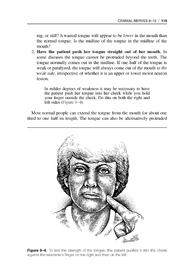

120 / CHAPTER 9
and withdrawn back into the mouth rapidly. This “trombone” movement of
the tongue is normal and becomes slower and abnormal in some diseases.
Ask the patient to protrude her tongue and then wiggle it
from side to side.
When you know how most people do this, you will quickly recognize ab-
normal tongue movements.
Ask the patient to rapidly repeat the sound “la-la-la-la.”
Parkinsonism, cerebellar lesions, or weakness and wasting of the tongue
will make the sound abnormal.
Diseases of the Twelfth Cranial Nerve
Most bilateral lesions of the tongue are the result of amyotrophic lateral
sclerosis. (See also the lower cranial nerves described above.)

The Upper Limb
1 0
The arm is between the shoulder and the elbow; the forearm is between
the elbow and the wrist.
Assessment of upper limb function may be directed when the patient
says, for example, “I have a pain in my right neck and shoulder and numb-
ness in my fingers and I drop things from my right hand.” He must have a
disease of the spinal cord, canal, root, or brachial plexus. The nature of his
complaint thus will direct your thinking.
When there are direct symptoms, try to convert these into defects of func-
tion. If the patient says, “My hand is numb,” then you will want to ask,
“Does bath [dish] water feel equally hot on both your hands? Can you put
your hand in your pocket [purse] and bring out a key or a coin and know
what it is without first looking at it? Has your handwriting changed? Can
you tie your necktie [put your earrings or contact lenses in] as well as you
used to? Can you do up buttons, zippers, or hooks and sew, type, play the
guitar, and work your personal computer as well as you always could?”
Also, remember that numbness implies a sensory disorder when doctors
use the word, but not so with patients. Patients with pure motor lesions,
basal ganglion disorders, and other nonsensory lesions sometimes describe
the affected part as “numb.”
In contrast is the undirected assessment of upper limb function. The pa-
tient’s family, or perhaps his employer, says, “He cannot do his office work
as well as he could a year ago. He seems to shuffle a bit when he walks, and
he does not stand up straight anymore.” What has this to do with the arms?
While you are taking the patient’s history, you notice that his right arm has
not lifted off the armrest of the chair once in 30 min. When he emphasizes
spoken speech with a gesture, only the left hand “does the talking.” All the
normal, small, useless movements such as adjusting the knot in the necktie,
sliding the glasses up onto the nose, or rubbing the cheek with a finger are
done with the left hand. The patient has parkinsonism. The most obvious
physical sign is the immobility of the right upper limb. Once you have
thought of the diagnosis, you make your inquiries from a different perspec-
tive, and yes, the patient, on reflection, admits that his handwriting is differ-
ent in the past 2 years and, yes, the newspaper does appear to tremble on oc-
casion when he is tired and holding it up in front of him. But the presenting
121

122 / CHAPTER 10
complaints were a gait disorder and inability to perform on the job; the right
upper limb was supposedly symptom-free.
The most important things to look at during history taking in any patient
are the face, the stance, and the upper limbs. While the patient answers ques-
tions, watch him. Similarly, do not let anyone else go to get your patient
from the waiting area. Get him yourself, then watch the patient as you intro-
duce yourself and as he gets out of the chair, goes with you to the consulting
room, sits, and gets ready to tell his story.
If he gets out of the chair by pushing down on the chair arms or on his
knees with his hands, his quadriceps are weak. If he seems to rock back and
forth in the chair two or three times before “launching” himself to stand, he
is stiff because of parkinsonism, medication, or some other cause. Do his
arms swing as he walks? Does his right arm come forward synchronously
with his left leg? Are there excessive, random, restless, small movements of
the hands and arms, suggesting chorea? Is there a repetitive, stereotyped
hand-and-arm movement such as wiping the lips every 15 s, as one might
see in a patient with tardive dyskinesia?
POSTURAL MAINTENANCE
When the examination of cranial nerves and visual fields is complete and
the patient is still sitting on the examining table with his legs hanging over
the side, do the following:
• Ask him to hold his arms out in front of him at shoulder level (loosely, not
rigid) with a few degrees of flexion at the elbow, his fingers separated, and
the palms uppermost.
• Then ask him to close his eyes (
Figure 10–1
) and watch him for 10 or 15 s.
If one arm begins to drift down toward the floor or down and out, or occa-
sionally up, this is evidence of organic disease. The test is not specific but it
is objective. The disease may be in the sensory system (ipsilateral or con-
tralateral), the contralateral basal ganglia, upper motor neuron system, or ip-
silateral cerebellum.
If both arms drift toward the floor, the test is uninformative. The patient
does not understand or he is tired, obtunded, on drugs, or ill in a generalized
way.
If neither arm drifts, gently tap first one arm and then the other to the side
and then toward the floor. As you do this, ask the patient to keep his arms in
their original position and not allow you to dislodge them. If one arm is
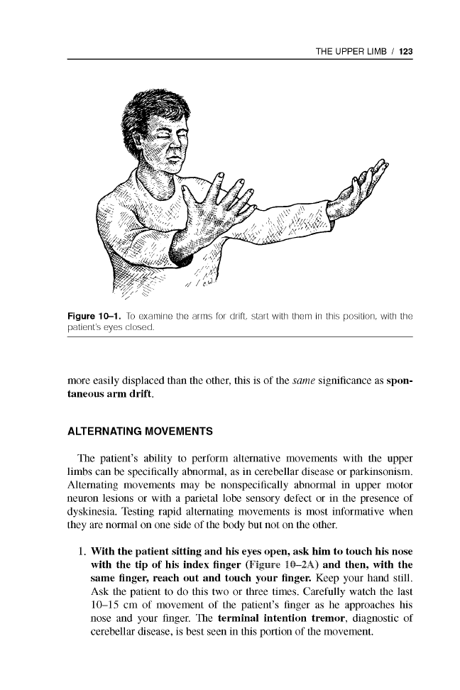
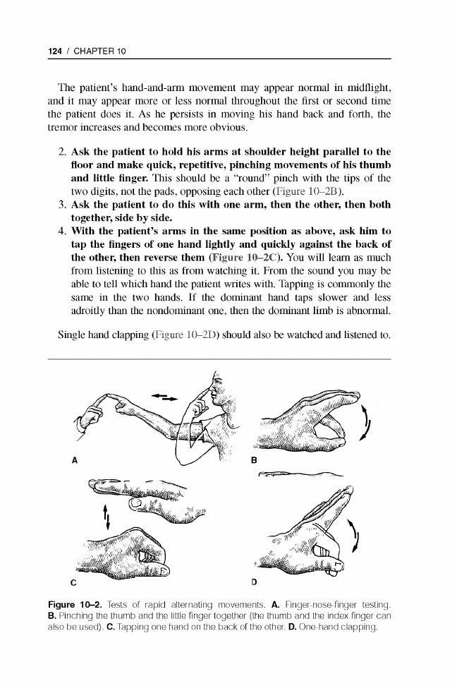
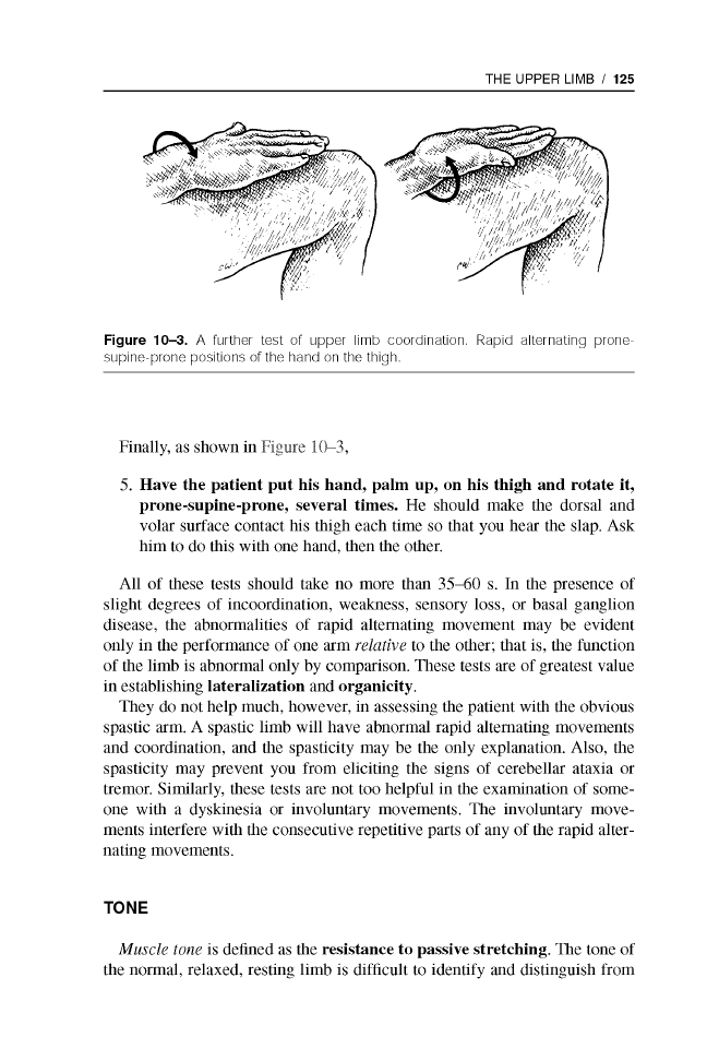

126 / CHAPTER 10
that of the pathological hypotonic limb. However, after you have examined
several patients with flaccid, hypotonic limbs that feel like the arms and legs
of a rag doll, you will know the tone of normal limbs.
Increased tone is not difficult to identify. The resistance may be greatest at
the start of stretching and then vanish as the muscle is lengthened, as with a
clasp knife. This is spasticity. In cerebral lesions it is greatest (and earliest
in a progressive lesion) in the flexor muscles of the upper limb and the ex-
tensor muscles of the lower limb, but not confined to these muscle groups.
When the lesion is spinal, the distribution is more variable.
Rigidity is the term used for increased tone resulting from disease of the
basal ganglia. It may be plastic, in which the resistance is uniform through-
out stretching, that is, like bending a piece of soldering wire, or cogwheel. In
the latter, as the muscle is stretched the examiner feels resistance, relaxation,
resistance, and so on, throughout the stretch. Rigidity is not more evident in
one muscle group than in another and, unlike spasticity, is greatest or greater
in the trunk than in the limbs.
Increased tone from any cause can often be easily exhausted by several
consecutive stretches. If the muscle being examined is then put at rest, the
increased tone can be felt again. In the upper limb it is important to assess
tone in the fingers and at the wrist, elbow, and shoulder.
1. Take the patient’s hand in your hand with her palm toward the
floor, your thumb in the middle of her palm, and your fingers on the
dorsum of her hand. With your other hand, gently and quickly ex-
tend all of her fingers together at the metacarpal phalangeal joints.
2. When the patient’s fingers have been extended as far as they can go
comfortably, let go of them. They will naturally resume a flexed po-
sition. Then stretch them again. Do this several times. There are no
diseases in which increased tone is manifest in the finger extensors
more than or earlier than the finger flexors. Therefore, you need not
stretch the extensors.
3. To test at the wrist, hold the forearm in one hand and passively ex-
tend the patient’s wrist as you did the fingers. After each extension
let the hand fall back into a flexed position, and repeat the passive
stretching quickly several times. You may elicit wrist clonus by do-
ing this (see the section on “Clonus” in Chapter 13).
There is another method to get the sense of the tone in the patient’s fore-
arm muscles.
4. Hold the supine patient’s forearm vertically, with her elbow flexed
to 90 degrees and her fingers toward the ceiling. Let your fingers

THE UPPER LIMB / 127
and thumb surround the patient’s forearm about halfway between
her wrist and elbow. Ask the patient to relax the wrist as much as
possible. Then shake her forearm from side to side. In a person with
normal tone the hand flops from side to side on the end of the forearm.
The first time you try this on a patient with parkinsonism or an upper
motor neuron lesion, you will at once recognize an increase in tone.
5. At the patient’s elbow, stretch the flexors starting from a position of
extreme elbow flexion. Stretch the flexors by rapidly extending the
forearm several times. Remember, at metacarpophalangeal and inter-
phalangeal joints and at the wrist and elbow, increases in tone are earli-
est and most evident in the flexors as opposed to the extensor muscles.
In an early upper motor neuron lesion, an increase in tone in the upper limb
may occur first in the pronator muscles of the forearm. Test for it this way:
6. As shown in
Figure 10–4
, have the patient lie on his back, with his
elbow flexed at a right angle and his forearm across his trunk. If
you are standing on the right side of the examining table (as shown
in the figure), take the patient’s left hand in your left hand while his
forearm is prone across his chest. Rapidly supinate and pronate his
forearm several times while keeping his elbow flexed and his fore-
arm across his trunk. The stretching of the pronator muscles may
elicit pronator clonus, and if there is increased tone, it is unmistakable
when examined this way.
SIZE
1. Measure the circumference of the patient’s forearm and arm, right
and left, where the circumference is greatest and record the mea-
surements in your notes. Have the arm in full extension.
Figure 10–5
shows the positions. Do not pick arbitrary distances distal and proximal
to the elbow and then measure the circumference at these points. Many
people have a larger forearm and arm on the dominant side, but the dif-
ference is usually not more than 5–10 mm.
2. Look and feel the small muscles of the patient’s hands when look-
ing at the thenar and hypothenar eminences. Always adduct all fin-
gers and the thumb. This “rounds” the thenar eminence and shows it
to its best advantage.
3. Look at the dorsum of the hand, noting the prominence of the ex-
tensor tendons. These will be more obvious in one hand than the other
if there is small muscle wasting in the former.
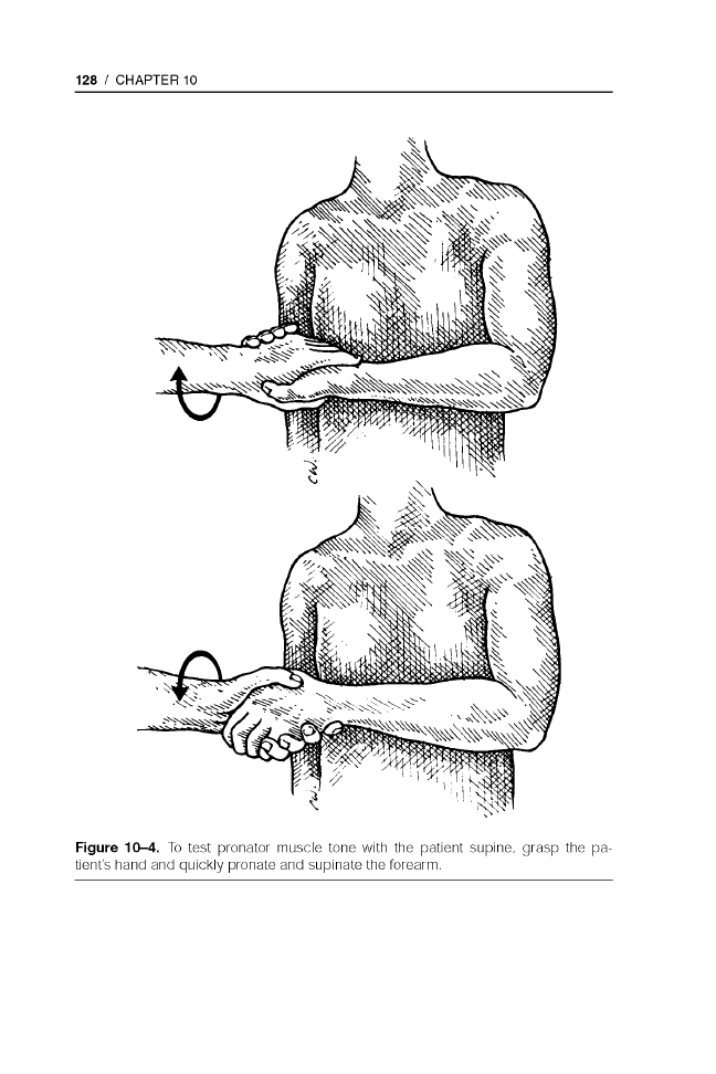
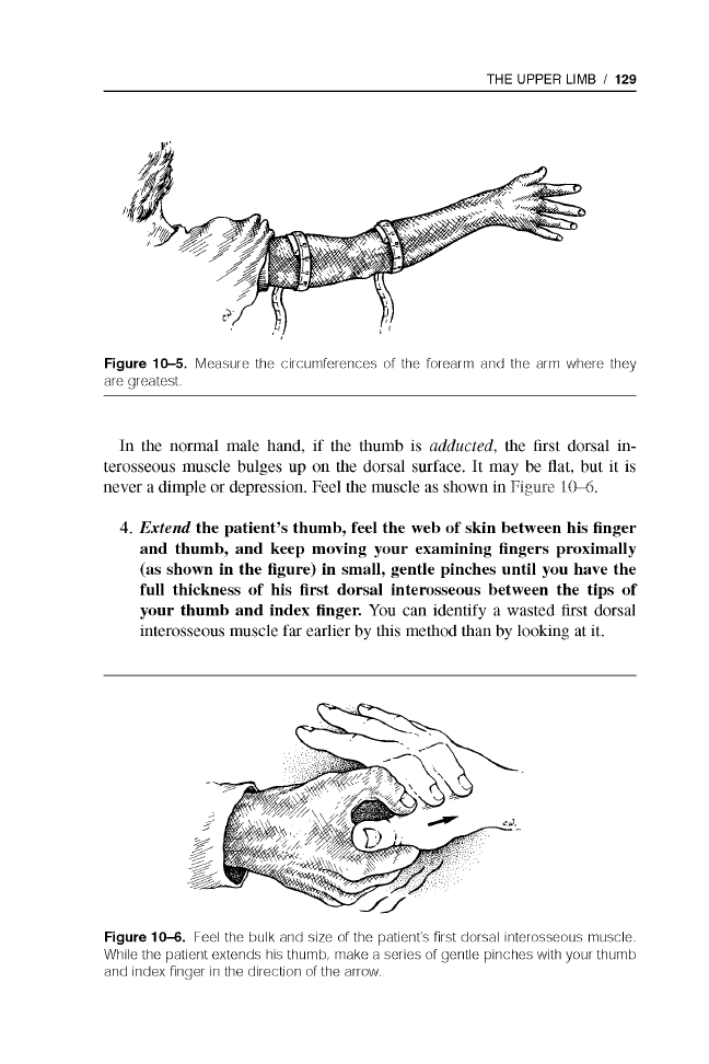

130 / CHAPTER 10
Upper limb size can be assessed when testing arm drift (
Figure 10–1
).
Look down the length of the patient’s arms from the fingertips toward the
chest. Small differences in arm size will be apparent.
EXAMINATION
The following are the minimal number of muscles you must examine.
With practice you can do them all and compare right and left in minutes. The
headings are by function rather than by the names of individual muscles.
Abduction of the Fingers
Dorsal Interossei: Ulnar Nerve, T1
With the palm of the patient’s hand on a flat surface, ask him
to spread his fingers against resistance.
The first dorsal interosseous (see step 4 of the previous section and
Figure
10–6
) is easy to see and feel as the index finger is abducted. The abductor of
the little finger is just as distinctive on the ulnar border of the hand.
Abduction of the Thumb
Abductor Pollicis Longus: Radial Nerve, C7; Abductor Pollicis Brevis: Me-
dian Nerve, T1
Put the dorsum of the patient’s hand on a flat surface. Ask
him to raise the thumb vertically. The surface of the pad of
the thumb is at right angles to the surface of the palm of the
hand.
The brevis is part of the thenar eminence, and the tendon of the longus
can be seen and felt on the lateral side of the wrist.
Opposition of the Thumb
Opponens Pollicis: Median Nerve T1
As shown in
Figure 10–7
, ask the patient to push the pad of
his thumb against the pad of his little finger. Try to separate
them with your index finger, moving in the direction of the
arrow shown in the figure.
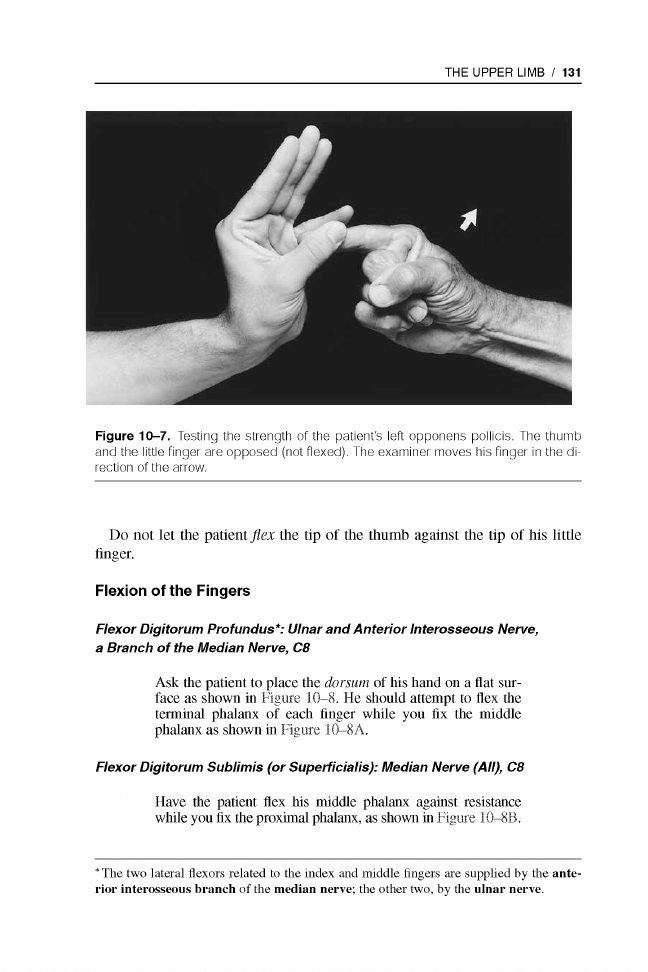
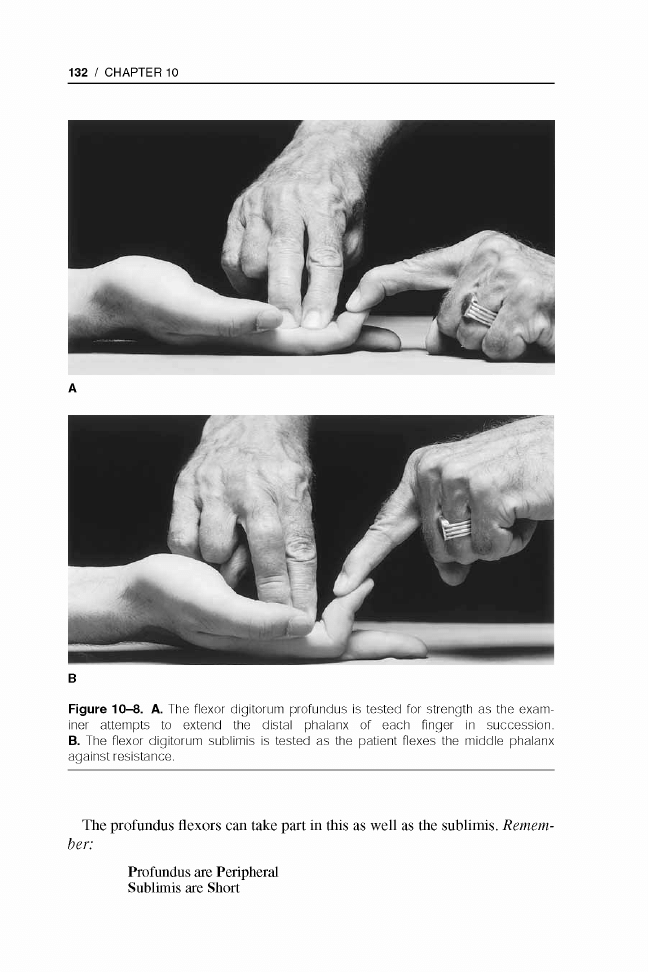

THE UPPER LIMB / 133
Flexion of the Wrist
Flexor Carpi Ulnaris: Ulnar Nerve, C8; Flexor Carpi Radialis: Median
Nerve, C8
Ask the patient to put the dorsum of his hand and forearm on
a flat surface and flex his wrist against resistance as shown
in
Figure 10–9
.
With wrist flexion plus a little abduction the tendon of the
flexor carpi radialis in the midline of the wrist is seen (arrow
in
Figure 10–9A
), while with flexion plus a little adduction
the flexor carpi ulnaris becomes palpable on the most medial
aspect of the wrist (
Figure 10–9B
).
The tendon of the flexor carpi ulnaris can also be seen and
felt on the medial wrist by abducting the little finger.
Flexion of the Forearm
Biceps: Musculocutaneous Nerve, C5 and C6; Brachioradialis: Radial
Nerve, C6
To test the biceps, have the patient’s forearm and hand on a
flat surface with the dorsal aspect of the limb touching the
surface. Fix his elbow and ask him to flex his forearm
against resistance.
To test the brachioradialis, start from the same position ex-
cept that the forearm is halfway between supine and prone;
that is, the ulnar edge of the hand and forearm is resting on a
flat surface and the thumb is uppermost. Ask the patient to
flex his forearm against resistance from this position.
These two powerful forearm flexors have the same root supply and differ-
ent nerve supply.
Flexion of the Thumb
Flexor Pollicis Longus: Anterior Interosseous Nerve, a Branch
of the Median Nerve, C8
Hold the proximal phalanx of the patient’s thumb between
your finger and thumb. This provides a fixed base. Then
have the patient flex the distal phalanx against resistance.
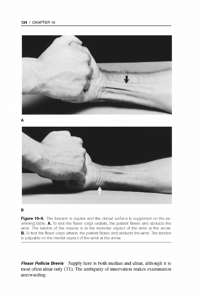
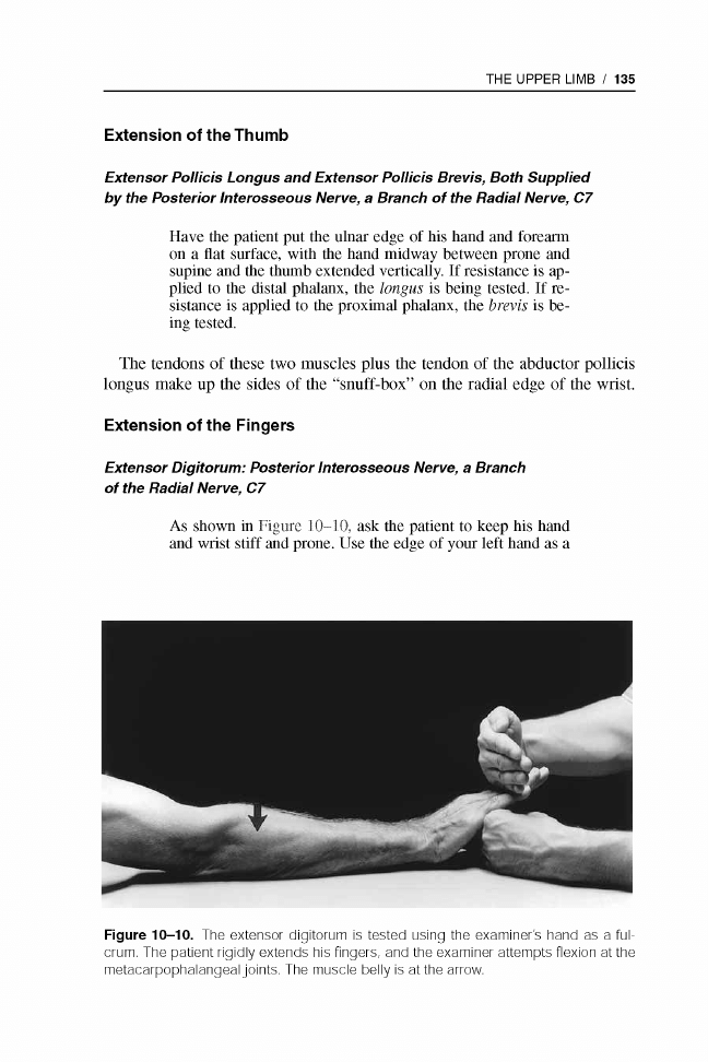

136 / CHAPTER 10
fulcrum in the middle of the patient’s palm. Attempt to flex
the patient’s fingers with your right hand.
The extensors function at the metacarpal phalangeal joints. Extension at
the more distal interphalangeal joints is performed by the interossei and lum-
bricales.
Extension of the Wrist
Extensor Carpi Radialis (Longus and Brevis): Radial Nerve, C7 and C8;
Extensor Carpi Ulnaris: Posterior Interosseous Nerve, a Branch
of the Radial Nerve, C7 and C8
The radialis is tested as in
Figure 10–11A
. Place the volar
aspect of the forearm on any flat surface. Allow some flex-
ion of the fingers at the interphalangeal and metacarpopha-
langeal joints in order to get the finger extensors relaxed and
out of the way. Then ask the patient to forcefully extend and
abduct the hand while you supply resistance to the dorsum
of the hand. The ulnaris is examined by having the patient
extend and adduct his hand (
Figure 10–11B
).
Extension of the Forearm
Triceps: Radial Nerve, C7
Allow the patient to flex his elbow to form about a 90-de-
gree angle at the elbow and then straighten his arm against
resistance.
Forearm extension is never as strong as forearm flexion. If you want to put
the triceps in its most disadvantageous position and elicit small amounts of
weakness, ask the patient to begin forearm extension from a position of max-
imum elbow flexion.
Abduction of the Arm
Supraspinatus: Suprascapular Nerve, C5; Deltoid: Axillary Nerve, C5
These two muscles work together but neither can do the job of the other.
The supraspinatus starts abduction.
Test the supraspinatus by standing behind the patient. He
should let his arms hang by his sides. Hold his elbow into
the side of his trunk while he attempts to abduct the arm.
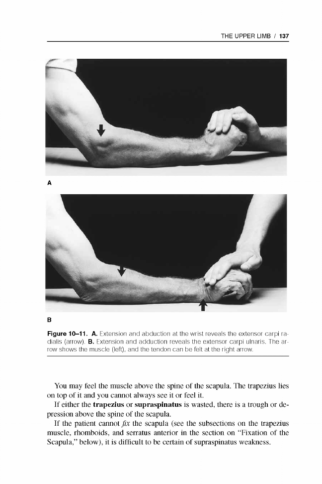

138 / CHAPTER 10
Test the deltoids by resisting abduction after the arm has
been abducted 45 degrees and more away from the trunk.
The deltoid is working most effectively when the arm is horizontal. The
muscle is easily seen and felt. When it is wasted, the shoulder is “square”
when looking at it from in front or behind. This is the principal abductor of
the arm.
Adduction of the Arm
Latissimus Dorsi: Thoracodorsal Nerve, C7
Have the patient hold his arm at shoulder height straight out
to one side, parallel to the floor. Ask him to put his hand on
your shoulder and push down toward the floor.
Examine the anterior and posterior axillary walls. The lower portion of the
posterior wall of the axilla is the latissimus dorsi.
You may also test it by holding it between your finger and
thumb while the patient has his arms at his side. Then ask
him to cough.
You will feel the edge of the latissimi contract between your fingers.
Teres Major: Subscapular Nerve, C7
Start with the patient’s arm in the same
position as above (arm parallel to the floor at shoulder height). You can see
this muscle only in the upper posterior axillary wall in thin people. It is off
the lateral edge of the scapula and posterior to the upper end of the latissimus.
Pectoralis Major: Clavicular Part—Lateral Pectoral Nerve, C5; Sternal Part—
Lateral and Medial Pectoral Nerve, C7, C8, and T1
Ask the patient to put one fist against the other and push as
in
Figure 10–12.
Fixation of the Scapula
A wing scapula and resulting weak shoulder are common complaints. The
three principal muscles concerned with fixation and movement of the
scapula have overlapping functions.
Trapezius Muscle: Spinal Accessory Nerve, C3 and C4
The trapezius pulls
the scapula upward. This was demonstrated in the “shoulder shrug” when
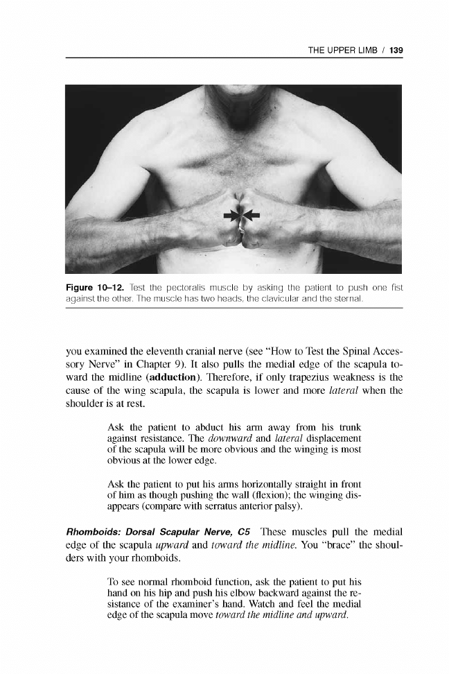

140 / CHAPTER 10
In rhomboid weakness this action produces winging, and the medial edge
of the scapula moves laterally and downward.
Serratus Anterior: Long Thoracic Nerve, C5 to C7
This muscle pulls the
scapula away from the midline and also forward.
When the serratus is weak, inspection of the back at rest shows the infe-
rior angle of the scapula is slightly winged and the medial edge is pulled to-
ward the midline.
Abduction of the arm against resistance reduces the wing-
ing. Pushing the horizontally flexed arm against the wall in-
creases the winging, and the inferior angle of the scapula is
lifted off the chest wall.
Remember: The best test to identify scapula winging resulting from
trapezius weakness is abduction of the arm against resistance; to test that
caused by serratus anterior weakness, use flexion (with the arm horizontal,
pushing forward against the wall).
UPPER LIMB ABNORMALITIES
The upper limb is of about the same importance in neurological assess-
ment as eye movements and vision. Diseases of the upper motor neuron,
parietal lobe, cerebellum, and basal ganglia may each produce characteristic
changes in the tone, power, posture, or function of the limb and are dealt
with elsewhere. Diseases of the spinal cord, nerve roots, brachial plexus, and
individual nerves present their own distinctive problems. Herewith are a few
comments on plexus and peripheral nerve lesions.
Brachial Plexus
Brachial plexus most commonly involves lesions caused by trauma (traction
injury to the arm, knife or ice pick wounds, or bullet penetration), metastatic
carcinoma from the breast or lung, or delayed radiation damage to the plexus
from treatment of the latter. Other causes are birth injuries, supraclavicular
pressure during anesthesia, intravenous drug abuse, serum and vaccination re-
actions, and paralytic brachial neuritis of unknown cause (possibly virus).
1. Upper trunk lesions (eg, Duchenne-Erb syndrome); usually a result of
perforating injuries
• Think of functions of the C5–C6 roots.
• There is weakness of the supraspinatus, infraspinatus, deltoid, biceps,
brachialis, brachioradialis, supinator, and extensor carpi longus and
brevis.

THE UPPER LIMB / 141
• The lateral aspect of the arm is numb.
• Biceps and supinator reflexes are absent.
• The rhomboids and serratus anterior are usually normal (as their
nerves—dorsal scapular and long thoracic, respectively—arise proxi-
mal to the common site of injury).
2. Lower trunk lesions (eg, Klumpke syndrome); common causes are
cervical rib, carcinoma of the lung apex, and metastatic carcinoma in
the axillary lymph nodes, as well as traction on the abducted arm
• Think of functions of the C8 and T1 roots.
• There is weakness of the flexor carpi ulnaris, flexor digitorum, in-
terossei, and thenar and hypothenar muscles; the hand is flat and
simian.
• The ulnar edge of the arm, forearm, and hand is numb.
• The triceps reflex is absent.
• Horner’s syndrome is commonly present.
3. Lateral cord lesions
• Think of functions of the musculocutaneous nerve and the lateral part
of the median nerve.
• There is weakness of the biceps, brachialis, coracobrachialis, and
pronator teres.
• Intrinsic hand muscles are normal (the medial part of the median
nerve).
• The radial edge of the forearm may be numb.
• The biceps reflex is absent.
4. Medial cord lesions
• Think of functions of the ulnar nerve and the medial part of the me-
dian nerve.
• There is weakness of the flexor carpi ulnaris, all flexor digitorum pro-
fundus, all interossei, all lumbricales, and all thenar and hypothenar
muscles.
• There is numbness, usually in the ulnar distribution.
5. Posterior cord lesions
• Think of functions of the radial and axillary nerves.
• There is weakness of the deltoid, teres minor, brachioradialis and
supinator, triceps, extensor carpi (all three), extensor digitorum mus-
cles, and long abductor pollicis.
• There is numbness in the upper outer aspect of the arm just below the
deltoid muscle.
6. Radial nerve lesions; the nerve is derived from C6 to C8 (mostly C7)
• These are caused by the pressure of a crutch, a gunshot, stab wounds,
fracture of the humerus, and on the medial aspect of the lower arm
from pressure during sleep while intoxicated or sedated (“Saturday
night palsy”).

142 / CHAPTER 10
• A lesion in the axilla causes weakness of the triceps, brachioradialis, the
three extensor carpi, and finger extensors; the triceps reflex is absent.
• The area of numbness (if any) is about a 3-cm circular area overlying
the first dorsal interosseous muscle; patients rarely have sensory
symptoms.
• The apparent weakness of finger flexion in the “wrist-dropped” hand
is not real; if the wrist is passively extended, the flexors are found to
be normal.
• Lesions at the midhumerus usually result in a normal triceps muscle.
• Lesions of the posterior interosseous nerve (the final branch of the ra-
dial nerve) produce weakness of the extensors of the wrist, index fin-
ger, and thumb and are usually caused by entrapment of the nerve
where it perforates the supinator muscle.
7. Median nerve lesions; the nerve is derived from C5 to T1 (mostly C6)
• These lesions are caused by a dislocated shoulder, stab or gunshot
wounds to the proximal nerve, and, more commonly, distal compres-
sion in the carpal tunnel at the wrist as part of gout, amyloidosis,
acromegaly, arthritis, or an occupational hazard.
• Lesions in the arm cause loss of pronation of the forearm, wrist flexion
to the radial side, paralysis of flexion of the thumb and index finger,
weakness of flexion of all the other fingers (flexor digitorum sublimis),
and paralysis of opposition of the thumb with sensory disturbance
over the lateral two thirds of the volar surface of the hand.
Lesions at the wrist cause thenar wasting and weakness with the character-
istic sensory loss. The common early complaint is a painful, numb hand that
disturbs sleep. Symptoms are relieved by shaking the hand. Pain and numb-
ness are often said to be in all fingers and pain is often proximal to the wrist
well up into the forearm and arm. While this cannot be explained anatomi-
cally, it is heard so often from so many well-motivated patients that it cannot
be denied. Patients always have sensory symptoms.
8. Ulnar nerve lesions; the nerve is derived from C8 and T1
• These are caused by perforating wounds, fractures of the lower end of
the humerus, and fractures and dislocation of the olecranon or head of
the radius. Symptoms may occur long after an elbow injury. Appar-
ently, minor pressure on the nerve at the elbow as an occupational
hazard or in patients confined to full bed rest will produce an ulnar
palsy.
• Repeated trauma to the heel of the hand on the ulnar side will trauma-
tize the nerve. The nerve divides into superficial and deep branches in
the hand, and the signs of a lesion here are usually entirely motor.

THE UPPER LIMB / 143
• Lesions at the elbow usually do not affect the flexor carpi ulnaris or
flexor digitorum profundus 3 and 4, but there is weakness of the ab-
ductors and adductors of the fingers, the adductor of the thumb, and
lumbricales 3 and 4, and the interossei (particularly the first dorsal)
and hypothenar muscles are wasted.
• A complete lesion reveals: half a claw hand with small muscle wast-
ing, the fifth finger held away from the fourth finger, hyperextension
at the metacarpophalangeal joints, and flexion at the interphalangeal
joints, most marked (the flexion) in the fourth and fifth fingers.
• Sensory disturbance is on the ulnar edge of all of the fifth finger, the
ulnar edge or tip of the fourth finger, and the ulnar edge of the palm.
Sensory symptoms are never proximal to the wrist (see below for dif-
ferentiation of a T1 lesion from an ulnar nerve lesion). Patients with
ulnar nerve lesions at the elbow always have sensory symptoms.
Froment’s sign of ulnar palsy can also be a useful indicator. When the pa-
tient holds a piece of paper between her thumb and index finger (ie, pinches
the piece of paper and pulls), in ulnar palsy the terminal phalanx of the
thumb will flex. The adductor pollicis is paretic, and the flexor pollicis
longus (median nerve) is functioning in its place.
How to tell the difference between
1. A C5 root lesion and an axillary nerve lesion: A C5 lesion abolishes
all abduction of the arm (deltoid and supraspinatus) and causes some
weakness of the clavicular head of the pectoralis major, biceps, and
brachioradialis muscles. An axillary nerve lesion abolishes only the
later part of abduction of the arm (deltoid).
2. A C6 root lesion and a musculocutaneous or radial nerve lesion:
• A C6 root lesion will cause weakness of the biceps, brachialis, and
brachioradialis (no forearm flexion) and the extensor carpi radialis.
• A musculocutaneous root lesion will weaken the biceps and brachialis
only. Forearm flexion is carried out by the brachioradialis when the
forearm is half supinated. The extensor carpi radialis is normal.
• A radial nerve lesion will weaken the brachioradialis and extensor
carpi radialis. The biceps and brachialis will flex the supine forearm.
3. A C7 root lesion and a radial nerve lesion:
• A C7 root lesion will cause weakness of arm adduction (latissimus
dorsi), forearm extension (triceps), hand extension (extensor carpi ul-
naris), finger extension (extensor digitorum), thumb abduction and
extension (abductor pollicis longus and extensor pollicis longus and
brevis), and hand flexion (flexor carpi radialis).
• A radial nerve lesion will produce all of the above except that there
will be no weakness of the latissimus (thoracodorsal nerve) or of the

144 / CHAPTER 10
flexor carpi radialis (median nerve). In addition, the brachioradialis
will be paretic.
4. A T1 root lesion and an ulnar nerve lesion:
• A T1 root lesion will cause weakness and wasting of all the small
muscles of the hand.
• An ulnar nerve lesion will produce a wasted hand, but the 4
1
/
2
mus-
cles supplied by the first thoracic root and the median nerve will be
preserved. These are the abductor pollicis brevis, opponens pollicis,
first and second lumbricales, and half the flexor pollicis brevis (which
may be supplied entirely by the ulnar nerve).

The Lower Limb
1 1
The best screening test of lower limb function is watching the patient
walk. Gait and its disorders are discussed in detail in Chapter 12. Go to the
waiting area yourself to get your patient, and watch him get out of the chair
and walk with you to the examining room. Then watch him sit down.
The leg is between the knee and the ankle; the thigh is between the hip
and the knee. The detailed examination of the lower limb is subdivided un-
der the headings of coordination, size, tone, and power.
COORDINATION
All of the tests of coordination are better lateralizers, but are less helpful
when the legs are equally abnormal.
When the patient is not sure whether one or both legs are abnormal and his
history is rather vague—for example, “I cannot walk as well as before” or “I
seem to stumble and fall down a lot”—the results of the following tests will
be abnormal if the problem is weakness, spasticity, sensory loss, or a cere-
bellar disorder.
Supine Leg Raising
With the patient supine on the examining table, ask her to lift her leg, with
the knee extended, off the table (thigh flexion) as high as she can and then
slowly put it down. Have her do this several times with one leg. Then ask
her to repeat the movement with the other leg.
If one leg is weak or spastic or has an involuntary movement or a proprio-
ceptive sensory defect, this simple leg-raising test will help you identify it. It
is a useful lateralizer, if not a localizer, and is similar to, but less sensitive
than, arm drift.
Rapid Alternating Movements
1. With the patient supine, ask him to tap the heel of one foot on the mid-
dle of the tibia of the opposite leg, as in
Figure 11–1A
. The tapping
foot should rise 30 cm each time. Watch the rhythm, rate, and regularity
145
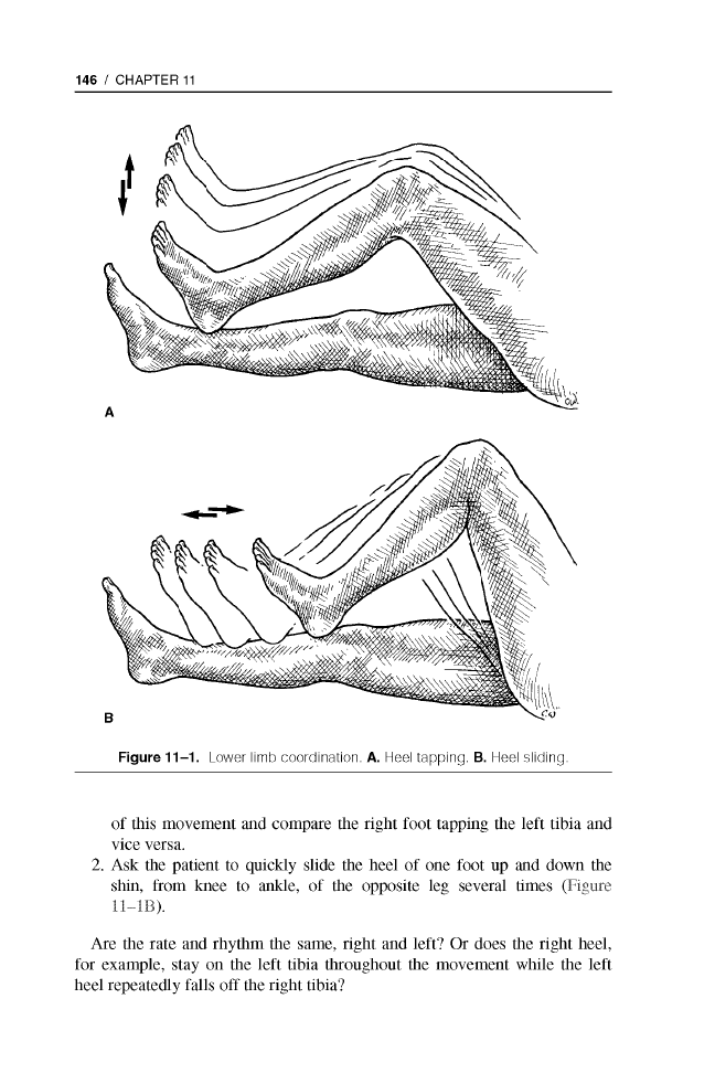

THE LOWER LIMB / 147
3. Stand at the foot of the examining table. Put the palm of your hand flat
against the sole of the patient’s foot, and ask him to tap your hand (ie,
move your hand away about 2–3 cm) with his foot repetitively and as
quickly as he can. You can do the same thing with the patient sitting by
asking him to tap the floor with his foot.
SIZE
Look at the patient’s bare legs while he is standing and you are squatting
about 2 m from him. Look at him front and back while he stands flat-footed,
on his toes, on his heels, and with his kneecaps drawn up (ie, standing at at-
tention).
When you are looking at the back of his legs and he goes up on his toes, a
minor amount of calf muscle wasting in one leg will become obvious.
Then, on the examining table, again in the supine position, measure the
circumference of the thigh and calf of each leg and write the measurements
in the notes.
Measure the leg at its greatest circumference, which is at about the junc-
tion of the upper and middle thirds. Do not measure the circumference at an
arbitrary distance below the lower edge of the patella. The greatest circum-
ference of the thigh is usually at the top of the thigh. If you always measure
the leg and thigh at their greatest circumference, there will be no confusion
on follow-up examination 6 months or 1 year later.
TONE
Reread the remarks on tone in Chapter 10 on the upper limb and in Chap-
ter 16 on the corticospinal system. When the tone in the legs is increased,
watching the patient walk is most revealing.
The bed-bound patient and the patient with slightly increased or decreased
tone is more difficult to assess.
At times, older patients have an involuntary, normal inability to let the
legs relax. Their legs are stiff. They have normal tendon reflexes, plantar re-
sponses are down, there is no clonus, and they do not walk with a spastic
gait. You may implore such a patient to “just relax” without any change in
this resistance of his lower limbs. This phenomenon, called gegenhalten,
precludes assessment of lower limb tone.
Assess the tone of the lower limbs as follows:
1. With the patient supine and his legs straight on the examining
table, put your hand in his popliteal fossa. Quickly lift your hand

148 / CHAPTER 11
toward the ceiling, that is, passively flex the knee. If the quadriceps
has normal tone, the heel will usually stay on the examining table as
the knee goes up. If the quadriceps is spastic, the heel will lift off the
table.
2. With the patient supine on the table, flex the right hip to a right an-
gle. Hold the thigh in this position with your left hand. Flex the
knee to a right angle. The leg is now parallel to the table. Hold the
patient’s heel in your right hand. Wait a few seconds until he re-
laxes, then drop the foot while you continue to support the thigh. If
the quadriceps tone is normal, the foot will drop and usually stop just
before it hits the table. If the patient is fully relaxed, it will hit the table.
If the tone is increased, the foot will drop in a jerky, cogwheel way and
never reach the table.
3. Flex the hip and knee each to about 45 degrees. Externally rotate
the hip so that the lateral aspect of the lower limb is resting on the
examining table. Rapidly dorsiflex the foot. If the tone is increased
in the posterior calf muscles, you will feel the resistance and you
may invoke clonus (see Chapter 13).
POWER
The examination of lower limb power is described with the patient supine
and then prone on the examining table. There is also a quick assessment of
leg strength with the patient on her feet.
The lower limb muscles are so powerful that minor weakness will not be
detectable unless you put the muscle in its most disadvantageous position.
For example, when you test the tibialis anterior with the patient supine on
the table, ask her to actively bend her foot at the ankle and keep her toes up
toward her chin while you try to forcefully plantar flex the foot. All you do is
slide her down the examining table—you cannot overcome her dorsiflexion.
Now have the patient stand up, hold onto a chairback if she needs support,
and stand on her heels, first with one leg, then the other. The toes of the
foot that she is complaining about come off the floor (if at all) by 2 cm. The
toes of the normal foot clear the floor by 6 cm.
You now have objective evidence of weakness of dorsiflexion at that an-
kle. You could not have found this with only the examination on the table.
The order of muscle testing is not anatomical. Start with the iliopsoas and
then examine the muscles, from above downward, with the patient supine
and then with the patient prone, examined from below upward.
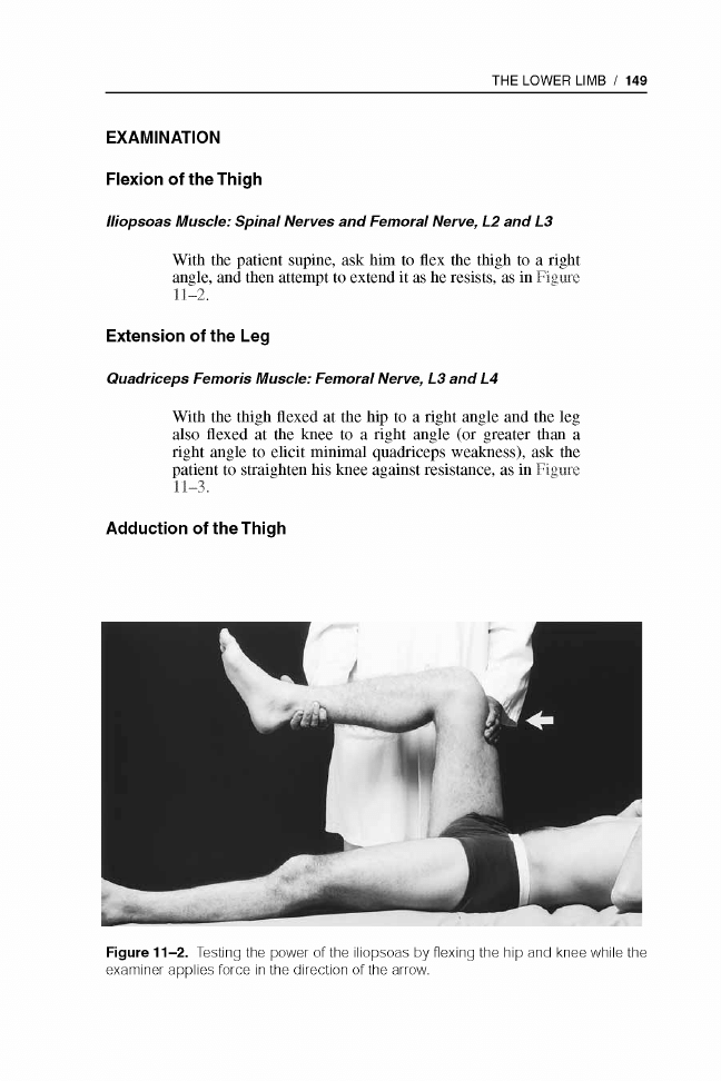
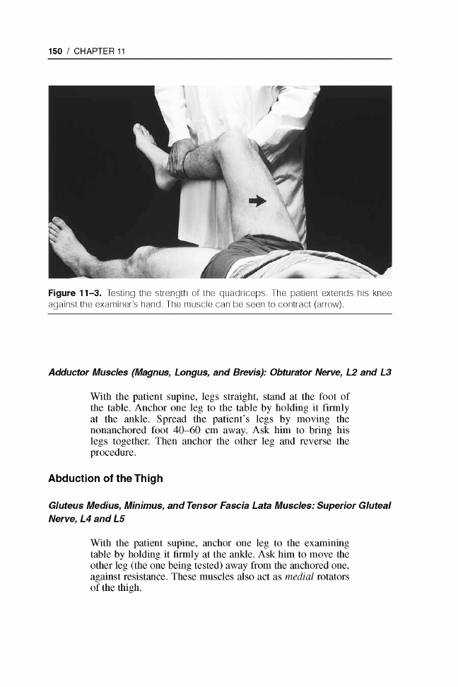
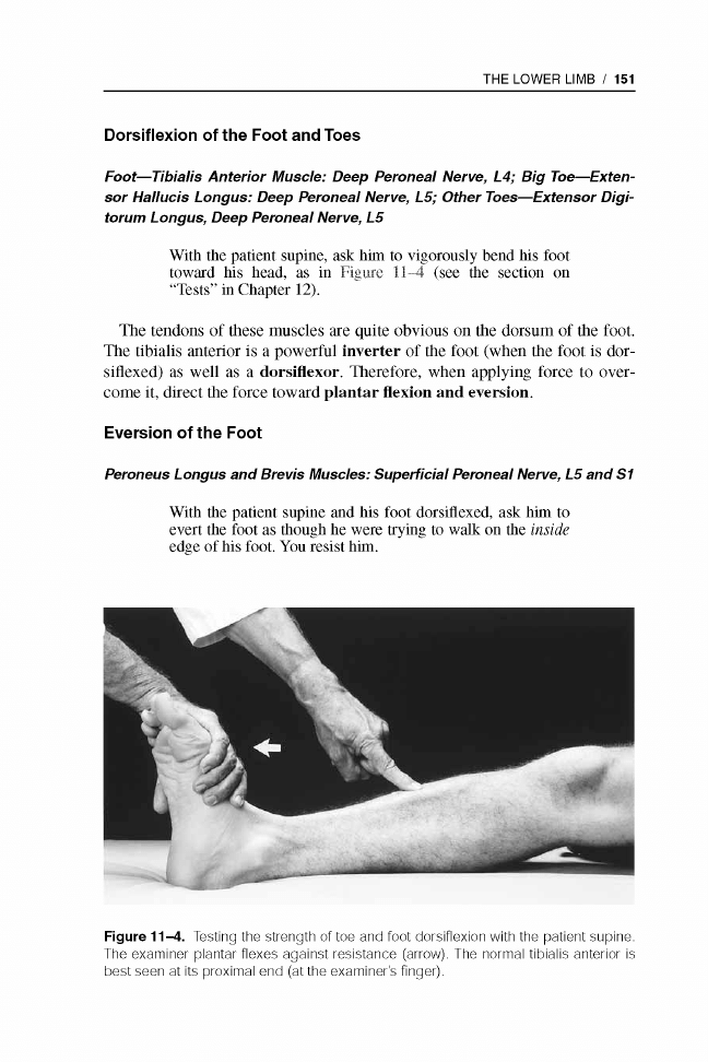

152 / CHAPTER 11
The tendons of the two muscles are seen inferior to the lateral malleolus.
These muscles also function as plantar flexors.
Inversion of the Foot
Tibialis Posterior Muscle: Tibial Nerve, L4 and L5
With the patient supine and his foot dorsiflexed, ask him to
invert his foot as though walking on the outside edge of his
foot. You resist this movement.
This muscle also acts as a plantar flexor.
Plantar Flexion of the Toes
Flexor Digitorum Longus and Flexor Hallucis Longus Muscles: Posterior
Tibial Nerve, S1 and S2
Ask the patient to curl his toes downward (plantar flexion)
while you resist the movement by pushing upward on the
terminal phalanges.
The patient now turns over, and the rest of the examination is conducted in
the prone position.
Plantar Flexion of the Foot
Gastrocnemius and Soleus Muscles: Tibial Nerve, S1 and S2
Gastrocnemius—With the patient prone and the knee ex-
tended, have him attempt to plantar flex his foot against re-
sistance.
This muscle also functions as a flexor of the knee.
Soleus—Have the patient do as above, except that the knee
must be flexed. Watch and feel the calf muscles while resist-
ing plantar flexion.
Small amounts of weakness of these two muscles may be evident only if
the patient is asked to stand on tiptoes, one foot at a time.
Remember, the peroneus (both), the tibialis posterior, and the plantar flex-
ors of the toes assist plantar flexion of the foot.

THE LOWER LIMB / 153
As you develop your routine of neurological examination, you may find
that immediately after testing the soleus is the best time to elicit the ankle re-
flexes (see Chapter 13).
Leg Flexion at the Knee
Hamstring Muscles (Biceps, Semitendinous, and Semimembranous): Sci-
atic Nerve, L5 and S1
With the patient prone, with the knee at a right angle, resist
further flexion as the patient attempts to move his heel to-
ward his buttock.
The tendons form the boundaries of the popliteal fossa, the biceps laterally
and the semitendinous medially. These muscles also act as extensors of the
thigh at the hip.
Thigh Extension at the Hip
Gluteus Maximus Muscle: Inferior Gluteal Nerve, L5 and S1
With the patient prone and the knee extended, ask him to
lift the straight leg off the examining table while you press
down on the lower posterior thigh (with a weak gluteus
maximus the patient will manage to get the leg extended
by rotating the pelvis). Make sure he keeps his pelvis on
the examining table. You can see and feel the buttock
tighten.
This muscle also acts as a lateral rotator of the thigh.
Assessment of Leg Strength When the Patient Is on His Feet
• Can he bring the leg forward as he walks?—iliopsoas
• Can he get out of a full knee bend without pulling himself up with his
arm?—quadriceps
• Can he get out of a chair without help from his arm?—quadriceps (Parkin-
sonian rigidity can mimic this.)
• Can he walk on his toes?—gastrocnemius and soleus
• Can he walk on his heels?—anterior tibial
• Does he abduct the leg from the hip and swing it outward and forward to
bring the limb ahead instead of the normal thigh flexion (ie, knee drop)?—
all thigh flexors

154 / CHAPTER 11
• Does he lift one foot much higher from the floor than the other with each
step (ie, a foot drop)?—anterior tibial and other muscles that are secondar-
ily dorsiflexors
SIGNS AND SYMPTOMS OF SINGLE ROOT LESION
• Second lumbar—Hip flexion weakness is usually the only defect on exam-
ination. Pain is in the upper anterior thigh.
• Third lumbar—Hip flexion, knee extension, and thigh adduction are all
weak. Pain is in the upper anterior thigh.
• Fourth lumbar—Symptoms of weakness of foot inversion (anterior and
posterior tibial muscles) and absent knee reflex. Pain and sensory loss are
evident on the medial leg above the malleolus.
• Fifth lumbar—There is weakness of dorsiflexion of the toes, particularly
the big toe. Sensory disturbance occurs on the dorsum of the foot; pain is
also here and on lateral calf.
• First sacral—Symptoms include weakness of eversion of the foot (per-
oneii) although innervation is not exclusively first sacral, weakness of
plantar flexion of the foot (gastrocnemius and soleus), weakness of leg
flexion (hamstring), absent ankle reflex, numbness on the lateral edge of
the foot, and pain here and on the back of the calf.
SIGNS AND SYMPTOMS OF NERVE LESIONS
• Lateral cutaneous nerve of the thigh (from L2 and L3)—Painful paresthe-
sias—burning, tingling discomfort in the anterolateral aspect of the
thigh—is the only symptom of a disease of this nerve, called meralgia
paresthetica. Symptoms are caused by entrapment or stretching of the
nerve under the lateral aspect of the inguinal ligament. The disease is com-
mon in people who are gaining or losing weight, during or after pregnancy.
Symptoms are often related to one posture only, such as sitting or standing.
Examination reveals hyperesthesia or, rarely, hypoesthesia in the anterior
lateral thigh. The thigh is strong, the knee reflex is normal, and disease is
benign.
• Obturator nerve (from L2, L3, and L4)—Lesions produce weakness of ad-
duction of the thigh and pain on the medial aspect of the thigh to the knee.
The nerve may be injured during delivery or labor or may be involved in
pelvic neoplasm.
• Femoral nerve (from L2, L3, and L4)—Lesions produce a wasted quadri-
ceps, weakness of leg extension, and, if the lesion is proximal, weakness of

THE LOWER LIMB / 155
thigh flexion (iliopsoas muscle) as well. The knee reflex is absent, and sen-
sory loss extends from the anteromedial thigh to the medial malleolus. Di-
abetes is the most common cause of femoral neuropathy, although pelvic
tumors, femoral hernia, and femoral artery aneurysms are also possible
causes. A retroperitoneal hematoma may compress the nerve, and drainage
of the hematoma is an emergency if the nerve is to be saved.
• Sciatic nerve (from L4 and L5 and S1 and S2)—The lesion will cause loss
of knee flexion (hamstrings) and no movement of any muscle below the
knee. Sensory loss will occur in all of the sole of the foot, the dorsum of
the foot, and the posterior and lateral leg. Causes are pelvic fractures; pen-
etrating injuries, including misplaced injections; and pelvic tumors. Lying
flat on a hard floor while in coma from any cause can produce a compres-
sion sciatic palsy.
• Peroneal nerve (the most common peripheral nerve lesion in the lower
limb that you will see)––The sciatic nerve divides into the tibial nerve and
the common peroneal nerve. The latter winds around the neck of the fibula,
a common site of lesions of this nerve. Diabetes, sitting with the legs
crossed for long periods, trauma, and sporting injuries (eg, in professional
figure skaters or skiers) as well as tightly applied plaster casts account for
most of the identifiable causes. There is weakness of dorsiflexion of the
toes and foot as well as eversion. The ankle reflex is normal, and although
the patient may say he has numbness over the dorsum of the foot, there are
usually no sensory abnormalities.
• Posterior tibial nerve (lesions are uncommon)––The nerve may be com-
promised in its tunnel on the inferomedial aspect of the calcaneus. Pain
and paresthesias over the sole of the foot in response to exercise are the
only complaints. There are no motor findings.

Stance, Gait, and Balance
1 2
Walking is an example of the superb integrative action of the nervous sys-
tem. It is not a learned series of consecutive motor acts but appears as an in-
nate, gestalt phenomenon when other motor skills are still quite primitive,
for example, a child may learn to walk at an age when he cannot yet feed
himself.
If walking has an anatomical “center,” the location is unknown. (Both the
cat and the dog can walk after bilateral hemispherectomy.) Walking can be
lost, and this may not be explained in terms of a discrete lesion. This is
called gait apraxia and is often associated with dementia and frontal lobe
signs. However, many demented people have normal gait, and gait apraxia
occurs with normal intelligence.
Watch people without gait disorders walk from the front, back, and side.
Considering that the entire weight of the moving body can be continu-
ously transferred from one foot to the other, every three quarters of a second,
in a perfectly rhythmic way, without conscious effort, and even while we are
preoccupied with some other matter, then walking must be one of the great
functions of the nervous system.
When one steps out with the right foot first, three things happen almost si-
multaneously to the right lower limb. The right hip flexes (the knee comes
away from the floor), the right knee flexes (the foot comes away from the
floor), and the right ankle plantar flexes in every step except the first one. All
the weight is on the left lower limb and, as the right leg comes forward and
passes the left, there is more right hip flexion and the beginning of right knee
extension and right ankle dorsiflexion in preparation for the transfer of
weight to the right heel and foot. While this swing-through of the right leg is
progressing, and starting just after the right leg passes the left one, the left
ankle plantar flexes in preparation for the push it is about to give to the floor.
Pay particular attention to the foot and ankle movements. The right heel
strikes the floor with the ankle dorsiflexed and the weight of the body on the
left foot. As the body comes forward, the right foot flexes and the right lower
limb, with the knee extended, takes the weight of the body. The left foot
plantar flexes and pushes off against the floor.
The arms reciprocate with the legs. As the left foot comes forward, so
does the right upper limb.
157

158 / CHAPTER 12
Ask the patient to get out of the chair, walk away from you
(6–7 m or so), turn quickly to his left, and without a pause
walk back toward you. When he reaches you, have him turn
quickly to his right and repeat the process.
The quick turn may evoke an ataxia not seen otherwise.
TESTS
If ordinary walking is normal, add a number of tests that may elicit an ab-
normality.
Have the patient walk a straight line, heel to toe. He should
put the right heel immediately in front of the left toes and
then the left heel immediately in front of the right toes and
so on.
Most people can walk this way across the examining room. If the patient
cannot, this itself is not a localizing sign, but it does tell you that something
is wrong.
• Have the patient walk on his tiptoes forward across the examining room.
• Ask him to walk on his heels backward across the examining room.
• Ask him to hop across the room on one foot and then the other.
Romberg Test
The Romberg test is named for Moritz Heinrich Romberg (1795–1873).
Tradition and your examiners demand that you be familiar with the Romberg
test. However, it is not a useful or specific way of evaluating ataxia or decid-
ing whether an ataxia is peripheral (sensory) or central (eg, cerebellum).
The basis of the test is the fact that a person with a defect in balance often
replaces the function of the diseased structure by use of his eyes. If the pa-
tient has an ataxic gait because of posterior column disease, peripheral neuri-
tis, or defective sensation for any reason, he may maintain a reasonably good
gait provided that he can see the horizon, the walls of the room, or some
landmark. The ataxia is more obvious if he closes his eyes or if he is com-
pelled to walk in the dark. The Romberg test is conducted in the following
manner:
The patient is asked to stand with his feet as close together
as possible while feeling comfortable and stable. He is then
asked to close his eyes. If he loses his balance, the Romberg
test is said to be positive.

STANCE, GAIT, AND BALANCE / 159
The tottering back and forth that goes on when a person closes his eyes is
not a positive Romberg. These small, normal movements occur because the
patient feels himself leaning toward one side and in correcting it, often over-
corrects it, then leans back a little too far the other way, and so forth. When
you see this happening, the patient is obviously very much aware of his posi-
tion in space and has excellent balance. Often, if you say to the patient
“Stand perfectly still,” the swaying will stop. The patient with a true positive
Romberg, on the other hand, is not aware of the beginning of a deviation of
his posture and falls over. He makes no attempt to correct it. If he keeps his
eyes open, he can maintain a posture; if he closes his eyes, he cannot.
The positive Romberg has been said to be indicative of a sensory, or affer-
ent, type of ataxia as opposed to a central, or cerebellar, type of ataxia. The
conclusion is not valid. Patients with all kinds of ataxia and with lesions in
many different parts of the nervous system will state that they walk reason-
ably well indoors, close to walls, in familiar situations. They are much worse
out on the street and cannot walk at all in the dark. Therefore, we cannot use
the Romberg test to suggest that any given ataxic patient has a sensory lesion
as opposed to a central cerebellar lesion.
When a patient complains of being unsteady or stumbling, for example,
and on limited examination no evidence of ataxia is found, you may elicit
some ataxia by having him stand with his feet together and his eyes shut. In
this way the test helps to elicit the physical signs that accompany his symp-
toms, but in no sense will it tell you where the lesion is.
COMMON DISEASES WITH ABNORMAL GAIT
Parkinsonism
In patients with parkinsonism, walking is slower than normal, cautious,
and contained. The steps get smaller and the patient will eventually shuffle.
In more advanced disease, she is flexed at the knees and hips and walks on
her toes while sliding her feet forward. Her arms are adducted and flexed at
the elbow, with her hands held in front of her thigh or abdomen.
Small unevenness of the floor or ground can trip her; her recovery of bal-
ance is poor and slow, with frequent falls.
A disturbance of walking or agility of the legs may be the first and only
complaint in this disease at a time when the patient has no tremor, no visible
akinesia, no rigidity, and nothing abnormal about her face or voice.
Cerebellar Disease
Patients with cerebellar disease are ataxic and usually have their feet wide
apart. Their gait is irregular in that there will be a short step followed by a

160 / CHAPTER 12
long step, a lurch to the right, a long step, a short step, and so on. They often
are helped by holding onto another person or a piece of furniture. Perhaps no
other gait disorder is improved as much by having an attendant for the pa-
tient to hold as he walks. A bilateral cerebellar lesion produces a gait disor-
der resembling the walk of a drunken person.
Disease of the cerebellar vermis can produce an abnormal gait and balance
while limb coordination may be normal.
Unilateral cerebellar hemisphere lesions produce ataxia of gait in the ipsi-
lateral limbs and incoordination toward the side of the lesion. The placement
of the ipsilateral foot and lower limb has all the random irregularities seen in
both legs in the patient with bilateral disease. The patient may complain that
he bumps into people when they walk with him on one side but not on the
other. The arm on the side of the cerebellar lesion does not swing. The more
acute, large, and recent the lesion, the greater will be the gait disturbance.
The cerebellum has great capacity to compensate when it is affected by le-
sions of slow onset and gradual progression.
Sensory System Disease
Patients with defective proprioception in the legs have an abnormal gait.
The lesion may be in the peripheral nerve, posterior root, posterior column,
medial lemniscus, or higher.
They also walk with the feet wide apart, watching the floor and landmarks,
but are less reeling, lurching, and wild than the patient with cerebellar ataxia.
They lift the foot unnecessarily high from the floor and often fling the foot
down again, sometimes stopping it before it reaches the floor and other times
slapping the floor too forcefully.
These patients are also much worse when walking out of doors, in the
dark, or if deprived of vision.
Upper Motor Neuron Disease
Unilateral
The hemiplegic gait is identified by arm and leg posture and per-
formance. The arm does not swing and the fingers, wrist, and elbow are
flexed. The arm is usually adducted, with the forearm across the abdomen.
The thigh is abducted at the hip, swung out and forward, with fixed plantar
flexion and inversion of the foot. There is weakness of both foot dorsiflexion
at the ankle and thigh flexion at the hip. For both of these reasons, the entire
lower limb is swung out from the hip and then brought forward, thus keep-
ing the toe from dragging on the floor.
Bilateral
In bilateral upper motor neuron disease the legs are stiff and the
steps are small with the knees adducted and little movement at the ankle, ir-

STANCE, GAIT, AND BALANCE / 161
respective of which foot is holding the weight or is coming forward. Walk-
ing requires a lot of effort, and the toes are often dragged on the floor. There
is often a compensating movement of the trunk or upper limbs with each la-
bored movement of the lower limbs.
The apt expression jiggling describes a mixed spasticity and cerebellar
ataxia and is most frequently seen in patients with multiple sclerosis. The in-
tention tremor of the lower limbs as each foot comes down to the floor plus
the stiffness results in a whole body movement that is a fine tremble or “jig-
gle,” mostly in the vertical dimension.
Weakness of the Hip Girdle, Lower Back, and Abdominal Muscles
This type of abnormal gait is usually a result of muscular dystrophy, al-
though myositis, poliomyelitis, and amyotrophic lateral sclerosis may be
causes.
The patient “waddles” from side to side and has a protuberant abdomen,
increased lumbar lordosis, and great difficulty in getting up from a chair.
Gait Apraxia
Gait apraxia is almost always of gradual onset and is slowly progressive.
In its early form the patient walks with her feet close together, takes small
steps (each step being less than the length of her foot), and walks with her
hips and knees flexed. There are frequent pauses followed by another series
of small steps.
The gait deteriorates over months or years until the patient cannot walk at
all. An attendant on either side of her or a mechanical walker in front of her
makes no difference. Her feet appear glued to the floor. If you move one of
her feet out in front of her at the usual distance of a normal stride and ask her
to bring the other foot forward, she will inch it forward in multiple small
slides up to, but not in front of, the formerly forward foot.
The obscurity of the disorder is compounded when, on examination, you
find the normal restless movements of the feet and see the patient cross and
uncross her legs in a normal way while sitting. There are no signs of upper
motor neuron, sensory system, basal ganglion, or cerebellar disease. These
patients can mimic normal walking movements or mimic pedaling a bicycle
while lying supine on the examining table.
This gait disorder may be associated with dementia and frontal lobe signs.
However, often the association is not present and many severely demented
patients have normal gait.
Normal-Pressure Hydrocephalus
The symptom triad suggesting normal-pressure hydrocephalus is demen-
tia, gait ataxia, and urinary incontinence. However, all these symptoms need

162 / CHAPTER 12
not be present and need not be equally severe. The ataxia, when present,
may be wide-based, slow, awkward walking. It is not the same in each pa-
tient with the disease and sometimes resembles the gait apraxia described in
the previous section.
Other Diseases to Consider When the Patient Is Ataxic
• Friedreich’s ataxia
• Hereditary cerebellar ataxia
• Olivopontocerebellar degeneration
• Cerebellar ataxia with conjunctival telangiectasia
• Parenchymatous cerebellar degeneration—As a nutritional disorder in
chronic alcoholics or as a remote nonmetastatic manifestation of carci-
noma
• Increased intracranial pressure
• Overdose of many different drugs

Reflexes
1 3
This chapter describes three types of reflexes:
• Tendon or muscle stretch reflexes
• Superficial reflexes
• Primitive reflexes
Reflexes are an important part of the neurological examination and require
some, but minimal, cooperation from the patient. They are objective evi-
dence of the state of the nervous system.
TENDON OR MUSCLE STRETCH REFLEXES
Examination of a tendon reflex is the examination of the reflex arc and a
number of suprasegmental systems that inhibit, condition, and modify the
quality of the reflex.
The segmental reflex arc is made up of an afferent and efferent system.
The afferent system has its cells in the posterior root ganglion, and its recep-
tors are the muscle spindles and Golgi tendon organ. The arc is monosynap-
tic, and the efferent fibers arise from the anterior horn cells. There are both
gamma (to the muscle spindles) and alpha (to the motor end plates) efferent
fibers, and both are governed by suprasegmental systems.
Some people have no tendon reflexes anywhere. A single absent tendon re-
flex (except for the jaw jerk) is an abnormal reflex. The disease is in the arc
and is most commonly in the nerve or root. Diseases of muscle, nerve, root,
or cord can abolish the tendon reflex.
Tendon reflexes in the lower limbs are easier to elicit and more active than
those in the upper limbs. Patients who are young, excitable, or embarrassed
often have more active tendon reflexes.
The bounds of just what is a normal or abnormal tendon reflex are not pre-
cisely defined. We often have to decide by evaluating other accompanying
physical signs and comparing the reflexes on the left half of the body with
those on the right or the reflexes in the upper limbs with those in the lower
limbs.
163

164 / CHAPTER 13
When a tendon reflex is abnormal (other than being absent), the following
synonymous terms are used to describe it: brisk, hyperactive, increased, or
pathological. An abnormal tendon reflex is never faster than a normal reflex.
(It looks faster, but it is not. It can become slower, but never faster than nor-
mal.)
The only guidelines to help identify an abnormal or increased tendon re-
flex are as follows:
1. The stimulus threshold: Is the amount of force used to stretch the ten-
don and evoke a contraction less than experience suggests is normal, or
less than is needed to evoke a response from the same reflex on the
other side of the body? If the same forceful muscle contraction is
elicited when using the handle of the reflex hammer on your middle
finger instead of the head of the hammer, then the stimulus threshold is
decreased and the reflex is increased and abnormal.
2. The reflexogenic zone: How big is the area from which the knee reflex
may be elicited? Usually, it is the patellar tendon only. If the reflex is
evoked by striking the middle of the tibia, then the reflexogenic zone is
increased and the reflex is abnormal. This is another manifestation of
reduced stimulus threshold.
3. The extent and duration of the response: Normally, only the quadri-
ceps contracts in response to patellar tendon stretching and not all of
the quadriceps. In an abnormal reflex more of the muscle contracts, it
lasts longer, and other muscles (adductors or even the opposite quadri-
ceps) may also contract. A normal knee reflex might be visible contrac-
tion of the quadriceps and no movement of the leg. The abnormal knee
reflex might consist of extension of the knee to a straight leg position
and a slow relaxation.
Summary
Diseases of the segmental reflex arc abolish tendon reflexes;
diseases of the suprasegmental system enhance them.
The following reflexes are examined routinely. They are described with
the patient lying down. It is helpful if the patient is relaxed and the limbs are
supported. Anxiety can increase tendon reflexes, but anxiety and contraction
of the antagonists can also abolish a reflex. Use a reflex hammer with a soft
head and enough weight to be effective. Do not poke. Use the hammer like a
golf club with a swing-through motion.
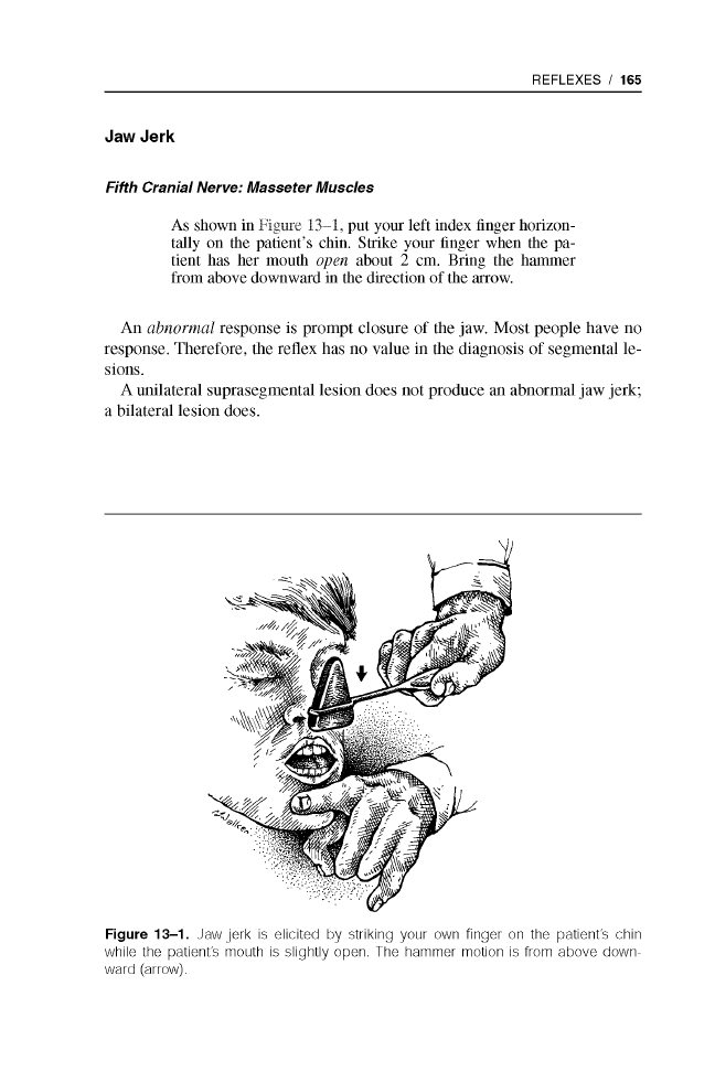
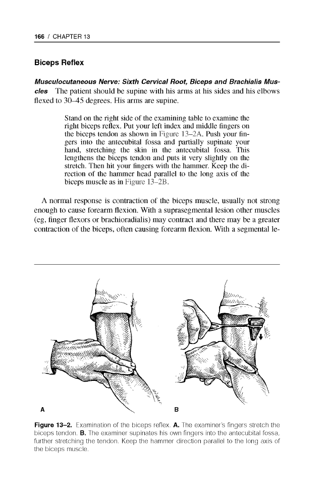

REFLEXES / 167
sion (C6 root) the biceps will not contract, but small contractions of the finger
flexors may be seen. This is known as inversion of the biceps reflex.
Supinator or Brachialis Reflex
Radial Nerve: Sixth Cervical Root, Brachioradialis Muscle
The patient is in the same position as for the biceps reflex
except that her forearm is midway between prone and
supine. Strike the radius just proximal to its styloid process.
A normal response is a visible and palpable contraction of the brachioradi-
alis muscle, usually not sufficiently strong to flex the forearm. With a
suprasegmental lesion the response is abnormal as described above, and with
a C6 segmental lesion the brachioradialis does not contract but the finger
flexors will (ie, an inverted supinator reflex).
Triceps Reflex
Radial Nerve: Seventh and Eighth Cervical Roots, Triceps Muscle
The patient should be supine. Stand on his right to examine
the left triceps reflex. Place his left forearm across his ab-
domen, supported by his body, and his elbow at about a right
angle. Gently pull his left hand toward you and strike the tri-
ceps tendon.
The normal response is less than the response of the biceps reflex and does
not cause extension of the forearm.
If the reflex cannot be obtained, alter the degree of flexion at the elbow
and use reinforcement (see the section on “Reinforcement,” later in this
chapter).
Finger Flexion Reflex
The finger flexion reflex has no diagnostic value at the segmental level. It
is absent in many people. The reflex is informative, however, when it is pre-
sent in one arm but not the other.
Hold the patient’s hand by the fingers as in
Figure 13–3A
.
Produce a little extension at the patient’s wrist and flexion at
both his metacarpophalangeal and interphalangeal joints. Ask
him to rest the weight of his arm and hand in your hand. Then
gently tap the dorsum of your fingers with the reflex hammer.
The patient’s fingers flex as they gently grasp your fingers.
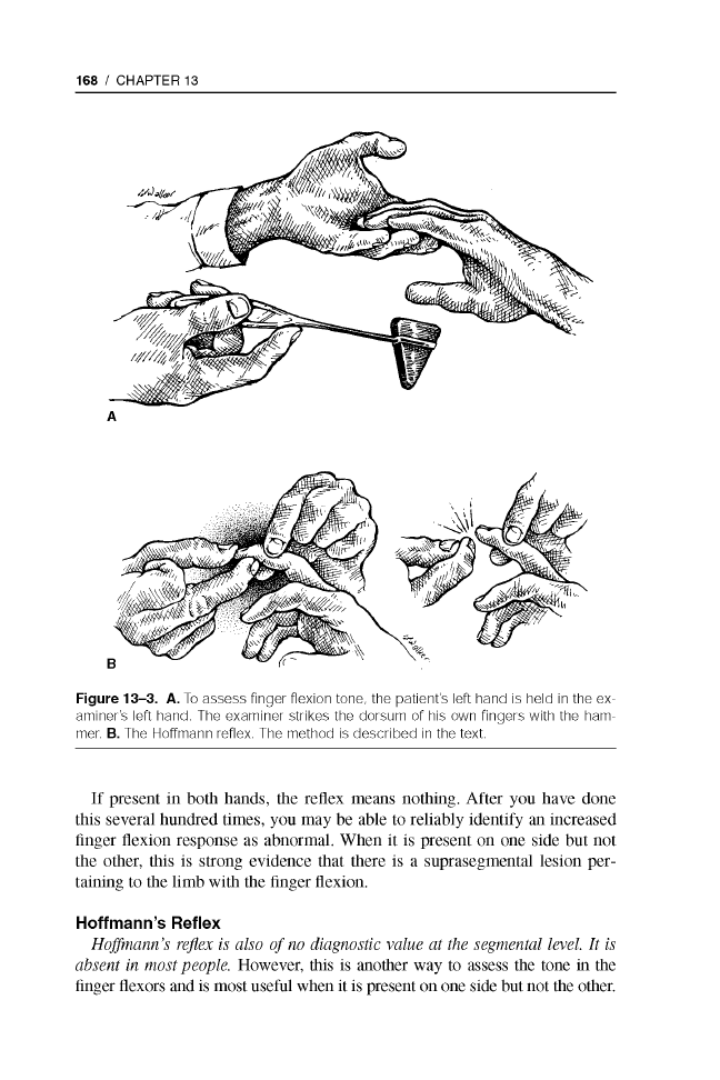

REFLEXES / 169
Hold the sides of the patient’s middle finger at the distal in-
terphalangeal joint between your thumb and index finger as
in
Figure 13–3B
. Forcefully and quickly flex the patient’s
middle finger distal phalanx, and immediately let go so that
the phalanx pops into extension.
This will stretch the profundus flexor of the patient’s middle finger. It and
the other finger and thumb flexors will then contract if their tone is in-
creased.
Knee Reflex
Femoral Nerve: Third and Fourth Lumbar Roots, Quadriceps Muscle
With the patient supine (or sitting), always flex the knee to
about a right angle. If the patient is supine, support his knee
with your hand or a pillow. As you compare the right and
left knee reflexes, make sure the amount of knee flexion
(and therefore quadriceps stretching) is the same in the two
legs. Strike the patellar tendon.
A normal response varies from a flicker of visible contraction of the
quadriceps to extension of the leg, lifting the foot off the bed or table.
Ankle Reflex
Tibial Nerve: First Sacral Root, Gastrocnemius and Soleus Muscles
There
are several methods to elicit this reflex. Unfortunately, the most convenient
and quickest are the least sensitive and least reliable.
• As in
, with the patient supine and his legs extended at the
knee, place the dorsum of your hand gently on the sole of the patient’s foot
and passively dorsiflex the foot. Then strike the palm of your hand with the
hammer. You have used the foot as a lever to stretch the Achilles tendon
and the gastrocnemius and soleus muscles. If there is no muscle contrac-
tion by this method, this does not mean that the reflex is absent; try the fol-
lowing method.
• As in
(this is the conventional method), place one foot on
the opposite shin, thereby flexing the knee. Apply gentle pressure to dorsi-
flex the foot and then strike the tendon. If there is no response try the fol-
lowing. (Note: When the patient was prone and strength in the hamstrings
and gluteus maximus muscles was being tested, there was an opportunity
to examine the ankle reflex.)
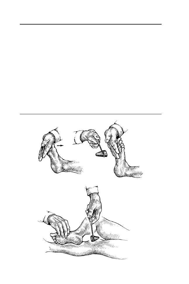
170 / CHAPTER 13
• Flex the knee as in
Figure 13–4C
. Put gentle pressure on the sole of the
foot to evoke some dorsiflexion and muscle stretching, then strike the ten-
don. If the reflex is absent by this method, there is disease in the segmental
arc.
• Having the patient kneel on the edge of the examining table with his back
to you is about as reliable as the previous method. As in
Figure 13–4D
,
passively dorsiflex the foot before you strike the tendon.
Clonus
Clonus has the same significance as pathological tendon stretch reflexes,
which were defined at the start of this section (ie, it is almost always indica-
tive of suprasegmental disease).
A
B
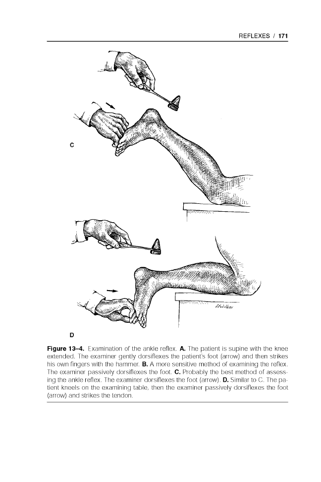
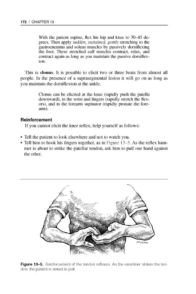

REFLEXES / 173
• If you cannot obtain an arm reflex, ask the patient to curl the fingers of the
other hand into a loose fist. As you are about to deliver with the reflex
hammer, ask him to make a tight, strong fist.
SUPERFICIAL REFLEXES
Corneal Reflex
Examination of the corneal reflex reveals information about the segmental
reflex arc and the suprasegmental system as high as the contralateral thala-
mus. (See the section on the fifth cranial nerve in Chapter 8.)
Pharyngeal Reflex
The pharyngeal reflex is often absent. (See the sections on the ninth and
tenth cranial nerves in Chapter 9.)
Abdominal Reflexes
Like the corneal reflex, the abdominal reflexes may be abolished by ipsi-
lateral segmental or contralateral suprasegmental lesions. The stimulus is
pain or touch, and the response is a simple abdominal muscle contraction.
The patient must be supine and relaxed. Stroke the skin of
the abdomen with a pin, pencil, or the pointed handle of the
reflex hammer, in the direction of the arrows shown in
Fig-
ure 13–6
. Keep each stimulus within the approximate der-
matome of an individual spinal nerve. Move the stimulus to-
ward the midline each time. The normal response is
contraction of the underlying muscle and movement of the
umbilicus toward the stimulus. Do not make the stimulus so
strong that it stretches the underlying muscle directly. The
stimulus is applied to the skin.
Lesions of spinal nerves or roots from T7 to T11 will abolish the reflexes.
The abdominal reflex is dependent on the integrity of a multisynaptic
suprasegmental system, part of which is the pyramidal tract. Thus, unilateral
absent abdominal reflexes can be an early and sensitive sign of recent acute
pyramidal tract disease.
Abdominal reflexes are usually absent in patients with lax, stretched ab-
dominal muscles and in the elderly.
Cremasteric Reflex
Stroke the inner upper aspect of the thigh with a pin or pen-
cil. The stimulus is of the same quality as was used to elicit

REFLEXES / 175
ion and adduction of the toes. It is important that you watch this series of
movements in every patient that you examine.
The normal response is called a flexor plantar response. The abnormal
response is extension of the big toe and abduction of the other toes. This is
the extensor plantar response, or Babinski response. The important thing to
watch is the big toe.
• The lower limb should be extended at the hip and knee (ie, with the patient
fully supine).
• The stimulus should be firm and noxious.
• Stroke the sole of the foot with the blunt handle of the reflex hammer.
• Start at the heel on the lateral aspect and move the stimulus toward the toes.
Stop the stimulus when a response is obtained. The big toe may extend af-
ter the stimulus has moved 3 cm or less. The total movement of the stimulus
can be up the lateral edge of the foot to the little toe and then across the sole
to the head of the metatarsal of the big toe. The response has usually ap-
peared before the stimulus has moved this far.
If the patient will not let you touch the sole of her foot, you may apply the
afferent stimulus at
• The posterolateral aspect of the foot, as a stroking motion from behind and
below the lateral malleolus, continuing it up the dorsolateral edge of the foot
• The edge of the tibia, by pressing on the bone at the tuberosity and running
your fingers firmly down the length of the shin to the ankle
• The calf, by squeezing the gastrocnemius and soleus muscles
All of these are less reliable methods than applying the stimulus to the
sole of the foot.
Primitive Reflexes
All of the primitive reflexes are present in healthy babies and are innate.
They disappear, however, as the higher cerebral centers develop with matu-
rity. They may reappear with disease or damage to these centers.
Grasp Reflex
The grasp reflex may be present bilaterally in patients who are semicon-
scious or fully alert. Like the extensor plantar response in the same clinical
setting, this is not very useful information.
Put your index and middle fingers or the handle of the reflex
hammer on the palm of the patient’s hand and slowly stroke
the palm toward the thumb or little finger.

176 / CHAPTER 13
The patient’s fingers flex and grasp the handle of the hammer. If you are
quick, you can remove the stimulus before it is caught. Sometimes a simple
nonmoving touch to the patient’s palm will evoke finger flexion. You may
repeat the stimulus indefinitely and provoke finger flexion each time. Tell the
patient “Do not squeeze this [handle] when I put it in your hand”—he can
overcome the reflex and not do it, usually once only. Once he has grasped it,
he usually cannot open his hand on command.
This reflex indicates contralateral frontal lobe disease. The reflex will van-
ish in the presence of corticospinal tract disease.
Sucking Reflex
Ask the patient to open his mouth 2 cm, then touch his lips
with a wet tongue blade, moving the stick from the lateral
aspect of the lips to the midline and then laterally again.
The response is a sucking motion of the upper and lower lips.
The response can also be obtained by gently tapping the up-
per or lower lip with the reflex hammer.
This reflex also indicates frontal lobe disease. It is also present, however,
when there is a bilateral corticospinal tract lesion above the midpons.
Palmar-Mental Reflex
Scratch the patient’s palm with the handle of the reflex ham-
mer. The scratch should be firm and the direction should be
from the fingers to the wrist or vice versa. A positive re-
sponse is a short-duration flicker or dimple in the skin of the
chin. This indicates contralateral frontal lobe disease; it will
vanish after a corticospinal tract lesion and its veracity and
usefulness are not proven.

Sensation
1 4
This part of the examination is difficult, and findings are not always repro-
ducible. The tests are crude, and the results are dependent on the cooperation
of and interpretation by the patient.
Sensory findings are always subjective. A painless ulcer on the sole of an
anesthetic foot or a cigarette burn on the edge of the middle finger are objec-
tive signs, but their significance rests on what the patient says about them.
Sensory symptoms depend on the vocabulary and intelligence of the pa-
tient. Descriptions such as “numb,” “tingling,” “prickling,” “asleep,” and
“like dental anesthetic coming out” are the most common. The rare patient
says “The part is dead; there is no feeling in it at all.” Others say that the part
(eg, the side of the face) is normal or unremarkable if it is not touched but
feels different when he shaves, for example.
More subtle sensory symptoms are descriptions such as “My fingers [or
toes] feel swollen like sausages,” “The skin is too tight,” or “I feel as though
I have tight gloves on all the time.”
Symptoms in the lower limbs may be described in this way: “I feel as
though I’m walking on cotton wool” (possibly a posterior column lesion) or
“My shoes are full of small stones; my feet are on fire” (a possible peripheral
nerve lesion) as well as the more usual descriptions of numbness, tingling,
and prickling. The proximal pain and peripheral paresthesia clue—“I
have pain in my shoulder and down the back beside my shoulder blade and
my little finger is asleep”—clearly tells you where the sensory signs should
be.
When you hear these symptoms, examine the part in more detail and, if
necessary, examine only the symptomatic area the next day.
The patient with no sensory complaints (including pain) and nothing in his
history to suggest a disturbance of sensation should have a sensory examina-
tion lasting 5 min, consisting of the following:
• Touch and pain sensation over the face (always the corneas), hands, feet,
and trunk
• Vibration sense and joint passive movement sense in the fingers and toes
177

178 / CHAPTER 14
Rules of Sensory Examination
Fatigue
Do not perform a sensory examination at the end of a lengthy ex-
amination process; the more tired the patient is, the more dubious the an-
swers. Reliable signs are more likely when the sensory system is examined
alone.
Suggestion
Do not interfere with the patient’s decisions by slanting your
instructions toward the abnormal. When you are comparing pinprick on the
two sides, say, “Does this [testing the right side] feel the same as this [testing
the left side]?” Do not ask the patient if the two sides feel different.
Demonstration
Demonstrate first what you are going to do. Touch the pa-
tient gently with the cotton several times in an area you know has normal
sensation before you examine the area of concern. Say to him, “Each time
you feel a touch, will you please say ‘yes.’ ”
Put the vibrating tuning fork in the middle of the patient’s sternum and tell
him that it is the vibration—not the sound of the tuning fork or the pressure
of the handle—you are concerned with.
Brevity
Brief instructions, brief answers, and brief examinations are advised.
Keep your instructions simple. Ask the patient to answer quickly “yes” for
each touch and “sharp” or “dull” when you touch him with the point or head
of the pin. You do not want a qualified answer.
Eyes Closed
Do not let the patient watch you do the sensory examination.
Have your assistant or the patient hold up the bedsheet to cut off the pa-
tient’s vision from the part being examined. If the examination is brief and
limited (eg, the sensory disturbance is a median nerve lesion at the wrist),
you can ask him to close his eyes.
Dementia, Delirium, and Confusional States
Patients in any of these cate-
gories cannot be examined for sensation except in the most crude way, such
as corneal reflexes or withdrawal of the limb from repetitive pinprick.
Examine from the Abnormal to the Normal
The patient with numb weak
legs should have his sensory level established by being examined from be-
low upward. The pin can be moved in short quick steps or more slowly
dragged over the skin.
Medicolegal Litigation
Patients who have been in accidents or who have
been injured at work or where a third party is involved require special men-

SENSATION / 179
tion. If they have sensory findings, draw a map of the limb or body part in-
volved and shade in the area of abnormality. Use slash marks for pain loss,
dots for touch loss, and so on. Draw the map immediately after the examina-
tion.
Definitions
Analgesia: Loss of appreciation of pain sensation
Anesthesia: Loss of appreciation of all forms of sensation
Dysesthesia: An unpleasant and abnormal response to an in-
nocuous stimulus
Hypalgesia: Decreased appreciation of pain sensation
Hyperesthesia: Abnormally increased sensitivity to any
stimulus
Hyperpathia: Abnormally increased sensitivity to a pain
stimulus
Hypoesthesia: Decreased appreciation of all forms of sensa-
tion
Paresthesia: An abnormal, spontaneous sensation (eg, tin-
gling or crawling)
TOUCH
The test object for touch is usually a piece of cotton wool. Pull the fibers
out so that you are using only a small amount of the cotton. Shield the pa-
tient’s vision. Tell him what you are going to do—touch him—and tell him
that each time you do this he should say “yes.” The force of the touch should
not deform the skin. A touch does not mean drag or wipe the cotton over the
skin. This is tickling and is anatomically related to pain conduction. A touch
is an end-point touch.
On most faces, 10 touches will result in 10 answers. On other parts of the
body some touches are normally ignored, but usually not more than one or
two out of 20. Is the number of ignored touches the same on the two halves
of the body? Hairy parts of the body are exquisitely sensitive to touch stimu-
lation. Bending a hair by touching it stimulates both superficial and deep
sensory systems. Lack of response to a touched hair is never normal.

180 / CHAPTER 14
Hallucinatory Answers
Apply the stimulus in an irregular rhythm so the patient does not antici-
pate it. Sometimes, after a pause of 5 s, during which no stimulus has been
applied, the patient says “yes.” This may represent a conduction delay in
which he is responding to the last touch. Usually, however, it is not. When
these “spontaneous” or hallucinatory responses occur as often on the right
half of the body as on the left, or as often from the normal areas as from the
symptomatic region, they are not diagnostic.
If responses occur on stimulation of one limb only or on one half of the
body only, that area is usually the abnormal one.
An occasional litigious patient with a numb arm will say “yes” to 95% of
the touches over all of his body except the arm. Each time you touch the
arm, he says “no.” This tells you two things about him. First, he is not think-
ing; second, his numbness is not organic.
Lesions
With peripheral nerve lesions the area of touch loss is greater than the
area of pain loss.
With spinal cord lesions if there is a loss of touch sensation, the patient
usually cannot walk. Touch is conveyed by several tracts in the cord.
With parietal lobe lesions many touches will not be perceived, while oth-
ers of the same intensity in the same area will be and there will be frequent
hallucinatory responses.
The most sensitive test of touch is in localization. Before touch sense is
absent, the patient will be unable to accurately locate the touched area. For
this reason the cotton used should be a few small, teased threads only.
SUPERFICIAL PAIN
The common testing object for superficial pain is a pin. Use a sterile com-
mon pin, which you will discard at the end of the examination. Tell the pa-
tient that you do not always do this with exactly the same amount of pres-
sure each time.
Touch the point of the pin to some part of the patient’s skin where you
know sensation is normal. Tell him that this is “sharp.” Touch the head on
the other end of the pin to the same area and tell him that this is “dull.” Then
test the patient’s pain perception on his face, hands and feet, arms, legs, and
trunk. The cornified skin on the normal palm and sole are not highly sensi-
tive to pain.
Test a large area, not a small spot. Repetitive stimuli on the same hypoes-
thetic spot may be felt as sharp or sharper than normal. Repetitive stimuli are

SENSATION / 181
cumulative and can overcome an elevated pain threshold. “Pain spots” per-
ceive the pin more acutely, and some touches with the pin will hit them.
Other areas are relatively insensitive, with fewer pain spots. Therefore, keep
the pin moving and use the point and the head with equal frequency, com-
paring the responses from a large area on one side to the responses from the
homologous area on the other side of the body.
If you find an abnormal area, are other sensations abnormal here as well?
Does the area conform to a spinal or peripheral nerve distribution? Does it
have a spinal cord configuration, and if so, is there a level? Is it present in
the homologous area of the trunk or limb of the opposite side?
Pain perception may be delayed. This is abnormal. Touch the dorsum of
the foot and ask the patient to say “yes” as soon as she feels it. She should
answer almost before you have lifted the pin from the skin.
Faulty pain localization is particularly useful in the diagnosis of cerebral
lesions. With the patient’s eyes closed, touch her with the pin and ask her to
point to the spot with one finger. This is normally more accurate in the distal
than in the proximal part of the limbs.
Hallucinatory responses occur with pain testing. They are of the same sig-
nificance as this type of response with touch testing.
Pain is cumulative. The last part of the body to be tested perceives the
identical pinprick more acutely than the first part tested. Therefore, reverse
the order of testing if this might explain why the patient says the last part
tested was more sensitive.
TEMPERATURE
Use large (250-mm-long) test tubes with rubber stoppers. Cold tap water
at about 20
°
C and hot water at about 45
°
C provide an adequate difference
to start.
Keep the outside of the tubes dry. There is no feeling of wetness, but a wet
tube is interpreted as a cold tube.
Apply the side of the tube, not the small bottom, to the skin. This sensa-
tion should be tested slowly. There is a normal longer latency between appli-
cation of the thermal stimulus and the response than with pain or touch stim-
ulation. Let the hot or cold tube, especially the hot one, stay on the skin for 2
s each time.
If the patient has paresthesias and testing for touch and pain yields normal
results, sometimes temperature testing gives useful information.
The purpose is to see whether the hot and cold tubes are as hot or as cold
in the area of concern as in a normal area. Once it has been established that
there is an area less sensitive than the normal area, the ability to differentiate

182 / CHAPTER 14
within that area may be defined by reducing the temperature difference in the
two tubes. When the water in the tube is about 30
°
C, the normal person can
differentiate a 1
°
difference in temperature. Slowly sliding the hot tube over
the skin (from abnormal to normal) will often define the boundaries of an
area of decreased sensation.
Expose the area to be examined to room temperature for 15 min before
starting. Reliable answers cannot be obtained from skin areas that are cold
and vasoconstricted.
Remember: Contact the skin with a large area of the tube for at least 2 s
per stimulus.
DEEP PAIN
Deep pain sensation may be intact even when the response to pinprick is
abnormal. The stimulus is squeezing the Achilles tendon or the calf or biceps
muscle. The normal response is an uncomfortable deep, slow, poorly local-
ized pain accompanied by nausea and protestations from the patient.
VIBRATION SENSE
Use a tuning fork that vibrates at 128 per second. A normal adult can feel a
vigorously vibrating tuning fork for 12–15 s at the ankle and for 15–20 s on
the distal phalanx of the index finger.
Instruct the patient as you place the handle of the vibrating fork on his
sternum or jaw. Tell him you want to know when the vibration stops.
Demonstrate this by grasping the vibrating tines of the fork with your other
hand while the handle is firmly against his sternum. This will stop the vibra-
tion.
Make the tuning fork vibrate by hitting it close to the base of the tines
with the heel of your hand. Put the handle on a distal bony prominence (eg,
the big toe or either malleolus at the ankle; if there is no sense of vibration,
you must move to a more proximal bony point). Ask the patient to tell you
when the vibration stops. Let the tuning fork “run down” on its own.
When the patient says that the vibrating has stopped at the right medial
malleolus, if you then quickly put the handle on the left medial malleolus
(without striking the fork again) the vibration is felt again for a few seconds.
In spite of what is stated in some textbooks, this is normal.
The decrease in vibratory sense may be gradual, being absent at the big
toe and ankle and felt for 2–3 s at the knee, 5 s at the iliac spine, and 15 s
over the spinous process of the first lumbar vertebra. These findings are con-

SENSATION / 183
sistent with a peripheral nerve lesion or a degenerative disease of the poste-
rior columns of the spinal cord.
If vibratory sense is absent at the ankle, knee, and pelvis and normal at
some spinous process, this is consistent with a transverse, compressive, or
destructive lesion of the spinal cord.
Vibratory sense and passive joint movements are each served by different
portions of the posterior columns of the spinal cord and may not be equally
abnormal. Apparently, otherwise normal older people have decreased or ab-
sent vibration sense at the ankles because of a segmental peripheral neuropa-
thy of unknown cause. Subacute combined degeneration of the spinal cord is
marked by a greater vibratory loss than passive joint movement loss. Tabes
dorsalis is the reverse. Vibratory sense is not impaired in cerebral lesions
above the thalamus, while defective sense of passive movement in one big
toe may be a critical physical sign of the parasagittal, parietal meningioma
(ie, it is a cortical sensation).
SENSE OF PASSIVE MOVEMENT
Ask the patient to watch you. If the patient does not receive a proper
demonstration and instructions before you start this test, the response may be
wrong half the time. Passive movement is tested at one joint only, starting at
the most distal toe or finger. Hold the proximal phalanx of the patient’s big
toe in your left hand and anchor it. Grasp the sides of the distal phalanx of the
big toe between your right thumb and index finger. Move the distal phalanx
of the toe slowly up (dorsiflexion) a random number of times, telling the pa-
tient that each of these movement is “up.” Do the same thing while moving
the toe down. Then, with his vision shielded, make random up-and-down
movements (eg, three up, two down, one up, four down), asking him to tell
you each time where it is. A wrong answer for the first move in a new direc-
tion is normal. Move the distal phalanx slowly, consistently, and a small dis-
tance each time.
POSITION SENSE
Does the patient know where his limbs are in space? A defect of this sen-
sation produces a major disability. With the patient sitting, his eyes closed,
and an arm held out in front of him (see
Figure 10–1
), an arm with defective
position sense will drift. This limb will often have slow, continuing, restless
movements called pseudoathetosis.
With the patient’s eyes closed, move the abnormal limb several times and

184 / CHAPTER 14
ask him to point to it with the other (normal) index finger. Place the abnor-
mal limb in a certain position and ask the patient, with his eyes closed, to
imitate the position with his normal limb. If position sense is defective, he
cannot do either. This sensation is defective in diseases of the posterior root,
posterior columns, and parietal lobe.
STEREOGNOSIS
Stereognosis can be assessed only when touch, pain, temperature, and vi-
bration sense in the hand are all normal. Can the patient, with his eyes
closed, identify common objects placed in his hand? He must be capable of
moving the object in his hand and quickly feeling it with his fingers and
thumb. A paralyzed hand cannot be examined for stereognosis. Use a key,
coin, pocketknife, and pen. All the objects, or all but one, may be identified
correctly after 15–20 s of examination in one hand and in the other after 3 s.
The former is abnormal. If the objects are not identified in either hand but
are recognized visually, only one lesion is present; the patient has tactile ag-
nosia but not astereognosis.
NUMBER WRITING
Number writing is also a test of cortical sensory integration. “Write” on
the patient’s palm random, single-digit numbers, first with her watching you,
then with her eyes closed. The bigger the drawing and the firmer and faster
the movement of the pencil, the easier it is for her to correctly guess the
number.
TWO-POINT DISCRIMINATION
The ability to tell one from two touches that are close together may be de-
fective in lesions of peripheral nerves, the posterior columns of the spinal
cord, and the cerebrum. A pair of dividers with dull points or an unwound
wire paper clip can be used to test this sensation.
The normal threshold on the lip is probably 1 mm, on the tip of the index
finger is 3–5 mm, and on the back is several centimeters. The two points of
the dividers are placed on the skin simultaneously with equal pressure. If the
sensation is defective in the index finger, two points at 5-mm separation will
be felt as one and may still be one at 9- to 10-mm separation, while the op-
posite (normal) side can identify two points at a separation of 3 mm. Some

SENSATION / 185
single-point touches on the abnormal side will also be interpreted as two.
BILATERAL SIMULTANEOUS STIMULATION
Sensory neglect of one half of the body or of one limb may be an isolated
sensory finding or part of a constellation of cerebral sensory signs.
If the patient has a right parietal lesion, for example, the sensory examina-
tion of his left side may be uninformative. However, if you touch the identi-
cal spot on the back of his right and left hands with cotton at the same mo-
ment and ask him, while his eyes are closed, to tell you where he has been
touched, he will consistently say “right”; that is, when stimulated simultane-
ously, he ignores the left side. When you touch his left side only, he always
says “left.” Similarly, touching the right side only always provokes a correct
answer. Sensory neglect revealed by bilateral simultaneous stimulation may
be the only or the earliest sensory abnormality.
SUMMARY
All bedside sensory findings are subjective except the corneal reflex.
Remember:
• Sensory examination should be of brief duration, with brief instructions,
and brief answers.
• In peripheral nerve lesions touch and superficial pain are most likely to be
abnormal.
• In posterior root lesions position sense, passive movement, and, to a lesser
extent, vibration sense will be abnormal.
• In spinal cord disease superficial pain and temperature, sense of passive
movement, and vibration will be affected, depending on what area of the
cord is diseased, with relative preservation of touch. When touch is lost be-
cause of cord disease, the patient usually cannot walk.
• In parietal lobe lesions discriminatory sensations are abnormal. These are:
sense of passive movement, number writing, two-point discrimination,
stereognosis, tactile or pain localization, and the recognition of two simul-
taneous stimuli on homologous body parts. It is not possible to say which
of these is most likely to be abnormal in any particular lesion.

The Cerebellum
1 5
The signs of cerebellar disease are more obvious when the lesion is unilat-
eral and disease in other parts of the nervous system is absent. The signs
vary, depending on whether the lesion is acute or chronic, bilateral or unilat-
eral, and hemispheric or midline.
MUSCLE TONE
Cerebellar lesions reduce muscle tone. Diseases of the hemispheres or the
vermis will produce hypotonia; with the latter, it will be more manifest in the
trunk.
The hypotonia is ipsilateral to the side of the lesion and more marked
when the lesion is acute. The affected limbs can be displaced into abnormal
postures with less sense of resistance, joints are hyperextendable, and the
range of limb movement is increased.
POSTURE
Cerebellar disease will cause the unsupported arm (held in front of the pa-
tient, with his eyes closed) to waiver and drift. It may be tremulous or may
have irregular, purposeless, repetitive, pseudoathetotic movements. These
are also seen when position sense is defective. While the patient holds the
limb still, as in testing for arm drift, if you gently tap the wrist to either side,
up or down, an abnormally large movement results. The same can be demon-
strated in the lower limb.
TENDON REFLEXES
In acute cerebellar lesions such as hemorrhage or trauma, all the tendon
reflexes may be absent for hours or days. After that they may be obtainable
but temporarily depressed.
In the less acute lesion some tendon reflexes are pendular.
187

188 / CHAPTER 15
Ask the patient to sit well forward on the edge of the exam-
ining table. Strike the patellar tendon as usual.
The leg will extend at the knee, then flex, and then extend again three or
four times, with a decreasing range each time. The braking action of the
quadriceps antagonists (the hamstrings) does not dampen the response after
the first contraction of the quadriceps.
Pendular reflexes are not an exclusive cerebellar sign and may be present
in any hypotonic condition.
TREMOR
Cerebellar tremor is an action or intention tremor and disappears when the
part is completely supported and at rest. It results from disease of the dentate
nucleus or its connections. The stationary outstretched upper limbs may
show a rhythmic constant tremor. This can be a flexion-extension tremor at
the wrist at three or four per second or of the whole arm at the shoulder,
somewhat slower and more coarse.
When the patient is sitting, a tremor of about the same frequency may in-
volve the head. It is usually an affirmative nodding tremor and often has pe-
riodicity to it. There will be 10 or 12 beats with more or less regular syn-
chrony, then a pause and no tremor for 5–10 s, and then the tremor resumes.
The pause is often associated with a minor change in head position or per-
haps the patient touched his chin or cheek with a finger. Often, however, the
tremor appears to stop and start spontaneously. It will disappear when the
patient is lying down and the head is fully supported.
The intention tremor of the unsupported, stationary upper limb becomes
less regular and less obvious when the limb starts to move. As the patient
brings the index finger from the fully outstretched position toward the tip of
the nose, there is clearly an irregularity and jerkiness to the passage, but in
the final few inches before the finger touches the nose the tremor will ex-
plode. It becomes faster and wilder, and the hand thrashes back and forth.
Immediately when the index finger contacts the nose, the whole thing damp-
ens down and the tremor is reduced entirely or by 90%. This burst of tremor
at the end of a voluntary movement also is not an exclusive cerebellar sign.
REBOUND, PAST POINTING, AND DYSMETRIA
To elicit rebound, have the patient flex his elbow to less than
a right angle. Resist him by pulling on the volar surface of

THE CEREBELLUM / 189
his wrist to extend his forearm. Stop suddenly by withdraw-
ing your hand.
A person with disease of the cerebellum cannot stop the released forearm
and may hit himself. The ability to quickly stop the flexion when the exten-
sor force is withdrawn is defective.
When the patient with cerebellar disease performs the rapid alternating
movements described in Chapter 10, abnormalities may become evident.
This is most useful when cerebellar tremor is minimal or absent. When the
patient attempts to touch the tip of his nose with his index finger, the finger
will stop before it gets to the nose. On the next attempt he will overshoot and
hit his nose too forcefully. When he is making rapid repetitive pinches of the
thumb and index finger or repetitive prone-supine-prone pats with his hand
on his thigh, you will see and hear the abnormality. The movements are of
uneven strength, speed, and frequency.
VOLUNTARY MOVEMENTS
With acute cerebellar disease there may be weakness and slowness of vol-
untary movements. The weakness is transient. There may also be slowness
of relaxation of voluntary movement.
NYSTAGMUS
A large, unilateral cerebellar hemisphere lesion will provoke coarse, slow
nystagmus on gaze to the side of the lesion and faster, finer nystagmus on
gaze to the other side.
Upbeat nystagmus may be seen in lesions of the vermis of the cerebellum,
although disease of other parts of the brain stem can cause this nystagmus.
Downbeat nystagmus is usually seen in diseases at the cervicomedullary
junction, including cerebellar herniation, Arnold-Chiari malformation, and
platybasia.
Ocular dysmetria is similar to past pointing or overshooting of the hand.
The patient attempts to fix gaze and the eyes overshoot, correct, overshoot to
a lesser extent in the opposite direction, and after several successively
smaller movements in either direction, will fix on the desired object. These
movements can be rotary or vertical. Ocular dysmetria, ocular flutter, and
opsoclonus are terms that may be used interchangeably.
Disorders of gait in cerebellar disease have been covered in Chapter 12.

190 / CHAPTER 15
SPEECH
The scanning, staccato speech of cerebellar disease is also called an ataxic
dysarthria. The patient’s speech sounds like a sobbing child who is trying to
talk after crying. The diaphragm rises in a series of irregular jerks, variable
in speed and amplitude. Multisyllabic words are broken into individual sylla-
bles and “Methodist” comes out as “Meth,” “o,” “dist.” In addition, words
are slurred. There is lack of lip, tongue, and pharynx coordination as well as
irregular air propulsion as a result of diaphragm ataxia.
Finally, it should be remembered that a unilateral frontal lobe lesion can
produce contralateral arm and leg ataxia, weakness, and an action tremor.
This diametric, diagnostic dilemma is rare, but it does occur.

The Corticospinal System
1 6
The most common abnormal signs encountered in neurological practice
are those of disease of the corticospinal system.
This system has several more or less synonymous names: the upper motor
neuron, pyramidal tract, and descending supraspinal pathway. The corti-
cospinal system is not a single tract. It includes the fibers arising from the
Betz cells in cortical area 4, the longest uninterrupted white matter tract in
the nervous system. It also includes the fibers of the rubrospinal, reticu-
lospinal, and vestibulospinal tracts as well as the effects of the basal ganglia
and cerebellum and their multisynaptic connections on the motor system.
All of these components are concerned with the facilitation, inhibition,
control, and modulation of movement. The sensory system is also an impor-
tant and integral contributor to normal movement.
The signs of disease of the corticospinal system depend on the level and
location of the lesion and the rate of its development and progression.
All upper motor neuron lesions have some features in common, although
one may cause spasticity and slowness of voluntary movement without
marked weakness, while another may produce hypotonia and profound pare-
sis.
The signs of the disease of the system may be considered under the head-
ings of power, tone, reflexes, and miscellaneous.
POWER
When the lesion is above the midpons, expect weakness in the contralat-
eral face, arm, and leg.
In Chapters 8 and 9 on the seventh and eleventh cranial nerves, the weak-
ness of the lower face with relatively normal upper face strength and the de-
tails of the shoulder shrug versus head turning strength were discussed.
Watch the speed and range of movement of the corner of the
mouth when the patient shows his teeth on command.
However dense the paresis of the lower face may be, emotional facial
movements will usually be normal on the paretic side.
191

192 / CHAPTER 16
Jaw opening and chewing will be normal, while half the tongue will be
transiently paretic and the tongue will point to the paretic side when pro-
truded. Although a lesion may produce permanent face, arm, and leg paresis,
the tongue weakness is usually transient. Palate and pharynx movements are
not affected by unilateral corticospinal system lesions.
Weakness of the upper limb is variable in amount but consistent in distribu-
tion. It is most marked and earliest (in a progressive lesion) in the extensors
and abductors of the fingers. Extensors of the wrist and forearm will be af-
fected, and eventually so will flexors. The common quick assessment of up-
per limb strength—“Squeeze my fingers”—is particularly inappropriate. By
the time the finger flexors are involved, the weakness can be diagnosed by the
posture of the limb. Minimal upper limb weakness may appear as slowness
and awkwardness in fine, discrete movements. The best tests to demonstrate
this are the rapid alternating movements described in Chapter 10. The maxi-
mum upper limb weakness is permanent loss of all useful movement.
In the lower limb, early and minimal weakness is first seen in the flexors
of the thigh and dorsiflexors of the foot and toes. Tripping over the edge of
a carpet, a curb, or a stair riser is a common early symptom.
Depending on the age of the lesion (whether it is spinal or cerebral and, if
the latter, the level), the paretic leg may assume a flexed or extended posi-
tion.
Following resolution of acute intracerebral lesions, the leg almost always
regains some power.
TONE
Increased tone from disease of the corticospinal system is usually called
spasticity. Rigidity usually implies extrapyramidal disease.
With a unilateral corticospinal system lesion above the brain stem, there
are no changes in tone evident in the head and neck.
In the upper limb the increased tone is in the flexors of the fingers, wrist,
and elbow. It is greatest in the early part of stretching. It can be abolished
temporarily by repeated stretches and will return after the muscle has been
put at rest. The posture of the upper limb is the consequence of flexor mus-
cle spasticity and extensor muscle weakness.
In the lower limb, spasticity is first and greatest in the quadriceps, gastroc-
nemius, and soleus. The footdrop posture is also a combined manifestation,
the foot being dropped down by dorsiflexor weakness and pulled down by
plantar flexor spasticity.
A hemiparesis resulting from a cortical lesion (a very rare occurrence)
will be flaccid and will remain this way. Many acute intracranial and all

THE CORTICOSPINAL SYSTEM / 193
acute spinal, corticospinal tract lesions present initially with flaccid weak-
ness. In the latter case this is called spinal shock. After days or weeks the
limbs become spastic. An acute lesion in one cerebellar hemisphere will pre-
sent with a flaccid, ipsilateral weakness, ataxia, and often absent reflexes.
REFLEXES
Corticospinal system lesions increase tendon reflexes and abolish superfi-
cial reflexes. The qualities of an increased reflex are listed in Chapter 13. A
unilateral corticospinal system lesion above the midpons will not produce a
jaw jerk; a bilateral lesion will.
An acute right-sided corticospinal system lesion above the decussation of
the pyramids or a left-sided lesion below the decussation but above the sev-
enth dorsal cord segment will abolish the left abdominal reflexes. In more
chronic diseases such as amyotrophic lateral sclerosis and cerebral palsy, the
abdominal reflexes are usually retained in spite of obvious corticospinal sys-
tem involvement. The cremasteric reflex will disappear if the appropriate
corticospinal lesion is present.
The plantar response is the most evanescent and difficult sign of corti-
cospinal system disease. Almost no one likes the sole of his foot to be
touched, tickled, or stroked. The normal withdrawal at the hip and knee di-
verts the examiner’s attention away from the toes. One may scratch a pa-
tient’s foot only a limited number of times.
The hip and knee should be in full extension, and the first attempt to elicit
the response is usually the best attempt.
A tired patient admitted late in the day may have easily obtained and
clearly extensor plantar responses. The same patient, after a good night’s
sleep, examined by the same person, may have flexor responses the next
morning.
The plantar response seems to lend itself to equivocation in the patient’s
records. The reflex is often recorded as 0 or with two arrows, one pointing
up and the other down. Make a decision; the examination is not over until
this is decided. An aphoristic view of the equivocal plantar response sug-
gests that if you cannot decide whether it is up or down, it is probably down.
This is correct more often than not.
MISCELLANEOUS
The gait of the patient with a corticospinal system lesion in the internal
capsule is distinctive. The arm and fingers are flexed and adducted and the

194 / CHAPTER 16
leg is circumducted in an arc at the hip, while the foot is plantar flexed and
inverted. There is little, if any, knee flexion with each step.
The posture of the acute hemiparetic patient in bed is abnormal. He may
be lying on his paretic arm and not be concerned; the leg will be extended
and externally rotated at the hip. Even when supine, there is more plantar
flexion at the ankle on the paretic side than on the normal side.
Clonus may be elicited at the ankle, knee, or wrist. The method is de-
scribed in Chapter 13. It is never the only sign of corticospinal tract disease
and is not a useful physical sign.
CONCLUSIONS
When diseased, this huge, complex system has simple signs: weakness,
slowness, and clumsiness of purposeful movement; changes in muscle tone,
stretch, and superficial reflexes; and the addition of new reflexes.
There is a quantitative relationship between only two of these signs. If
muscle tone is increased, the tendon reflexes will be increased. This is al-
ways true, unless the muscle is so stretched that striking its tendon can
stretch it no further. The opposite relationship is not true: tendon reflexes
may be pathologically brisk, while muscle tone is normal.
A minimal corticospinal system syndrome might consist of unilateral ab-
sent abdominal reflexes or an extensor plantar response. A larger constella-
tion might consist of flaccid weakness of the arm and leg, pathologically
brisk tendon reflexes, normal superficial reflexes, and a flexor plantar re-
sponse.
The signs vary and are unrelated to each other in a quantitative way, ex-
cept as mentioned above. The variations are dependent on site, size, and rate
of progress of the lesion. A proper examination includes verification of all
the possible manifestations of a corticospinal system lesion.
PSEUDOBULBAR PALSY
Pseudobulbar palsy is caused by bilateral corticobulbar system disease
above the brain stem—most commonly, bilateral strokes that have occurred
at different times. As the lesion is not in the bulb but above it, a more accu-
rate title might be suprabulbar palsy.
The signs of bulbar dysfunction are
• Dysphagia—These patients have trouble chewing and swallowing. Food
falls out of the mouth while eating or enters the larynx without provoking

THE CORTICOSPINAL SYSTEM / 195
coughing or remains in the mouth between the cheek and the teeth for
hours after a meal.
• Dysarthria—Speech lacks resonance and variation in tone. It sounds stran-
gled and is usually high-pitched and weak.
• Jaw jerk, snout, and sucking reflexes are usually present.
• The tongue cannot protrude beyond the teeth by more than 1 in, and rapid
alternating movements of the tongue are not possible.
• Emotional control is defective. Words, situations, and events that are not
sad will provoke uncontrollable crying. These patients also may laugh in-
appropriately but I have never witnessed the latter.
• Some signs of bilateral hemiparesis are present in the limbs.

Higher Cortical Functions:
1 7
Intelligence and Memory
The majority of patients seen by the neurologist do not require testing of
intelligence. In the course of history taking the patient’s memory and intelli-
gence are revealed and this is often sufficient.
However, a more formal examination must be carried out in the following
situations:
• Where the symptoms are vague, circumstantial, and attributed to some-
one else—for example, “She says [pointing to his wife] I keep losing my
glasses and my keys and last week I forgot her name.”
Doctor: “Did you?”
Patient: “Did I what?”
Doctor: “Forget her name.”
Patient: “I’m not sure. I don’t think so.”
• Chronic substance abuse—In addition to using the popular street drugs,
young people who cannot afford them will try sniffing gas, glue, or solvent
from a plastic bag over their heads. They may die of asphyxiation if they
lose consciousness before pulling the bag off. You may be asked to see one
of these people after their first seizure. A short assessment of the patient’s
intellect will often reveal a wipeout of memory and thinking ability.
• Patients seen in a psychiatric ward or hospital or sent to you by a psychiatrist
• Patients who are said by a family member, themselves, or another physi-
cian to be depressed
• Patients with a major change in behavior, personality, dress, or speech
Irritability and frequent altercations with neighbors, coworkers, family, or
the police over inconsequential issues, as well as faulty concentration, gross
exaggeration in relating day-to-day activities, and a disinhibited attitude in a
person with a previously rather careful approach to life may all signal the de-
menting process.
You will probably never see an unaccompanied patient who says, “Doctor,
I’m losing my mind.” It is almost axiomatic that as dementia advances, in-
sight vanishes. The demented patient usually has no personal history and no
physical signs.
197

198 / CHAPTER 17
Therefore, the history is dependent on the family member who spends the
most time with the patient. The details needed include the patient’s educa-
tion level, occupation, hobbies, other illnesses, previous head injuries,
seizures, and prescription and other drugs being taken.
Testing is done under the following headings: orientation, general infor-
mation, memory, thinking (both abstract and logical), and calculation.
Orientation
Does the patient know the day, date, month, year, and time (within half an
hour)? What is the name of the building he is now in, the city, province or
state, and country? How did he get here—by car, bus, train, or trolley; from
where and how long did it take? What is his full name, age, date of birth, and
regimental number, if any? If he is an old soldier and the regimental number
is forgotten, he is in serious trouble.
General Information
Every person carries a volume of information, some of which is a reflec-
tion of his own personal likes, hobbies, and habits. Ask the patient the name
of the capital city of his country, how many provinces or states there are,
who is the prime minister of Canada or the president of the United States,
and the names of the past four prime ministers and the past four presidents—
most Canadians seem to remember three Canadian prime ministers and five
American presidents (it was ever thus). You can make up your own list of
appropriate questions, having knowledge of the patient’s premorbid occupa-
tion and interests as well as whatever is currently in the news.
Memory
There are three categories or stages of memory.
Immediate
Tell the patient you are about to give him
• A man’s name
• The name of a flower
• A street address
Ask him to repeat them aloud as soon as he hears them. In about 3–5 min
ask him to repeat the three items. Small degrees of dementia will interfere
with this test.
Intermediate
Can the patient remember events that occurred yesterday or 1
week ago?

HIGHER CORTICAL FUNCTIONS: INTELLIGENCE AND MEMORY / 199
Remote
Does the patient know the names, ages, and dates of birth of his
children; his own wedding date; his mother’s maiden name; and such things
as the dates of the beginning and end of World Wars I and II?
Thinking
Abstract
The interpretation of proverbs is a quick way to assess the ability
of patients to think figuratively. Dementing patients think in literal, concrete
ways. For example,
Ask the patient the meaning of “A stitch in time saves nine.”
The dementing person will talk about clothing, sewing, mending, and so
on. The normal answer will be something about small amounts of early
maintenance preventing major repairs at a later date.
Logical
Logical thinking can be tested with a variety of short, simple ques-
tions:
• What is similar between an orange and a lemon?
• What is similar between a pair of scissors and a knife?
• If you found a sealed, addressed, stamped envelope on the street, what
would you do with it? Correct: Pick it up and drop it in the next mailbox.
Wrong: Open it, find the name of the sender or the intended recipient, and
try to contact one or the other, etc.
Calculation
Calculation is an inevitable part of all examinations of higher cerebral
function. Difficulties with numbers, words, and concentration are among
the most common presenting symptoms in dementia.
Number Recall
Ask the patient to immediately repeat back to you a series of
random, single-digit numbers.
Start with three numbers (eg, 7, 4, and 9), then four, and continue. Repeti-
tion by most people reveals recollection of seven digits forward and five
backward. (This is a test more of concentration than of numerical skill, but it
is a good way to start. Tests consisting of numbers seem to intimidate most
people.)

200 / CHAPTER 17
100 Minus 7
Ask the patient to subtract 7 from 100, to tell you the result,
and then subtract 7 from 93, 7 from 86, and so on.
Most people can do this in about 90 s with no errors.
Miscellaneous Numerical and Concentration Tests
You can make your own tests. Generally, you are testing things learned by
rote and the ability to manipulate information. Ask the patient,
• “How many 4¢ stamps can you get for a quarter [of a dollar]?”
• “What are 9 plus 3, 21 minus 8, 7 times 8, and 90 divided by 3?”
• “What is 7 times 2? Add 6 to the answer, divide that answer by 2, and sub-
tract 3 from the answer. What number did you end with?”
A similar but less difficult test of concentration is to ask the patient to per-
form a multistep, consecutive act.
Tell the patient “Stand up, pick up this piece of paper, face
toward the window, and fold the paper.”
The entire command is given before the patient starts.
Spatial and Constructional Ability
Defects in these function may occur from disease in any part of the cerebrum
on either side of the midline. However, parietal lobe lesions are most often the
site, particularly if the rest of the cortical functions are more or less normal.
Present the patient with consecutive drawings, one at a time,
of two-dimensional or three-dimensional drawings and ask
him to reproduce them.
If the patient has normal vision and a functioning dominant upper limb, he
should be able to reproduce drawings like those in
Figure 17–1
, taking 1–1.5
min for each.
For more detailed methods of testing higher cerebral function, see The
Mental Status Examination in Neurology (3rd ed., R.L. Strub and F.W.
Black, editors. Davis, Philadelphia, 1993).
MOOD
Profound changes in mood are not a common part of organic neurological
disease. There are exceptions. The dementing patient with insight will often
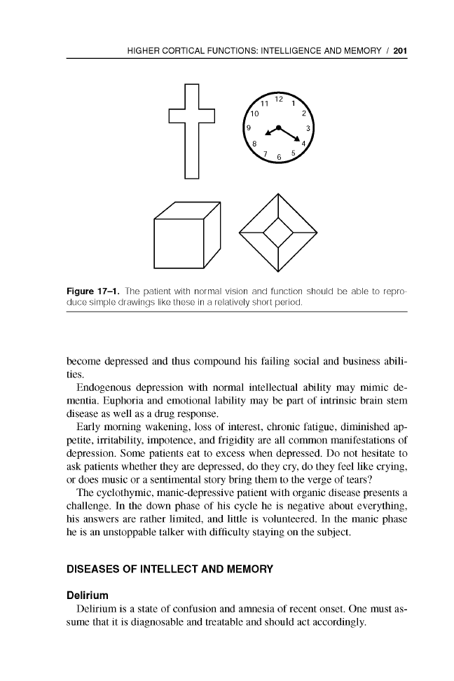

202 / CHAPTER 17
The patient is confused. This may be minor, in that he does not know the
date or the time, or severe, with no knowledge of his name, the place, or any
other identifying facts.
He may be drowsy, stuporous or overactive, tremulous, and agitated. He
may be hallucinating at either level of alertness. Eating, rest and sleep, wash-
ing, and the details of bodily functions are ignored. Epileptic seizures may
occur.
There are many causes of delirium; several of them originate outside the
central nervous system.
• Infections: Commonly in the pulmonary and genitourinary tracts
• Drugs: Barbiturates,
L
-dopa, anticholinergics, steroids, and anesthetic
agents
• Hypoxia: Obstructive lung disease, pulmonary embolus, myocardial in-
farct, and congestive heart failure
• Metabolic: Lung, liver, or kidney failure; hyponatremia; hyperglycemia;
alcohol withdrawal; and postoperative electrolyte imbalance
• Central nervous system diseases: Postictal state, subarachnoid hemor-
rhage, head injury, cerebral thromboses, encephalitis, meningitis, and brain
abscess
The diagnosis and management of acute delirium call for energetic and
wide-ranging thinking by the attending doctor.
Meningitis does not always cause a stiff neck in the elderly; pneumonia
may not raise body temperature or change the white blood cell count.
The mildly demented elderly man may have quite a passable existence at
home with familiar surroundings, pets, and family. One night in the hospital
for a single cataract operation under local anesthetic may change him into a
frightened, hallucinating, overactive, delirious person. When he returns to
his preadmission state within 1 day of being home but was amnesic for all of
his hospital stay, we realize how fragile is the stability of the central nervous
system.
Dementia
Dementia is failure of the intellect, which often starts as a subtle decay in
social graces and business abilities. The conduct and behavior changes may
combine or alternate with impulsiveness or apathy. Judgment, initiative, and
decision making become blunted. Memory deficits eventually become the
dominant problem. These deficits are initially for new information and re-
cent memory, but the amnesic process extends backward into the earlier
parts of the patient’s life. Confabulatory stories often fill the patient’s con-

HIGHER CORTICAL FUNCTIONS: INTELLIGENCE AND MEMORY / 203
versation. A stranger can be completely taken in with the glib and resource-
ful verbal inventions of the demented.
Speech disturbances, word-finding difficulties, anomia, and other dyspha-
sic signs are common. Depression and agitation are equally common. The
demented person may forget or refuse to eat, spend the night pacing the
floor, and ignore personal hygiene.
The process can begin at age 40. Inability to remember the names of
friends is so common at age 40–50 that it is normal. The rate of progression
is variable although difficult to assess accurately because the onset is subtle
and not clearly identified. Most demented patients incapable of caring for
themselves are in their sixth or seventh decade. Relatives usually say that the
patient’s memory and intellect have been slipping for the previous 4–5 years,
but it has been much more obvious in the immediate 6 months. The disease
does not accelerate, but when basic skills needed for dressing, washing, eat-
ing, and other activities of daily living have vanished, the process appears to
be more acute.
Dementia is always organic, is almost always progressive, and is rarely
treatable. The common causes are
• Alzheimer’s disease—Diagnosed by exclusion of other diseases; can be
proven only histologically
• Normal-pressure hydrocephalus—See Chapter 12.
• Huntington’s chorea, supranuclear palsy, parkinsonism, spinocerebellar de-
generation, and Jakob-Creutzfeld disease
• Meningioma, glioma, abscess, subdural hematoma, postmeningitis or en-
cephalitis, and general paresis
• Hypoxia, hypoglycemia, head injury, and multiple cerebral infarctions
• Pernicious anemia, hypothyroidism, chronic renal failure, chronic dialysis,
Wilson’s disease, pellagra, and Wernicke-Korsakoff syndrome
As in the management of a patient with acute delirium, the physician must
be thorough and thoughtful in the investigation of the demented patient. The
family needs help and support, and the patient needs protection. If a treatable
lesion cannot be found, this must be faced and the inevitability of the disease
process must be explained. If the patient can be cared for at home without
exhausting the spouse, this is probably the best arrangement. Eventually, in-
stitutional care will be necessary.

Disorders of Speech
1 8
Aphasia is a loss or impairment of language caused by disease of the cere-
brum. In this sense, language means “speaking, writing, reading, and listen-
ing.” The words aphasia and dysphasia are used interchangeably.
The parkinsonian patient with low-amplitude, monotone, arrhythmic
speech or the patient with cerebellar disease and staccato speech are not
aphasic; they are dysarthric. The patient with bilateral vocal cord paresis is
aphonic. Stammering or stuttering is not aphasia. Some psychotic or de-
mented patients repeat whatever is said to them. This is echolalia, and in
these patients it is not part of aphasia.
The acute schizophrenic may have a fluent, unintelligible jumble of words
and neologisms. This is part of his thought disorder, not aphasia.
Finally, the patient who is mute because of psychosis or other reasons
cannot be said to be aphasic. He may be, but you cannot say so.
Anatomy
Language is a function of the dominant cerebral hemisphere. Most people
are right-handed, and their left hemisphere is dominant. Of the 5–10% who
are left-handed, the majority also have a dominant left hemisphere.
• Broca’s area: This is Brodmann’s area 44 at the posterior end of the third
frontal convolution and is concerned with the motor aspects of language.
• Wernicke’s area: This is Brodmann’s areas 41 and 42 at the posterior end
of the superior temporal gyrus. Deep to Wernicke’s area in the insula are
the transverse gyri of Heschl. These are the primary auditory cortices.
These areas are concerned with understanding spoken language.
• Angular gyrus: This is Brodmann’s area 39 and surrounds the terminal
vertical part of the superior temporal sulcus. It is part of the important in-
ferior parietal lobule. It is concerned with the understanding of written
language.
• Supramarginal gyrus: Brodmann’s area 40 surrounds the terminal vertical
end of the lateral sulcus and is the other part of the inferior parietal lobule.
It is also concerned with the comprehension of language.
These cortical areas are connected to each other and to other cortical areas
by the arcuate fasciculus. They also have connections to the thalamus and
205

206 / CHAPTER 18
the nondominant cerebral hemisphere. These anatomical areas concerned
with language each have an associated type of speech disorder, for example,
Broca’s area and an expressive aphasia, or Wernicke’s area and a receptive
aphasia. This is simplistic. Clinically, patients do not display sharply defined
and distinctive dysphasias that are exclusively defects of expression or de-
fects of reception. A Broca’s aphasia, for example, if the result of a small su-
perficial lesion, can present with defects of spoken and written speech only.
A lesion in the same cortical area, but deeper, will produce the same defects
plus defects of comprehension.
All areas of the brain concerned with language are supplied by the middle
cerebral artery. There are many reports of normal language with disease in
these areas, although the reverse is not true. The areas are not cytologically
distinctive, and electrical stimulation of them in the awake patient has not re-
vealed great information.
Language should be thought of as a combined function of some primary
sensory or motor areas in concert with their appropriate association areas
plus the white matter fasciculi connecting them.
EXAMINATION OF THE APHASIC PATIENT
Spontaneous Speech
Fluency is the most important thing to note about the patient’s sponta-
neous speech.
Fluent Speech
In fluent speech the words pour forth, paraphasia, neologisms, and errors
included. Nonfluent means that the patient has few words; he labors with
pursed lips and much frustration, then brings out three words after 10 s of
trying. Also note the content, comprehensiveness, and errors.
Repetition
Ask the patient to repeat words, numbers, and sentences. Keep your
speech slow, regular, and loud. The two most helpful tests in assessing an
aphasic patient are fluency and ability to repeat.
Comprehension
Ask the patient to perform some act that verifies his understanding of spo-
ken speech, for example, “Point to the door,” “Touch your ear,” “Stand up,”
“Sit down,” or “Is today Wednesday?” (the patient’s head nod or shake being
used as a “yes” or “no” response).

DISORDERS OF SPEECH / 207
Some conflicts may occur here—a defect of body image or an apraxia may
prevent the patient from pointing to his ear or another part of his body, con-
fusion may cause the wrong answer to the day-of-the-week question, and a
disorder of language is not the cause of their error. Also, if your questions
are too close together, the answer to the last question may be used for the
next question. This is perseveration and is not diagnostic, being present to a
greater or lesser extent in almost all types of aphasias.
Naming
Ask the patient to name familiar, everyday objects. Go slowly and start
with big, whole objects and then the smaller parts of objects. A patient may
have normal naming ability for “watch,” “strap,” and “buckle,” but when
you point to the stem, hands, or numerals of the watch, he is lost and tries to
describe these smaller parts by their function, for example, “the thing you
wind it with.”
Reading
Give the patient some simple thing to read aloud. Write short, one-step
commands that you wish him to read and obey in order to test his compre-
hension for written language. Some types of aphasia show excellent compre-
hension of written material but a major defect in reading aloud.
Writing
Ask the patient to write a spontaneous sentence or dictate a sentence for
her. Her signature is not suitable.
TYPES OF APHASIA
Broca’s Aphasia
This lesion is in Broca’s area and its deep white matter connections. This
is a disorder of language output.
The patient is nonfluent and cannot repeat, name, read aloud, or write. He
usually can understand written and spoken language, although, if stressed by
repeated commands given quickly, defects can be shown here also. The pa-
tient may have only two or three words that are used over and over or he
may burst out with profanities while attempting to speak. He looks and acts
frustrated by his illness. Weakness and sensory loss of the right lower face,
arm, and leg are frequent accompaniments. In recovery, the speech remains
slow and agonizing, lacks rhythm and finesse, and is made up mostly of
nouns or isolated verbs.

208 / CHAPTER 18
Wernicke’s Aphasia
This lesion is in Wernicke’s area, and this is a sensory or receptive disor-
der.
The patient is fluent and cannot repeat, name, read aloud, comprehend
writing, or write. Unlike those with Broca’s dysphasia, the patient with the
posterior lesion is a relaxed, comfortable, voluble talker. The sentences are
devoid of meaning. They are filled with neologisms and paraphasias. The
rhythm is more or less normal, and the length of phrases and sentences often
seems normal. There is some hesitation and searching for words, but nothing
like the lockjawed consternation of the patient with Broca’s lesion.
The patient cannot hear himself and usually shows no concern over his
jargon of scrambled words and near-words. Some patients have a greater de-
fect in understanding spoken speech as opposed to written speech, while the
reverse may be present in others.
A hemiparesis is an uncommon accompaniment of this type of aphasia, as
is a cerebral sensory defect. Quadrantanopic field defects are more likely to
be found.
Conduction Aphasia
This lesion is in the arcuate fasciculus that connects Broca’s and Wer-
nicke’s areas.
The patient is fluent and can comprehend what he reads and hears, but
cannot repeat, name, read aloud, or write. There are many pauses and word
searches, and the fluency is less than in Wernicke’s dysphasia. The patient
can hear his errors and paraphasias and often has better performance once he
has started or is into a sequence of words (eg, counting). The patient under-
stands what he hears and reads but can repeat neither.
Anomic Aphasia
This is also known as amnesic and nominal aphasia. It is not related to a
single local lesion.
The patient is fluent, but has trouble finding words. She has pauses, substi-
tutions, and much circuitous, meaningless speech. This is evident in sponta-
neous speech and in naming. All other aspects of language except writing
may be normal.
Anomic aphasia is present, to a greater or lesser degree, in all aphasias. It
is the most common residual defect after any aphasia.
Global Aphasia
This lesion is large, involving both Broca’s and Wernicke’s areas. All as-
pects of language function are abnormal. The patients are nonfluent, and all

DISORDERS OF SPEECH / 209
tests of language are abnormal. Global aphasia is commonly accompanied
by hemiparesis, hemisensory loss, and a hemianopia.
OTHER LANGUAGE DISORDERS
Sensory and motor transcortical aphasias, pure word deafness, pure word
blindness (alexia) with and without agraphia, pure word muteness (aphemia),
and thalamic and striatal aphasia are outside the scope of this work.
Suggested Additional Reading
Rose, F. Clifford and Whurr, Renata: Aphasia. Whurr Pub-
lishers Ltd, United Kingdom, 2000.
Basso, Anna: Aphasia and Its Therapy. Oxford University
Press, Oxford, New York, 2003.
Zaidel, Eran and Iacoboni, Marco: The Parallel Brain. MIT
Press, Cambridge, MA, 2003.
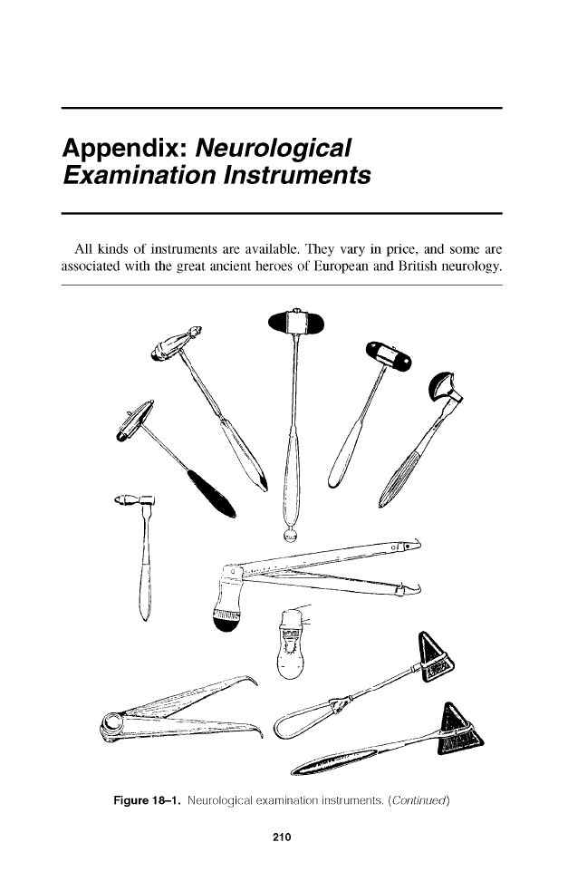
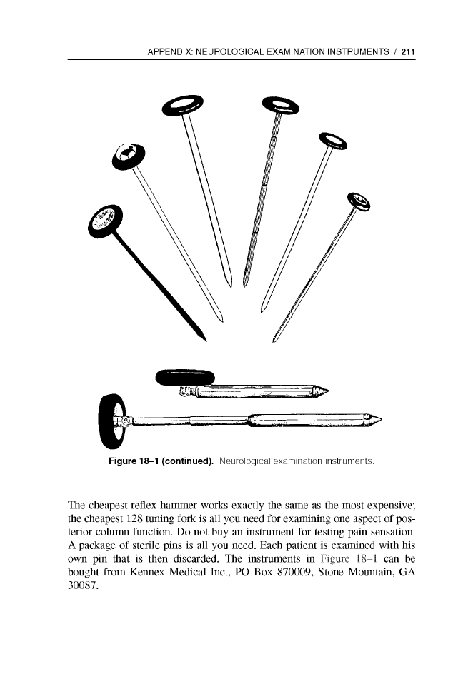

Abdominal muscles, weakness of, gait
abnormalities and, 161
Abdominal reflexes, 173, 174f
corticospinal system lesions affecting,
193
Abducens nerve (cranial nerve VI)
lateral rectus muscle and, 49
lesions of, 57–59
Abduction
of arm, 136–138
of eye, 45
lateral rectus muscle in, 49–50
superior and inferior oblique muscles
in, 54
of fingers, 130
of thigh, 150
of thumb, 130
Abductor pollicis brevis muscle, 130
Abductor pollicis longus muscle, 130
Abetalipoproteinemia, CPEO in, 69
Accommodation, 66–67
in Adie’s syndrome, 65
ACG. See Acute angle-closure glaucoma
Acoustic nerve (cranial nerve VIII),
107–111. See also Cochlear nerve;
Vestibular nerve
cochlear nerve, 107–109
diseases of, 110–111
tinnitus and, 111
vestibular nerve, 109–110
Acoustic neuroma, tinnitus caused by, 111
Acuity, visual, 19
distance vision, 11, 12
in latent nystagmus, 12, 73
in papilledema, 39
Acute angle-closure glaucoma (ACG), 26
Adduction
of arm, 138, 139f
of eye, 46
defects in, convergence and, 67
medial rectus muscle in, 50
superior and inferior rectus muscles
in, 53–54
Adductor muscles (magnus, longus,
brevis), 150
Adie’s syndrome (tonic pupil), 65
Afferent pupil defect, 64
Aging. See Elderly
Alcohol use and abuse, nystagmus caused
by, 71
Alternating movements
lower limb coordination tested with,
145–147, 146f
upper limb coordination tested with,
123–125, 124f, 125f
Altitudinal field defect, 17f
Amblyopia, 10
American Medical Association test card,
for near vision testing, 13
Amsler grid, in macular degeneration diag-
nosis, 28
Amyotrophic lateral sclerosis, jaw weak-
ness in, 96
Analgesia
definition of, 179
facial, trigeminal nerve disorders caus-
ing, 96
Anesthesia, definition of, 179
Angiography, fluorescein, in macular de-
generation diagnosis, 28
Angle-closure glaucoma, acute (ACG), 26
Angular gyrus, 205
Anisocoria, 62, 65
Ankle clonus, in corticospinal system dis-
ease, 194
Ankle reflex, 169–170, 170–171f
Index
NOTE: Page numbers in bold face type indicate a major discussion. A t following
a page number indicates tabular material and an f following a page number
indicates a figure.
213

214 / INDEX
Anomic aphasia, 208
Anosmia, 87–88
central, 88
unilateral, 87
Anterior chiasmal lesions, visual field de-
fects caused by, 29–30, 29–30f
Anterior fossa tumors, anosmia caused by,
88
Anterior (saccadic) gaze center, 80–81
Anterior interosseous nerve
flexor digitorum profundus and, 131
flexor pollicis longus and, 133
Anterior ischemic optic neuropathy, 25
Anterior tibial muscle, 148, 151, 151f, 153,
154
Aphakia, 10
Aphasia, 205–211
anomic, 208
Broca’s, 206, 207
comprehension, 206–207
conduction, 208
definition of, 205
examination of patient with, 206–207
fluent speech in, 206
global, 208–209
naming in, 207
reading in, 207
repetition in, 206
spontaneous speech in, 206
types of, 207–209
Wernicke’s, 208
writing in, 207
Aphonia, 205
Apraxia, gait, 157, 161
Arcuate fasciculus, 205
Arcuate scotoma, 20
in glaucoma, 26
Area
Broca’s, 205
Brodmann’s 17–19 (visual cortex), 21
lesions of, 35–36
field defects caused by, 35, 35f
gaze palsy and, 80–81
Brodmann’s 17 (striate cortex), 21
Brodmann’s 18 (parastriate cortex), 21
lesions of, gaze palsy and, 80–81
Brodmann’s 19 (peristriate cortex),
21
Brodmann’s 39 (angular gyrus), 205
Brodmann’s 40 (supramarginal gyrus),
205
Brodmann’s 41 and 42 (Wernicke’s
area), 205
Brodmann’s 44 (Broca’s area), 205
centralis. See Posterior pole
Argyll-Robertson pupils, 64
Arm, 121. See also Upper limb
abduction of, 136–138
adduction of, 138, 139f
circumference measurement of, 127,
129f
Arm drift, 122–123, 122f
Arnold-Chiari malformation, downbeat
nystagmus in, 73
Arteriovenous “nicking,” retinal, in hyper-
tension, 43
Arteritis, giant cell, of central retinal
artery, 25
Artery
carotid, occlusion of, pupils and, 66
retinal
branch, occlusion of, 27
central
giant cell arteritis of, 25
occlusion of, 27
in hypertension, 43
occlusion of, 27
ophthalmoscopic examination of, 6,
7f
temporal, giant cell arteritis of, 25
Aspirin, tinnitus and, 111
Astigmatism, 10
examiner’s, eyeglass use during oph-
thalmoscopic examination and, 3
patient’s, lens for ophthalmoscopic ex-
amination and, 10
Ataxia. See also Gait
in cerebellar disease, 159–160
contralateral arm and leg, frontal lobe
lesion causing, 190
miscellaneous diseases causing, 162
in normal-pressure hydrocephalus,
161–162
tests of, 158–159
Ataxic dysarthria, speech in cerebellar dis-
ease and, 190

INDEX / 215
Atheroma, carotid artery, pupils in, 66
Atropine, factitious big pupil caused by, 66
Axillary nerve
deltoid muscle and, 136, 138
lesion of, C5 root lesion differentiated
from, 143
Babinski response (extensor plantar re-
sponse), 174, 175
in corticospinal system lesions, 193
Back muscles, weakness of, gait abnormal-
ities and, 161
Balance, 157–162. See also Gait
Bardet-Biedl syndrome, retinitis pigmen-
tosa and, 26
Basal ganglia disease, rigidity in, 126
Bassen-Kornzweig syndrome
CPEO and, 69
retinitis pigmentosa and, 26
Bell’s palsy, 104
Bell’s phenomenon, 82–83
Betz cells, 191
Biceps muscle, 133
in biceps reflex, 166–167
Bilateral homonymous hemianopsia (corti-
cal blindness), 36
Bilateral simultaneous stimulation, in sen-
sory examination, 185
Binocular vision, testing, 47, 48f
Birmingham Optical Group test card, for
near vision testing, 13
Bitemporal hemianopic central scotoma,
17f, 20–21
Bitter, as taste substance, 104
Bjerrum’s screen, for central visual field
testing, 16
Blepharospasm, 61
ptosis differentiated from, 61
Blind spot (physiological scotoma), 20
finding, 20
Blindness
cortical (bilateral homonymous hemi-
anopsia), 36
glaucoma causing, 26
night, in retinitis pigmentosa, 25
Blinking, 99–102
pattern of in myasthenia gravis, 68
Botulinin toxin, eye affected by, 67
Brachial plexus lesions, upper limb af-
fected in, 140–144
lateral cord, 141
lower trunk, 141
medial cord, 141
posterior cord, 141
upper trunk, 140–142
Brachialis muscle, in biceps reflex,
166–167
Brachialis reflex, 167
Brachioradialis muscle, 133
in brachialis reflex, 167
in supinator reflex, 167
Brain stem lesions
caloric testing and, 85
doll’s eye test and, 85
gaze palsy in, 80
nystagmus in, 73–74
signs and symptoms associated with,
74
Broca’s aphasia, 206, 207
Broca’s area, 205
Brodmann’s areas 17–19 (visual cortex), 21
lesions of, 35–36
anterior, field defects caused by, 35,
35f
gaze palsy and, 80–81
Brodmann’s area 39 (angular gyrus), 205
Brodmann’s area 40 (supramarginal
gyrus), 205
Brodmann’s areas 41 and 42 (Wernicke’s
area), 205
Brodmann’s area 44 (Broca’s area), 205
C5 root lesion, axillary nerve lesion dif-
ferentiated from, 143
C6 root lesion, musculocutaneous or radial
nerve lesion differentiated from, 143
C7 root lesion, radial nerve lesion differen-
tiated from, 143–144
Calcarine sulcus, 21
Calculation ability, 199–200
Caloric testing, 85
in gaze palsy, 80
in unconscious patient, 85, 110
of vestibular function, 109–110

216 / INDEX
Carotid artery occlusion, pupils in, 66
Cataract surgery, corneal reflex absent af-
ter, 93
Cataracts, visual acuity affected by, 12
near vision, 13
Cecocentral scotoma, 20
Central nervous system disease, delirium
caused by, 202
Central retinal vein occlusion, 26–27
Central scotoma, 19–20, 29
occipital lobe lesion causing, 23
Central vision (macular vision), 11–13,
19
Cerebellar disease, 187–190
dysmetria in, 188–189
ocular, 189
gait abnormalities in, 159–160
gaze palsy and, 81
muscle tone in, 187
nystagmus in, 73–74, 189
signs and symptoms associated with,
74
past pointing in, 188–189
posture in, 187
rebound in, 188–189
speech in, 190
tendon reflexes in, 187–188
tremor in, 188
voluntary movements in, 189
Cerebellar–pontine angle tumor, seventh
(facial) nerve palsy and, 105
Cerebral lesions, gaze palsy in, 80
CF. See Counting fingers, in vision loss
Chiasm, 22
lesions of, 28–33
anterior, 29–30, 29–30f
of body, 31–32, 31–32f
field defects caused by, 17f, 22,
28–33, 29–33f
lateral, 33, 33f
posterior, 32, 32f
Choked disc. See Papilledema
Choking, in pharyngeal paresis, 113
Chronic open-angle glaucoma (OAG),
26
Chronic progressive external ophthalmo-
plegia, 68–69
ptosis in, 61, 68
Clonus, 170–172
ankle, 194
in corticospinal system disease, 194
finger, 172
forearm, 172
knee, 172, 194
wrist, 126, 172, 194
Clostridium boutlinum, toxin of, eye af-
fected by, 67
Cochlear nerve, 107–109
deafness caused by lesions of, 107–109
Cochlear nucleus, 107
Cogwheel rigidity, 126
Concentration tests, miscellaneous, 200
Concomitant strabismus (nonparalytic stra-
bismus), 56
Conduction aphasia, 208
Conductive hearing loss, 108, 109
Confrontation testing, of visual fields,
13–15, 14f, 18
Confusional states. See also Delirium; De-
mentia
sensory examination precluded by,
178
Congenital nystagmus
optokinetic nystagmus absent in, 77
pendular, 72
Conjugate deviation of eyes
forced, 79
downward, 82
supratentorial gaze “centers” and,
80–81
upward, 82
“wrong way,” 81
Conjugate gaze palsy, 79
supratentorial gaze “centers” and, 80–81
Constrictor muscles, of pharynx, vagus
nerve supplying, 113
Constructional ability, 200, 201f
Contact lenses, corneal reflex prevented
by, 92
Convergence, 66–67
excess, 67
forced downward gaze and, 82
paralysis/paresis of, 67, 85–86
supranuclear, 86
Convergence nystagmus, 75

INDEX / 217
Convergence palsy, 67, 85–86
supranuclear, 86
Convergence-retraction nystagmus, 75
Coordination, tests of
lower limb, 145–147, 146f
upper limb, 123–125, 124f, 125f
Corneal reflex, 91–93, 91f, 92f, 173
absence of
after cataract surgery, 93
in elderly, 93
in ophthalmic nerve lesions, 92
in thalamic lesions, 93
contact lenses preventing, 92
methods of elicitation of, 91f
Cortex, visual, 21
frontal, gaze palsy and, 80
lesions of, 35–36
anterior, field defects caused by, 35,
35f
field defects caused by, 35, 35f
gaze palsy and, 80–81
occipital
division of, 21
gaze palsy and, 80, 81
vision represented in, 22
parastriate (area 18), 21
gaze palsy and, 80–81
peristriate (area 19), 21
striate (area 17), 21
Corti, organ of, 107
Cortical blindness (bilateral homonymous
hemianopia), 36
Cortical functions, higher, 197–203
Corticobulbar system disease, bilateral,
pseudobulbar palsy caused by, 194
Corticospinal system disease (upper motor
neuron lesions), 191–195
clonus in, 194
facial weakness in, 97, 191–192
gait abnormalities in, 160–161, 193–194
jaw weakness in, 97, 192
miscellaneous manifestations of,
193–194
muscle tone in, 192–193
power in, 191–192
reflexes in, 193
sternomastoid and trapezius weakness
in, 118
Cotton-wool spots/exudates
in central retinal vein occlusion, 26–27
in papilledema, 38
Coughing, in pharyngeal paresis, 113
Counting fingers, in vision loss, 12
Cover test, 46
CPEO. See Chronic progressive external
ophthalmoplegia
Cranial nerves. See Nerve(s), cranial
Cremasteric reflex, 173–174
corticospinal system lesions affecting,
193
CRVO. See Central retinal vein occlusion
Cup (physiologic), ophthalmoscopic exam-
ination of, 7f, 8–9
Cutaneous nerve of thigh, lateral, signs and
symptoms of lesions of, 154
Cyclophoria, 47
Deafness, 107–109
cochlear nerve lesions causing, 107–109
conductive, 108, 109
nerve, 108, 109
Deep peroneal nerve
extensor digitorum longus and, 151
extensor hallucis longus and, 151
tibialis anterior and, 151
Delirium, 201–202
in central nervous system disease, 202
drug use and abuse causing, 202
hypoxia causing, 202
infection causing, 202
metabolic disorders causing, 202
sensory examination precluded by, 178
Deltoid muscle, 136, 138
Dementia, 197–198, 202–203
anosmia and, 88
depression resembling, 201
sensory examination precluded by, 178
Demonstration, in sensory examination,
178
Denervation supersensitivity, pupillary, in
Adie’s syndrome, 65
Depression, dementia resembled by, 201
Depression of eye. See Ocular depression
Descending supraspinal pathway,
191–195. See also Corticospinal sys-
tem disease

218 / INDEX
Deviations, ocular. See Ocular deviations
Diencephalon, lesions of, nystagmus in, 74
Digastric muscle, anterior belly of,
mandibular branch of trigeminal
nerve supplying, 89
Diopters, 37
Diphtheria, eye changes in, 67
Diplopia/double vision, 45–49
eye patch and, 57
head posture and, 57
identifying muscles responsible for,
47–49, 48f
in inferior oblique muscle dysfunction,
53
in inferior rectus muscle dysfunction, 51
in lateral rectus muscle dysfunction,
49–50
in medial rectus muscle dysfunction, 50
monocular, 57
in paresis of convergence, 67
in superior oblique muscle dysfunction,
53
in superior rectus muscle dysfunction, 51
in thyroid ocular myopathy, 68–69
Direct pupil response to light, 63
Disc hyperemia, in papilledema, 37
Distance vision, 11–13, 11f
Doll’s eye test, 85
in gaze palsy, 80
in unconscious patient, 85, 110
Dorsal interosseous muscle, 129, 129f, 130
Dorsal midbrain syndrome (Parinaud’s
syndrome), 83
defective downward gaze in, 82, 83
defective upward gaze in, 83
Dorsal scapular nerve, rhomboid muscles
and, 139–140
Dorsiflexion, foot and toes, 151, 151f
Double vision. See Diplopia
Downbeat nystagmus, brain stem and cere-
bellar lesions causing, 73, 189
Downward gaze, 82
forced, 82
paralysis of, 82
Drift, arm, spontaneous, 122–123, 122f
Drug overdose
caloric testing in, cautions for use of, 85,
110
doll’s eye test in, cautions for use of, 85,
110
Drug use and abuse
delirium caused by, 202
nystagmus caused by, 71
Duchenne-Erb syndrome, upper trunk le-
sions in, 140–142
Dysarthria
aphasia differentiated from, 205
ataxic, speech in cerebellar disease and,
190
in pseudobulbar palsy, 195
Dysesthesia, definition of, 179
Dysmetria, cerebellar disease causing,
188–189
ocular, 189
Dysphagia
in pharyngeal paresis, 113
in pseudobulbar palsy, 194–195
Dysphasia. See Aphasia
E chart, for visual acuity testing, 12
Echolalia, 205
Eighth cranial nerve. See Nerve(s), cranial,
VIII
Elderly
accommodation loss in, 66
anosmia in, 88
corneal reflex absence in, 93
decreased vibration sense in, 183
facial asymmetry in, 99
lower limb tone in, 147
pupil size in, 62
taste sensation in, 104
upward gaze affected in, 82
Elevation of eye. See Ocular elevation
Eleventh cranial nerve. See Nerve(s), cra-
nial, XI
End-point nystagmus, 75
Equatorial division, 21
Esophoria, 46
Esotropia, 46
Eversion, of foot, 151–152
Exotropia, 46
Extension
of fingers, 135–136, 135f
of forearm, 136

INDEX / 219
of leg, 149, 150f
of thigh at hip, 153
of thumb, 135
of wrist, 136, 137f
Extensor carpi radialis (longus and brevis)
muscles, 136, 137f
Extensor carpi ulnaris muscle, 136, 137f
Extensor digitorum longus muscle, 151,
151f
Extensor digitorum muscle, 135–136, 135f
Extensor hallucis longus muscle, 148, 151,
151f
Extensor muscle weakness, upper limb, in
corticospinal system lesions, 192
Extensor plantar response (Babinski re-
sponse), 174, 175
in corticospinal system lesions, 193
Extensor pollicis brevis muscle, 135
Extensor pollicis longus muscle, 135
External ocular muscles
in concomitant strabismus, 56
diseases causing weakness of, 67–69
convergence assessment in, 67
functions of, 49–55
in paralytic strabismus, 56–57
Exudates
macular, in papilledema, 38
retinal
cotton-wool
in central retinal vein occlusion,
26–27
in papilledema, 38
in hypertension, 43
Eye
muscles of. See External ocular muscles;
Eyelid muscles
normal, 39, 40f
in papilledema, 39, 41f
Eye closing muscles, in myasthenia gravis,
68
Eye movements, 45–49
in chronic progressive external ophthal-
moplegia, 68
external ocular muscles and, 49–55
lateral, testing, 47, 48, 48f
monocular, in concomitant strabismus,
56
terminology related to, 45–47
vertical
lateral rectus muscle in, 50
testing, 48
Eye opening muscles, in myasthenia
gravis, 68
Eye patch, double vision and, 57
Eyeglasses, during ophthalmoscopic exam-
ination
examiner’s, 3
patient’s, 10
Eyelid muscles
diseases causing weakness of, 67–69
in myasthenia gravis, 68
Facial nerve (cranial nerve VII), 97–105
anatomy of, 97
autonomic functions of, 102–104
blinking and, 99–102
diseases of, 104–105
examination of, 98–99, 100–101f
motor functions of, 97–98
palsy of, 105
facial weakness differentiated from,
97–98, 98–99, 100–101f, 105
parkinsonism and, 105
saliva as function of, 102
sensory root of, 102–104
taste as function of, 102
testing, 103–104, 103f
tears as function of, 102
Facial numbness, trigeminal nerve disor-
ders causing, 96
Facial pain, trigeminal nerve disorders
causing, 95–96
Facial weakness, 105
appearance at rest and, 99
in corticospinal system disease (upper
motor neuron lesions), 97, 191–192
in parkinsonism, 105
seventh nerve lesion differentiated from,
97–98, 98–99, 100–101f, 105
Factitious big pupil, 66
Fatigue, sensory examination results af-
fected by, 178
Femoral nerve
iliopsoas muscle and, 149
in knee reflex, 169

220 / INDEX
Femoral nerve (cont.)
quadriceps femoris muscle and, 149
signs and symptoms of lesions of,
154–155
Fields of vision. See also Visual field de-
fects
central (macular), 11–13, 19
equatorial division and, 21
gaze palsy and, 80–81
in glaucoma, 26
organization of, 21–22
peripheral, testing, 13–18
confrontation testing for, 13–15, 14f,
18
perimetry for, 16
string test for, 14f, 16
Fifth cranial nerve. See Nerve(s), cranial, V
Finger clonus, 172
Finger flexion reflex, 167–168, 168f
Fingers. See also Upper limb
abduction of, 130
extension of, 135–136, 135f
flexion of, 131–132, 132f
weakness of, in corticospinal system le-
sions, 192
First cranial nerve. See Nerve(s), cranial, I
First dorsal interosseous muscle, 129, 129f,
130
Flame hemorrhages, in papilledema, 38
Flexion
of fingers, 131–132, 132f
reflex and, 167–168, 168f
of forearm, 133
of leg at knee, 153
plantar
of foot, 151, 152, 152–153
of toes, 152
of thigh, 149, 149f, 153
of thumb, 133–134
of wrist, 133, 134f
Flexor carpi radialis muscle, 133, 134f
Flexor carpi ulnaris muscle, 133, 134f
Flexor digitorum longus muscle, 152
Flexor digitorum profundus muscle, 131,
132f
Flexor digitorum sublimis/flexor digitorum
superficialis muscles, 131–132, 132f
Flexor hallucis longus muscle, 152
Flexor muscle spasticity, upper limb, in
corticospinal system lesions, 192
Flexor plantar response, 175
Flexor pollicis brevis muscle, 134
Flexor pollicis longus muscle, 133
Fluent speech, in aphasia, 206
Fluorescein angiography, in macular de-
generation diagnosis, 28
Flutter, ocular, cerebellar disease causing,
189
Following (smooth pursuit) gaze center, 81
Foot. See also Lower limb
dorsiflexion of, 151, 151f
dorsum of, sensory loss of in sciatic
nerve lesions, 155
eversion of, 151–152
inversion of, 151, 152
plantar flexion of, 151, 152, 152–153
sole of, sensory loss of in sciatic nerve
lesions, 155
Foramen
infra-orbital, maxillary branch of
trigeminal nerve in, 89
mental, mandibular branch of trigeminal
nerve in, 89
ovale
jaw weakness in disorders of, 97
mandibular branch of trigeminal
nerve in, 89
rotundum, maxillary branch of trigemi-
nal nerve in, 89
superior orbital, ophthalmic branch of
trigeminal nerve in, 88
Forearm, 121. See also Upper limb
circumference measurement of, 127, 129f
extension of, 136
flexion of, 133
Forearm clonus, 172
Fourth cranial nerve. See Nerve(s), cranial,
IV
Fovea
anatomic. See Macula
ophthalmoscopic examination of, 7f,
9–10
Foveola. See Fovea
Freezing, coma caused by
caloric testing in, cautions for use of, 85
doll’s eye test in, cautions for use of, 85

INDEX / 221
Froment’s sign, 143
Frontal cortex, lesions of, gaze palsy and,
80
Frontal (saccadic) gaze center, 80–81
Frontal gyrus, middle, lesions of, ocular
manifestations of, 80
Frontal lobe lesions, contralateral arm and
leg ataxia caused by, 190
Frontalis muscle, in facial weakness vs.
seventh nerve lesion, 97, 100f
Fundus oculi
in hypertension, 43
ophthalmoscopic examination of, 2–10
technique for, 2–6, 4f, 5f
Gag reflex
glossopharyngeal nerve and, 112
paresis of pharynx and, 113
vagus nerve and, 112–113
Gait, 157–162
in abdominal weakness, 161
abnormal, diseases with, 159–162
in cerebellar disease, 159–160
in corticospinal (upper motor neuron)
disease, 160–161, 193–194
hemiplegic, 160
in hip girdle weakness, 161
jiggling, 161
in lower back weakness, 161
in miscellaneous disorders, 162
in normal-pressure hydrocephalus,
161–162
in parkinsonism, 159
Romberg test in evaluation of, 158–
159
in sensory system disease, 160
tests of, 158–159
Gait apraxia, 157, 161
Gastrocnemius muscle, 152, 153
in ankle reflex, 169–170
Gaze
downward, 82
forced, 82
paralysis of, 82
primary position of, nystagmus and, 72
upward, 82
paralysis of, 82
Gaze “centers,” supratentorial, 80–81
anterior (saccadic), 80–81
following (smooth pursuit), 81
frontal (saccadic), 80–81
occipital (smooth pursuit), 81
posterior (smooth pursuit), 81
volitional (saccadic), 80–81
Gaze-evoked nystagmus, 72
Gaze palsy, 79–86
cerebellar, 81
in cerebral lesions, 80
conjugate, 79
optokinetic nystagmus absent in, 76
supratentorial gaze “centers” and,
80–81
optokinetic nystagmus absent in, 76
parastriate cortex lesions and, 80–81
pursuit, 79
reflex, 79
voluntary, 79
Gegenhalten, 147
Geniculate body, lateral, 22
Geniculate ganglion
herpes affecting, seventh (facial) nerve
involvement and, 105
taste cells in, 102
Gennari’s line, 21
Giant cell arteritis, of central retinal artery,
25
Glasses. See Eyeglasses
Glaucoma, 26
acute angle-closure (ACG), 26
chronic open-angle (OAG), 26
nasal field defects caused by, 33
Global aphasia, 208–209
Glossopharyngeal nerve (cranial nerve
IX), 102, 111–112
diseases, 115–116
evaluation of, 111–112, 114
Glossopharyngeal neuralgia, 115–116
Gluteal nerve
inferior, gluteus maximus and, 153
superior, gluteus medius and minimus
and tensor fascia lata muscles and,
150
Gluteus maximus muscle, 153
Gluteus medius muscle, 150
Gluteus minimus muscle, 150

222 / INDEX
Grasp reflex, 175–176
Groucho Marx maneuver, in seventh (fa-
cial) nerve evaluation, 98, 100f
Guillain-Barré syndrome (infectious
polyneuritis), seventh (facial) nerve
involvement in, 104
Gyrus
angular, 205
middle frontal, lesions of, ocular mani-
festations of, 80
supramarginal, 205
Hallucinatory answers, in touch evalua-
tion, 180
Hamstring muscles (biceps, semitendi-
nous, semimembranous), 153
Hand. See also Upper limb
visual examination of, 127–128, 129f
Hand movements only, visual acuity test-
ing and, 12
Head injury, anosmia caused by, 87–88
Head posture, abnormal, double vision
and, 57
Hearing evaluation, 107–109
Heel sliding, lower limb coordination
tested with, 146, 146f
Heel tapping, lower limb coordination
tested with, 145–146, 146f
Hemianopia, 20
bilateral homonymous, 36
homonymous, 17f, 20
with macular loss, 21
with macular sparing, 17f, 21
occipital lobe lesion causing, 23
with macular splitting, 17f, 20–21
Hemifacial defect, 99
Hemiparesis, cortical lesion causing, 192
Hemiplegic gait, 160
Hemorrhages, retinal
flame-shaped, 38
globular subhyaloid, 38
in hypertension, 43
in papilledema, 38
splinter, 38
Herpes simplex infection, seventh (facial)
nerve involvement and, 105
Heterophoria (latent strabismus), 46
High myopia, ophthalmoscopic examina-
tion in patient with, 10
Hip girdle, weakness of, gait abnormalities
and, 161
Hippus, 62
History
dementia and, 197–198
in sensory disturbances, 177
in upper limb dysfunction, 121–122
in visual loss, 24
HM. See Hand movements only
Hoffmann’s reflex, 168–169
Holmes-Adie syndrome, 65
Homonymous field defects
hemianopia, 17f, 20
hemianopia, bilateral (cortical blind-
ness), 36
optokinetic nystagmus absent in, 76
Horizontal field defect, 17f
Horner’s syndrome, 62, 65
pupils in, 62, 65
Hydrocephalus, normal-pressure, gait af-
fected in, 161–162
Hypalgesia, definition of, 179
Hyperemia, optic disc, in papilledema,
37
Hyperesthesia
definition of, 179
facial, trigeminal nerve disorders caus-
ing, 96
Hyperopia (hypermetropia), 10
lens for ophthalmoscopic examination
and, 10
Hyperpathia, definition of, 179
Hyperphoria, 47
Hypertension, fundus changes in, 43
Hypertropia, 46
Hypoesthesia, definition of, 179
Hypoglossal nerve (cranial nerve XII),
118–120
diseases of, 120
examination of, 118–120, 119f
Hypophoria, 47
Hypothenar eminence, 127
Hypotonia, cerebellar lesions causing,
187
Hypotropia, 46
Hypoxia, delirium caused by, 202

INDEX / 223
Iliopsoas muscle, 149, 149f, 153
Inattention field defect, 15
optokinetic nystagmus absent in, 76
Increased intracranial pressure, optic nerve
head swelling caused by. See Pa-
pilledema
Infections, delirium caused by, 202
Infectious polyneuritis (Guillain-Barré
syndrome), seventh (facial) nerve in-
volvement in, 104
Inferior gluteal nerve, gluteus maximus
and, 153
Inferior oblique muscle, 53
Inferior rectus muscle, 51, 53–54
Infra-orbital foramen, maxillary branch of
trigeminal nerve in, 89
INO. See Internuclear ophthalmoplegia
Instruments, for neurological examination,
210, 210–211f
Intelligence
diseases of, 201–203
testing, 197–203
Intention tremor
cerebellar, 188
terminal, 123–124
Intermediate nerve (sensory root of sev-
enth cranial nerve), 102–103
Internuclear ophthalmoplegia, 84
Interosseous nerve
anterior
in finger flexion, 131
flexor pollicis longus and, 133
posterior
extensor carpi ulnaris and, 136
extensor digitorum and, 135
extensor pollicis longus/extensor pol-
licis brevis and, 135
Intracranial pressure, increased, optic
nerve head swelling caused by. See
Papilledema
Intraocular pressure, elevated, in glau-
coma, 26
Inversion, of foot, 151, 152
Jaeger’s test type, for near vision test-
ing, 13
Jaw jerk, 165, 165f
in pseudobulbar palsy, 195
Jaw weakness
in amyotrophic lateral sclerosis, 96
in corticospinal system disease (upper
motor neuron lesions), 97, 192
in foramen ovale disorders, 97
in myasthenia gravis, 96
in poliomyelitis, 96
in trigeminal nerve disorders, 96–97
Jerk nystagmus, 71. See also Nystagmus
brain stem and cerebellar lesions caus-
ing, 73
latent, 72–73
primary eye position and, 72
Jiggling gait, 161
Jugular foramen syndrome, 116
Kearns-Sayre syndrome
CPEO in, 69
retinitis pigmentosa and, 26
Klumpke syndrome, lower trunk lesions
in, 141
Knee, leg flexion at, 153
Knee clonus, 172
in corticospinal system disease, 194
Knee reflex, 169
difficulty eliciting, 172
Language, 205. See also Speech
Larynx, 114, 115f
paralysis of, 113, 114
vagus nerve supplying, 113–114
Latent nystagmus, 72–73
visual acuity affected by, 12, 73
Lateral chiasmal lesions, field defects
caused by, 33, 33f
Lateral cutaneous nerve of thigh, signs and
symptoms of lesions of, 154
Lateral geniculate body, 22
Lateral rectus muscle, 49–50
Lateralization, tests of, 125
Latissimus dorsi muscle, 138
Leber’s optic neuropathy, 28
Leg, 145. See also Lower limb
circumference measurement of, 147
extension of, 149, 150f
flexion of at knee, 153
strength of, 148, 153–154

224 / INDEX
Leg and arm ataxia, contralateral, frontal
lobe lesions causing, 190
Leg circumference, measuring, 149, 150f
Leg raising test, supine, 145
LGB. See Lateral geniculate body
Light reaction (pupillary), 63
direct and consensual, 63
Limbs. See Lower limb; Upper limb
Litigation, medicolegal, sensory examina-
tion and, 178–179
Logical thinking, evaluation of, 199
Long thoracic nerve, serratus anterior mus-
cle and, 140
Lower limb, 145–155
coordination evaluation and, 145–147,
146f
examination of, 149–154
muscle tone and, 147–148
corticospinal system lesions affecting,
192
nerve lesions affecting, signs and symp-
toms of, 154–155
power (strength) of, 148, 153–154
corticospinal system lesions affecting,
192
single root lesions affecting, signs and
symptoms of, 154
size of, 147
Lower motor neuron lesions, facial weak-
ness caused by, 97
Lumbar root lesions, signs and symptoms
of, 154
Macula, 22
anatomic. See Posterior pole
ophthalmoscopic examination of, 7f,
9–10
Macular degeneration, 28
Macular exudates, in papilledema, 38
Macular fibers, 22
nasal, 22
Macular loss, hemianopia with, 21
Macular sparing, hemianopia with, 17f, 21
occipital lobe lesion causing, 23
Macular splitting, hemianopia with, 17f,
20–21
Macular vision (central vision), 11–13, 19
Mandibular branch of trigeminal nerve, 89,
89f
motor function mediated through,
94–95, 95f
Marcus Gunn pupil, 64
Masseter muscle
in jaw jerk, 165
mandibular branch of trigeminal nerve
supplying, 89, 94, 95f
Maxillary branch of trigeminal nerve, 89,
89f
Mecholyl, pupillary response to, in Adie’s
syndrome, 65
Medial longitudinal fasciculus lesions
internuclear ophthalmoplegia caused by,
84
“one-and-a-half” syndrome caused by,
84
Medial rectus muscle, 50
in thyroid ocular myopathy, 69
Median nerve
abductor pollicis brevis and, 130
flexor carpi radialis and, 133
flexor digitorum profundus and, 131
flexor digitorum sublimis/flexor digito-
rum superficialis and, 131
flexor pollicis brevis and, 134
flexor pollicis longus and, 133
lesions of, upper limb involvement in,
142
opponens pollicis and, 130
Medicolegal litigation, sensory examina-
tion of patient and, 178–179
Memory, 198–199
diseases of, 201–203
immediate, 198
intermediate, 198
remote, 199
Meningioma, anosmia caused by, 88
Mental foramen, mandibular branch of
trigeminal nerve in, 89
Metabolic disorders, delirium caused by,
202
Metacarpal phalangeal joints, 136
Midbrain lesions, pupils in, 66
Middle frontal gyrus, lesions of, ocular
manifestations of, 80
Miosis, pontine, 66

INDEX / 225
MLF. See Medial longitudinal fasciculus
Monocular diplopia, 57
Mood, 200–201
Motor functions
of fifth cranial (trigeminal) nerve,
94–95, 95f
of seventh cranial (facial) nerve, 97–98
Motor neurons
lower, lesions of, facial weakness
caused by, 97
upper, lesions of, 191–195
clonus in, 194
facial weakness caused by, 97
gait abnormalities in, 160–161,
193–194
jaw weakness and, 97
miscellaneous manifestations of,
193–194
muscle tone affected by, 192–193
power affected by, 191–192
reflexes affected by, 193
sternomastoid and trapezius weakness
caused by, 118
Movements, alternating
lower limb coordination tested with,
145–147, 146f
upper limb coordination tested with,
123–125, 124f, 125f
Multiple sclerosis
CPEO in, 69
gait abnormalities in, 161
Muscle(s)
abdominal, weakness of, gait abnormali-
ties and, 161
abductor pollicis brevis, 130
abductor pollicis longus, 130
anterior tibial, 148, 151, 151f, 153, 154
biceps, 133
in biceps reflex, 166–167
brachialis, in biceps reflex, 166–167
brachioradialis, 133
in brachialis reflex, 167
in supinator reflex, 167
constrictor, of pharynx, 113
deltoid, 136, 138
digastric, anterior belly of, mandibular
branch of trigeminal nerve supply-
ing, 89
dorsal interosseous, 129, 129f, 130
extensor carpi radialis (longus and bre-
vis), 136, 137f
extensor carpi ulnaris, 136, 137f
extensor digitorum, 135–136, 135f
extensor digitorum longus, 151, 151f
extensor hallucis longus, 148, 151, 151f
extensor pollicis brevis, 135
extensor pollicis longus, 135
external ocular
in concomitant strabismus, 56
diseases causing weakness of, 67–69
convergence assessment in, 67
functions of, 49–55
in paralytic strabismus, 56–57
first dorsal interosseous, 129, 129f, 130
flexor carpi radialis, 133, 134f
flexor carpi ulnaris, 133, 134f
flexor digitorum longus, 152
flexor digitorum profundus, 131, 132f
flexor digitorum sublimis/flexor digito-
rum superficialis, 131–132, 132f
flexor hallucis longus, 152
flexor pollicis brevis, 134
flexor pollicis longus, 133
frontalis, in facial weakness vs. seventh
nerve lesion, 97, 100f
gastrocnemius, 152, 153
in ankle reflex, 169–170
gluteus maximus, 153
gluteus medius, 150
gluteus minimus, 150
hamstring (biceps, semitendinous, semi-
membranous), 153
iliopsoas, 149, 149f, 153
inferior oblique, 53
inferior rectus, 51, 53–54
lateral rectus, 49–50
latissimus dorsi, 138
masseter
in jaw jerk, 165
mandibular branch of trigeminal
nerve supplying, 89, 94, 95f
medial rectus, 50
in thyroid ocular myopathy, 69
mylohyoid, mandibular branch of
trigeminal nerve supplying, 89

226 / INDEX
Muscle(s) (cont.)
oblique
inferior, 53
superior, 51–53, 54, 54f
opponens pollicis, 130–131, 131f
orbicularis oculi, in facial weakness vs.
seventh nerve lesion, 97–98
pectoralis major, 138, 139f
peroneus brevis, 151–152
peroneus longus, 151–152
platysma, 99, 101f
posterior tibial, 152
pronator, testing tone of, 127, 128f
pterygoid
jaw weakness and, 97
mandibular branch of trigeminal
nerve supplying, 89, 94
quadriceps femoris, 147–148, 149, 150f,
153
in knee reflex, 169
rectus
inferior, 51, 53–54
lateral, 49–50
medial, 50
in thyroid ocular myopathy, 69
superior, 50–51, 52–53f, 53–54
rhomboid, 139–140
serratus anterior, 140
soleus, 152, 153
in ankle reflex, 169–170
sternomastoid, 116, 117f
spinal accessory nerve supplying, 116
upper motor neuron lesion affecting,
118
stylopharyngeus, glossopharyngeal
nerve supplying, 111
superior oblique, 51–53, 54, 54f
superior rectus, 50–51, 52–53f, 53–54
supraspinatus, 136–137
temporalis, mandibular branch of
trigeminal nerve supplying, 89, 94
tensor fascia lata, 150
teres major, 138
of thigh, adductor magnus/longus/bre-
vis, 150
tibialis anterior, 148, 151, 151f, 153, 154
tibialis posterior, 152
trapezius
testing, 116–118, 137, 138–139
upper motor neuron lesion affecting,
118
triceps, 136
in triceps reflex, 167
upper limb flexor, spasticity of, 192
yoke, in paralytic strabismus, 56–57
Muscle stretch (tendon) reflexes, 163–173.
See also specific type and Reflexes
cerebellar disease affecting, 187–188
corticospinal system lesions affecting,
193
pendular, 187–188
reinforcement of, 172–173, 172f
Muscle tone, 125–126
cerebellar lesions affecting, 187
corticospinal system lesions affecting,
192–193
lower limb
corticospinal system lesions affecting,
192
testing, 147–148
upper limb
corticospinal system lesions affecting,
192
testing, 125–127, 128f
Muscular dystrophy
hip/back/abdominal weakness in, gait
abnormalities and, 161
sternomastoid muscle affected in, 118
Musculocutaneous nerve
biceps muscle and, 133
in biceps reflex, 166–167
lesion of, C6 root lesion differentiated
from, 143
Mute patient, 205
Myasthenia gravis
CPEO in, 69
jaw weakness in, 96
ocular manifestations of, 67–68
sternomastoid muscle affected in, 118
Mylohyoid muscle, mandibular branch of
trigeminal nerve supplying, 89
Myopathy
myotubular, CPEO in, 69
ocular, thyroid, 68–69

INDEX / 227
Myopia, 11
high, ophthalmoscopic examination in
patient with, 10
lens for ophthalmoscopic examination
and, 10
Myotonic dystrophy, 68
temporalis and masseter muscles af-
fected in, 96
Myotubular myopathy, CPEO in, 69
Nasal field, optic inversion and, 21
Nasal macular fibers, 22
Nasopharyngeal carcinoma, anosmia
caused by, 88
Near vision, 11f, 13
Nerve(s)
abducens. See Nerve(s), cranial, VI
acoustic. See Nerve(s), cranial, VIII
anterior interosseous
flexor digitorum profundus and, 131
flexor pollicis longus and, 133
axillary
deltoid muscle and, 136, 138
lesion of, C5 root lesion differentiated
from, 143
cochlear, 107–109
deafness caused by lesions of,
107–109
cranial
I (olfactory nerve), 87–88
II (optic nerve), lesions of, 24–34. See
also under Optic
field defects caused by, 22, 24–34
III (oculomotor nerve)
inferior oblique muscle and, 53
lesions of, 59–60
medial rectus muscle and, 50
palsy of
ptosis in, 62
pupils in, 62–63, 63, 65
superior rectus muscle and, 51
IV (trochlear nerve)
lesions of, 60
superior oblique muscle and, 51
V (trigeminal nerve), 88–97
corneal reflex and, 91–93, 91f, 92f
diseases of, 95–97
facial numbness (analgesia/hyper-
esthesia/paresthesia) and, 96
facial pain and, 95–96
jaw jerk and, 165
jaw weakness and, 96–97
mandibular branch of, 89, 89f
motor function mediated
through, 94–95, 95f
maxillary branch of, 89, 89f
motor function and, 94–95, 95f
ophthalmic branch of, 88–89, 89f
lesions of, corneal reflex absent
in, 92
pain perception and, 90–91
peripheral anatomy of, 88–90, 89f
sensory testing and, 90–93
spinal nucleus and tract of, 93, 94f
temperature sensation and, 93
VI (abducens nerve)
lateral rectus muscle and, 49
lesions of, 57–59
VII (facial nerve), 97–105
anatomy of, 97
autonomic functions of, 102–104
blinking and, 99–102
diseases of, 104–105
examination of, 98–99, 100–101f
motor functions of, 97–98
palsy of, 105
facial weakness differentiated
from, 97–98, 98–99,
100–101f, 105
parkinsonism and, 105
saliva as function of, 102
sensory root of, 102–104
taste as function of, 102
testing, 103–104, 103f
tears as function of, 102
VIII (acoustic nerve), 107–111. See
also Cochlear nerve; Vestibular
nerve
cochlear nerve, 107–109
diseases of, 110–111
tinnitus and, 111
vestibular nerve, 109–110
IX (glossopharyngeal nerve), 102,
111–112, 114
diseases affecting, 115–116

228 / INDEX
Nerve(s) (cont.)
cranial (cont.)
X (vagus nerve), 112–116
diseases affecting, 115–116
evaluation of, 114
larynx and, 113–114
palate and, 112–113, 113f
pharynx muscles and, 113
vocal cord function and, 114
XI (spinal accessory nerve), 116–118
diseases of, 118
testing, 116–118, 117f, 138–139
trapezius muscle and, 138–139
XII (hypoglossal nerve), 118–120
diseases of, 120
examination of, 118–120, 119f
deep peroneal
extensor digitorum longus and, 151
extensor hallucis longus and, 151
tibialis anterior and, 151
dorsal scapular, rhomboid muscles and,
139–140
facial. See Nerve(s), cranial, VII
femoral
iliopsoas muscle and, 149
in knee reflex, 169
quadriceps femoris muscle and, 149
signs and symptoms of lesions of,
154–155
glossopharyngeal. See Nerve(s), cranial,
IX
gluteal
inferior, 153
superior, 150
hypoglossal. See Nerve(s), cranial, XII
inferior gluteal, gluteus maximus and,
153
intermediate (sensory root of seventh
cranial nerve), 102–103
interosseous
anterior
in finger flexion, 131
flexor pollicis longus and, 133
posterior
extensor carpi ulnaris and, 136
extensor digitorum and, 135
extensor pollicis longus/extensor
pollicis brevis and, 135
lateral cutaneous of thigh, signs and
symptoms of lesions of, 154
long thoracic, serratus anterior muscle
and, 140
mandibular, 89, 89f
motor function mediated through,
94–95, 95f
maxillary, 89, 89f
median
abductor pollicis brevis and, 130
flexor carpi radialis and, 133
flexor digitorum profundus and, 131
flexor digitorum sublimis/flexor digi-
torum superficialis and, 131
flexor pollicis brevis and, 134
flexor pollicis longus and, 133
lesions of, upper limb involvement in,
142
opponens pollicis and, 130
musculocutaneous
biceps muscle and, 133
in biceps reflex, 166–167
lesion of, C6 root lesion differentiated
from, 143
obturator
adductor muscles and, 150
signs and symptoms of lesions of, 154
oculomotor. See Nerve(s), cranial, III
olfactory. See Nerve(s), cranial, I
ophthalmic, 88–89, 89f
lesions of, corneal reflex absent in, 92
optic. See Nerve(s), cranial, II
pectoral, pectoralis muscle and, 138
peripheral, lesions of
pain sensation in, 181, 185
touch sensation in, 180, 185
peroneal
deep
extensor digitorum longus and, 151
extensor hallucis longus and, 151
tibialis anterior and, 151
signs and symptoms of lesions of, 155
posterior interosseous
extensor carpi ulnaris and, 136
extensor digitorum and, 135
extensor pollicis longus/extensor pol-
licis brevis and, 135

INDEX / 229
posterior tibial
flexor digitorum longus and flexor
hallucis longus muscles and, 152
signs and symptoms of lesions of, 155
radial
abductor pollicis longus and, 130
in brachialis reflex, 167
brachioradialis and, 133
extensor carpi radialis and, 136
extensor carpi ulnaris and, 136
extensor digitorum and, 135
extensor pollicis longus/extensor pol-
licis brevis and, 135
lesions of
C6 root lesion differentiated from,
143
C7 root lesion differentiated from,
143–144
upper limb involvement in,
141–142
in supinator reflex, 167
triceps and, 136
scapular, rhomboid muscles and,
139–140
sciatic
hamstring muscles and, 153
signs and symptoms of lesions of, 155
spinal, iliopsoas muscle and, 149
spinal accessory. See Nerve(s), cranial,
XI
subscapular, teres major muscle and, 138
superficial peroneal, peroneus longus
and brevis muscles and, 151–152
superior gluteal, gluteus medius and
minimus and tensor fascia lata
muscles and, 150
suprascapular, supraspinatus muscle
and, 136
thoracic, long, serratus anterior muscle
and, 140
thoracodorsal, latissimus dorsi and, 138
tibial
in ankle reflex, 169–170
gastrocnemius muscle and, 152
posterior
flexor digitorum longus and flexor
hallucis longus muscles and,
152
signs and symptoms of lesions of,
155
soleus muscle and, 152
tibialis posterior muscle and, 152
trigeminal. See Nerve(s), cranial, V
trochlear. See Nerve(s), cranial, IV
ulnar
dorsal interossei and, 130
flexor carpi ulnaris and, 133
flexor digitorum profundus and, 131
flexor pollicis brevis and, 134
lesions of
T1 root lesion differentiated from,
144
upper limb involvement in, 142–143
vagus. See Nerve(s), cranial, X
vestibular, 109–110
testing function of, 109–110
Nerve deafness, 108, 109
Neuralgia, glossopharyngeal, 115–116
Neuritis
optic, 25, 42
retrobulbar, 24–25, 42
pupils in, 64
Neurological examination instruments,
210, 210–211f
Neuroma, acoustic, tinnitus caused by, 111
Neuropathy, optic, 28
anterior ischemic, 25
Leber’s, 28
Neuroretinitis, 42
Night blindness, in retinitis pigmentosa, 25
Ninth cranial nerve. See Nerve(s), cranial,
IX
Normal-pressure hydrocephalus, gait af-
fected in, 161–162
Null position, 72
Number recall test, 199
Number writing, in sensory examination,
184
Numbness
facial, trigeminal nerve disorders caus-
ing, 96
patient’s perception of, 121
Nystagmus, 71–77
alcohol causing, 71
brain stem lesions causing, 73–74
signs and symptoms associated with,
74

230 / INDEX
Nystagmus (cont.)
cerebellar lesions causing, 73–74, 189
signs and symptoms associated with,
74
congenital, 72–73
optokinetic nystagmus absent in, 77
pendular, 72
convergence, 75
convergence-retraction, 75
diencephalon lesions causing, 74
downbeat, 73
brain stem and cerebellar lesions
causing, 73, 189
drugs causing, 71
end-point, 75
gaze-evoked, 72
in internuclear ophthalmoplegia, 84
jerk, 71
brain stem and cerebellar lesions
causing, 73
latent, 72–73
primary eye position and, 72
latent, 72–73
visual acuity affected by, 12, 73
lesion sites and, 73–74
optokinetic (railroad), 76–77
pendular, 71
acquired, 74
brain stem and cerebellar lesions
causing, 74
congenital, 72
diencephalon lesions causing, 74
primary eye position and, 72
periodic alternating, 75
primary eye position and, 72
rebound, 75
signs and symptoms associated with, 74
types of, 71–73
upbeat, brain stem and cerebellar lesions
causing, 73, 189
vestibular lesions causing, 74
signs and symptoms associated with,
74
voluntary, 75–76
OAG. See Chronic open-angle glaucoma
Oblique muscles
inferior, 53
superior, 51–53, 54, 54f
Obturator nerve
adductor muscles and, 150
signs and symptoms of lesions of, 154
Occipital cortex
division of, 21
lesions of, gaze palsy and, 80, 81
vision represented in, 22
Occipital (smooth pursuit) gaze center, 81
Occipital lobe, 21
lesions of, 35
field defects caused by, 23, 35, 35f
Ocular convergence, 66–67
excess, 67
forced downward gaze and, 82
paralysis/paresis of, 67, 85–86
supranuclear, 86
Ocular depression, 46
inferior rectus muscle in, 51
lateral rectus muscle in, 50
medial rectus muscle in, 50
superior oblique muscle in, 51–53,
54f
Ocular deviations
conjugate
forced, 79
downward, 82
supratentorial gaze “centers” and,
80–81
upward, 82
“wrong way,” 81
secondary, in paralytic strabismus, 56
skew, 84
Ocular dysmetria (ocular flutter/opso-
clonus), cerebellar disease causing,
189
Ocular elevation, 46
inferior oblique muscle in, 53
lateral rectus muscle in, 50
medial rectus muscle in, 50
superior rectus muscle in, 50–51, 52f
Ocular muscles. See External ocular mus-
cles
Ocular myopathy, thyroid, 68–69
Ocular near response, 64, 67
Ocular rotation, 46, 55
inferior oblique muscle in, 53
inferior rectus muscle in, 51
superior oblique muscle in, 51–53, 54f
superior rectus muscle in, 51, 53f

INDEX / 231
Oculomotor nerve (cranial nerve III)
inferior oblique muscle and, 53
lesions of, 59–60
medial rectus muscle and, 50
palsy of
ptosis in, 62
pupils in, 62–63, 63, 65
superior rectus muscle and, 51
Oculopharyngeal dystrophy, 68
Oculosympathetic palsy (Horner’s syn-
drome), 62, 65
pupils in, 62, 65
OKN. See Optokinetic nystagmus
Old age. See Elderly
Olfactory nerve (cranial nerve I), 87–88
“One-and-a-half” syndrome, 84
One hundred minus seven test, 200
Open-angle glaucoma, chronic (OAG), 26
Ophthalmic branch of trigeminal nerve,
88–89, 89f
lesions of, corneal reflex absent in, 92
Ophthalmoplegia
chronic progressive external, 68–69
ptosis in, 61, 68
internuclear, 84
Ophthalmoscope, 1, 2f
technique for fundus examination with,
2–6, 4f, 5f
Opponens pollicis muscle, 130–131, 131f
Opposition of thumb, 130–131, 131f
Opsoclonus, cerebellar disease causing,
189
Optic atrophy, 28
in papilledema, 28, 38
Optic chiasm. See Chiasm
Optic disc (optic nerve head). See also Op-
tic nerve
edema of, in hypertension, 43
in glaucoma, 26
hyperemia of, in papilledema, 37
normal, 39, 40f
ophthalmoscopic examination of, 7f, 8
in papilledema, 37, 38, 39, 41f. See also
Papilledema
Optic inversion, 21
Optic nerve (cranial nerve II). See also Op-
tic disc
lesions of, 24–34. See also specific type
field defects caused by, 22, 24–34
Optic nerve fibers, ventral nasal, 22
Optic neuritis, 25, 42
Optic neuropathy, 28
anterior ischemic, 25
Leber’s, 28
Optic radiation, 22
lesions of, 34
field defects caused by, 23, 34, 34f
optokinetic nystagmus absent in, 76
posterior, field defects caused by, 35,
35f
Optic tract, 22
lesions of, 33–34
field defects caused by, 23, 33–34,
33–34f
Optokinetic nystagmus, 76–77
Orbicularis oculi muscles, in facial weak-
ness vs. seventh nerve lesion, 97–98
Orbital fissure, superior, ophthalmic
branch of trigeminal nerve in, 88
Orbital foramen, superior, ophthalmic
branch of trigeminal nerve in, 88
Organ of Corti, 107
Organicity, tests of, 125
Orientation, 198
Pain
in Bell’s palsy, 104
deep, testing perception of, 182
facial, trigeminal nerve disorders caus-
ing, 95–96
sensation of, 90–91
cumulative, 181
delayed, 181
superficial, 180–181
Pain fibers, facial, 102
“Pain spots,” 181
Palate, vagus nerve supplying muscles of,
112–113, 112f
Palmar-mental reflex, 176
Palsy (paralysis)
Bell’s, 104
convergence, 67, 85–86
supranuclear, 86
downward gaze, 82
facial nerve, 105
facial weakness differentiated from,
97–98, 98–99, 100–101f, 105

232 / INDEX
Palsy (paralysis) (cont.)
gaze, 79–86
cerebellar, 81
in cerebral lesions, 80
conjugate, 79
optokinetic nystagmus absent in,
76
supratentorial gaze “centers” and,
80–81
optokinetic nystagmus absent in,
76
parastriate cortex lesions and, 80–
81
pursuit, 79
reflex, 79
voluntary, 79
inferior rectus muscle, 51
lateral rectus muscle, 49, 50
medial rectus muscle, 50
oculomotor
ptosis in, 62
pupils in, 62–63, 63, 65
oculosympathetic (Horner’s syndrome),
62, 65
pupils in, 62, 65
palatal, 113–114, 114f
pharyngeal, 113
pseudobulbar, 115, 194–195
Saturday night, 141
superior oblique muscle, 53
superior rectus muscle, 51
supranuclear convergence, 86
third nerve
ptosis in, 62
pupils in, 62–63, 63, 65
upward gaze, 82
Papilledema, 37–42
identification of, 38–39
optic atrophy and, 28, 38
visual acuity and, 39
Papillitis, 42
Paracentral scotoma, in glaucoma, 26
Paralysis. See Palsy
Paralytic strabismus, 56–57
Parastriate cortex (area 18), 21. See also
Visual cortex
lesions of, gaze palsy and, 80–81
Paresis. See Palsy
Paresthesia
definition of, 179
facial, trigeminal nerve disorders caus-
ing, 96
Parietal lobe lesions, 34
field defects caused by, 23, 34, 34f
sensation affected in, 185
touch sensation affected in, 180
Parinaud’s syndrome, 83
defective downward gaze in, 82, 83
defective upward gaze in, 83
Parkinsonism
facial weakness in, 105
gait abnormalities in, 159
upward gaze in, 82
Passive movement, sense of, 183
Passive stretching, resistance to, muscle
tone as, 125. See also Muscle tone
Past pointing, cerebellar disease causing,
188–189
Patch (eye), double vision and, 57
Paton’s lines (retinal wrinkling), in
papilledema, 38, 42
Pectoral nerve, pectoralis muscle and, 138
Pectoralis major muscle, 138, 139f
Pendular nystagmus, 71
acquired, 74
brain stem and cerebellar lesions caus-
ing, 74
congenital, 72
diencephalon lesions causing, 74
primary eye position and, 72
Pendular tendon reflexes, in cerebellar dis-
ease, 188–189
Pericentral retinitis pigmentosa, 26
Perimetry, 16
Periodic alternating nystagmus, 75
Peripheral nerve lesions
pain sensation in, 181, 185
touch sensation in, 180, 185
Peripheral vision, 19
testing, 13–18
confrontation testing for, 13–15, 14f,
18
perimetry for, 16
string test for, 14f, 16
Peristriate cortex (area 19), 21. See also
Visual cortex

INDEX / 233
Peroneal nerve
deep
extensor digitorum longus and,
151
extensor hallucis longus and, 151
tibialis anterior and, 151
signs and symptoms of lesions of, 155
Peroneus brevis muscle, 151–152
Peroneus longus muscle, 151–152
Perserveration, 207
Pharyngeal reflex, 112, 173
Pharynx
constrictor muscles of, 113
vagus nerve supplying muscles of, 113
Physiologic cup, ophthalmoscopic exami-
nation of, 7f, 8–9
Pilocarpine, pupillary response to, in
Adie’s syndrome, 65
Pinealoma, Parinaud’s syndrome caused
by, 83
Pinhole effect
examiner’s refractive error during oph-
thalmoscopic examination and, 3
vision testing and, 13
Plantar flexion
of foot, 151, 152, 152–153
of toes, 152
Plantar reflex, 174–175
corticospinal system lesions affecting,
193
Plastic rigidity, 126
Platysma muscle, 99, 101f
Poliomyelitis, jaw weakness in, 96
Polymyositis, sternomastoid muscle af-
fected in, 118
Polyneuritis
cranialis, 116
infectious (Guillain-Barré syndrome),
seventh (facial) nerve involvement
in, 104
Pontine lesions
gaze palsy in, 83–84
miosis caused by, 66
“one-and-a-half” syndrome caused by,
84
sternomastoid muscle affected in, 118
Position of gaze, primary, nystagmus and,
72
Position sense, evaluation of, 183–184
Posterior chiasmal lesions, field defects
caused by, 32, 32f
Posterior fossa, periodic alternating nys-
tagmus and, 75
Posterior (smooth pursuit) gaze center, 81
Posterior interosseous nerve
extensor carpi ulnaris and, 136
extensor digitorum and, 135
extensor pollicis longus/extensor polli-
cis brevis and, 135
Posterior pole (retinal), 22
ophthalmoscopic examination of, 7f,
9–10
Posterior root lesions, sensation affected
in, 185
Posterior tibial muscle, 152
Posterior tibial nerve
flexor digitorum longus and flexor hallu-
cis longus muscles and, 152
signs and symptoms of lesions of, 155
Posture (postural maintenance)
cerebellar disease affecting, 187
double vision and, 57
upper limb, 122–123, 123f
Power. See also Weakness
corticospinal system (upper motor neu-
ron) disease affecting, 191–192
lower limb, 148, 153–154
upper limb, 192
Presbyopia, 11
Pretectum, lesions of, upward gaze palsy
in, 82
Primary eye position, nystagmus and, 72
Primitive reflexes, 175–176. See also spe-
cific type
Pronator muscle tone, testing, 127, 128f
Proprioception, defective, gait abnormali-
ties in, 160
Proximal pain and peripheral anesthesia
clue, 177
Pseudobulbar palsy, 115, 194–195
Pseudoretinitis pigmentosa, 26
Pterygoid muscle
jaw weakness and, 97
mandibular branch of trigeminal nerve
supplying, 89, 94
Pterygopalatine ganglion, 102

234 / INDEX
Ptosis, 61–62
bilateral, 62
in myasthenia gravis, 68
blepharospasm differentiated from,
61
in chronic progressive external
ophthalmoplegia, 61, 68
Horner’s syndrome and, 62
in oculopharyngeal dystrophy, 68
third nerve palsy and, 62
Pupils, 62–66
abnormal, causes and types of, 64–66
in Adie’s syndrome, 65
afferent defect of, 64
Argyll-Robertson, 64
big, factitious, 66
in carotid artery occlusion, 66
consensual response to light, 63
denervation supersensitivity of, 65
direct response to light, 63
equality of, 62–63
examination of, 63–64
in Horner’s syndrome, 62, 65
light reaction of, 63
Marcus Gunn, 64
in midbrain lesions, 66
near reaction of, 64, 67
in newborn, 62
in oculomotor nerve lesions/third nerve
palsy, 62–63, 63, 65
in pontine lesions, 66
size of, 62
differences in (anisocoria), 62, 65
medications affecting (factitious big
pupil), 66
small. See Miosis
swinging flashlight test in evaluation of,
64
tonic, 65
Pursuit gaze palsy, 79
Pursuit movements, testing, 47–48, 48f
Pyramidal tract, 191–195. See also Corti-
cospinal system disease
Quadrantanopia, 17f
Quadriceps femoris muscle, 147–148, 149,
150f, 153
in knee reflex, 169
Radial nerve
abductor pollicis longus and, 130
in brachialis reflex, 167
brachioradialis muscle and, 133
extensor carpi radialis and, 136
extensor carpi ulnaris and, 136
extensor digitorum and, 135
extensor pollicis longus/extensor polli-
cis brevis and, 135
lesions of
C6 root lesion differentiated from,
143
C7 root lesion differentiated from,
143–144
upper limb involvement in, 141–142
in supinator reflex, 167
triceps and, 136
in triceps reflex, 167
Railroad nystagmus (optokinetic nystag-
mus), 76–77
RAO. See Retinal arteries, occlusion of
Rapid alternating movements
lower limb coordination tested with,
145–147, 146f
upper limb coordination tested with,
123–125, 124f, 125f
RD. See Retinal detachment
Rebound, cerebellar disease causing,
188–189
Rebound nystagmus, 75
Rectus muscles
inferior, 51, 53–54
lateral, 49–50
medial, 50
in thyroid ocular myopathy, 69
superior, 50–51, 52–53f, 53–54
Reflection, minimizing during ophthalmo-
scopic examination, 3
Reflex arc, 163
Reflex gaze palsy, 79
Reflex hammer, 209f, 211
Reflexes, 163–176. See also Responses
abdominal, 173, 174f
ankle, 169–170, 170–171f
Babinski, 174, 175, 193
biceps, 166–167
brachialis, 167

INDEX / 235
corneal, 91–93, 91f, 92f, 173
absence of
after cataract surgery, 93
in elderly, 93
in ophthalmic nerve lesions, 92
in thalamic lesions, 93
contact lenses preventing, 92
methods of elicitation of, 91f
corticospinal system lesions affecting,
193
cremasteric, 173–174
finger flexion, 167–168, 168f
gag
glossopharyngeal nerve and, 112
paresis of pharynx and, 113
vagus nerve and, 112–113
grasp, 175–176
Hoffmann’s, 168–169
knee, 169
difficulty eliciting, 172
palmar-mental, 176
pharyngeal, 112, 173
plantar, 174–175, 193
primitive, 175–176
sucking, 176
superficial, 173–175, 193
supinator, 167
tendon (muscle stretch), 163–173
cerebellar disease affecting, 187–188
corticospinal system lesions affecting,
193
pendular, 187–188
reinforcement of, 172–173, 172f
triceps, 167
upper motor neuron lesions affecting,
193
Reflexogenic zone, of tendon reflex, 164
Relative scotoma, 19
Responses. See also Reflexes
extensor plantar (Babinski), 174, 175
in corticospinal system lesions, 193
flexor plantar, 175
Retina. See also under Retinal
abnormal, 37–43
arteriovenous “nicking” of, 43
in hypertension, 43
lesions of, 24–34. See also specific type
field defects caused by, 22
ophthalmoscopic examination of, 2–10,
5f
organization of, 21
in papilledema, 37–42
posterior pole of, 22
ophthalmoscopic examination of, 7f,
9–10
Retinal arteries
branch, occlusion of, 27
central
giant cell arteritis of, 25
occlusion of, 27
in hypertension, 43
occlusion of, 27
ophthalmoscopic examination of, 6, 7f
Retinal detachment, 27
Retinal exudates
cotton-wool
in central retinal vein occlusion,
26–27
in papilledema, 38
in hypertension, 43
Retinal hemorrhages
flame-shaped, 38
globular subhyaloid, 38
in hypertension, 43
in papilledema, 38
splinter, 38
Retinal veins
branch, occlusion of, 27
central, occlusion of, 26–27
ophthalmoscopic examination of, 6–8,
7f
in papilledema, 37, 37–38
pulsation of, 6–7, 7f
Retinal wrinkling, in papilledema, 38, 39
Retinitis
central serous, 25
pigmentosa, 25
atypical, 26
Retinopathy, pigmentary, 26
Retrobulbar neuritis, 24–25, 42
pupils in, 64
Rhomboid muscles, 139–140
Rigidity, 126
cogwheel, 126
plastic, 126
Rinne test, 108

236 / INDEX
Romberg test, 158–159
Root lesion
C5, axillary nerve lesion differentiated
from, 143
C6, musculocutaneous or radial nerve
lesion differentiated from, 143
C7, radial nerve lesion differentiated
from, 143–144
lumbar, signs and symptoms of, 154
posterior, sensation affected in, 185
sacral, signs and symptoms of, 154
T1, ulnar nerve lesion differentiated
from, 144
Rotation (eye), 46, 55
inferior oblique muscle in, 53
inferior rectus muscle in, 51
superior oblique muscle in, 51–53, 54f
superior rectus muscle in, 51, 53f
RP. See Retinitis, pigmentosa
Saccadic center, 80–81
Saccadic movements, testing, 47–48, 48f
Saccule, testing of, 109–110
Sacral root lesions, signs and symptoms of,
154
Saliva, sensory cells/fibers in, 102
Salt, as taste substance, 104
Sarcoidosis, anosmia in, 88
Saturday night palsy, 141
Scapula, fixation of, 138–140
Scapular nerve, dorsal, rhomboid muscles
and, 139–140
Schizophrenia, speech in, 205
Sciatic nerve
hamstring muscles and, 153
signs and symptoms of lesions of, 155
Scotoma, 17f, 19
absolute, 19
arcuate, 20
in glaucoma, 26
bitemporal hemianopic central, 17f,
20–21
cecocentral, 20
central, 19–20, 29
occipital lobe lesion causing, 23
in glaucoma, 26
paracentral, 26, 30
physiological (blind spot), 20
finding, 20
relative, 19
Second cranial nerve. See Nerve(s), cra-
nial, II
Secondary deviation, in paralytic strabis-
mus, 56
Sectoral retinitis pigmentosa, 26
Segmental reflex arc, 163
Seizures, ocular manifestations of, 80
Semicircular canals, testing of, 109–110
Sensation, 177–185
disturbances of
gait abnormalities and, 160
terminology related to, 179
trigeminal nerve lesions causing, 88
loss of on dorsum of foot, in sciatic
nerve lesions, 155
of pain
deep, 182
superficial, 180–181
of passive movement, 183
of position, 183–184
of temperature, 181–182
testing, 90–93, 177–185
bilateral simultaneous stimulation
and, 185
demonstration in, 178
number writing and, 184
rules of examination and, 178–179
stereognosis and, 184
subjectivity of findings in, 177
two-point discrimination and, 184
of touch, 179–180
of vibration, 182–183
Sensory examination. See Sensation, test-
ing
Serratus anterior muscle, 140
Seventh cranial nerve. See Nerve(s), cra-
nial, VII
Shock, spinal, 193
Shoulder, weakness of, fixation of scapula
and, 138–140
Single hand clapping, 124–125, 125f
Sixth cranial nerve. See Nerve(s), cranial,
VI
Skew deviation, 84
Smell, 87–88

INDEX / 237
Smooth pursuit center, 81
Snellen card, for visual acuity testing,
11–12
Sole of foot, sensory loss of in sciatic
nerve lesions, 155
Soleus muscle, 152, 153
in ankle reflex, 169–170
Solitary nucleus, taste cells in, 102
Sour, as taste substance, 104
Spasticity, 126
in corticospinal system lesions, 192
upper limb flexor muscle, 192
Spatial ability, evaluation of, 200, 201f
Spectacles. See Eyeglasses
Speech
anatomic basis of, 205–206
comprehension and, 206–207
disorders of, 205–211. See also Aphasia
in cerebellar disease, 190
examination of patient with, 206–207
fluent, 206
nonfluent, 206
repetition and, 206
spontaneous, 206
Spinal accessory nerve (cranial nerve XI),
118
diseases of, 118
testing, 116–118, 117f, 138–139
trapezius muscle and, 138–139
Spinal cord, subacute combined degenera-
tion of, vibratory sense loss in, 183
Spinal cord
lesions of
pain sensation affected in, 181, 185
sensation affected in, 185
touch sensation affected in, 180, 185
subacute combined degeneration of, loss
of vibratory sense in, 183
Spinal nerves, iliopsoas muscle and, 149
Spinal shock, 193
Spinal trigeminal nucleus and tract, 93, 94f
Splinter hemorrhages, in papilledema, 38
Spontaneous arm drift, 122–123, 122f
Spontaneous speech, in aphasia, 206
Squint, 46–47
in paralytic strabismus, 56
Stance, 157–162. See also Gait
Stephens syndrome, CPEO in, 69
Stereognosis, in sensory examination,
184
Sternomastoid muscle, 116, 117f
spinal accessory nerve supplying, 116
upper motor neuron lesion affecting, 118
Stimulus threshold, for tendon reflex, 164
Strabismus, 47
accommodative, 56
concomitant (nonparalytic), 56
latent (heterophoria), 46
paralytic, 56–57
Strength. See also Weakness
corticospinal system (upper motor neu-
ron) disease affecting, 191–192
lower limb, 148, 153–154
upper limb, 192
Striate cortex (area 17), 21. See also Visual
cortex
String test, for visual field testing, 14f, 16
Stroke, ocular manifestations of, 80
Stylopharyngeus muscle, glossopharyngeal
nerve supplying, 111
Subacute combined degeneration of spinal
cord, vibratory sense loss in, 183
Subhyaloid hemorrhages, globular, in pa-
pilledema, 38
Submandibular ganglion, 102
Subscapular nerve, teres major muscle
and, 138
Sucking reflex, 176
Suggestion, sensory examination results
affected by, 178
Sulcus, calcarine, 21
Superficial pain sensation, evaluation of,
180–181
Superficial peroneal nerve, peroneus
longus and brevis muscles and,
151–152
Superficial reflexes, 173–175. See also
specific type
corticospinal system lesions affecting,
193
Superior gluteal nerve, gluteus medius and
minimus and tensor fascia lata mus-
cles and, 150
Superior oblique muscle, 51–53, 54, 54f
Superior orbital fissure, ophthalmic branch
of trigeminal nerve in, 88

238 / INDEX
Superior orbital foramen, ophthalmic
branch of trigeminal nerve in, 88
Superior rectus muscle, 50–51, 52–53f,
53–54
Superior salivatory nucleus, 102
Supinator reflex, 167
Supine leg raising test, 145
Supramarginal gyrus, 205
Supranuclear convergence palsy, 86
Suprascapular nerve, supraspinatus muscle
and, 136
Suprasegmental systems, reflex arc and,
163
Supraspinal pathway, descending, 191–
195. See also Corticospinal system
disease
Supraspinatus muscle, 136–137
Supratentorial gaze “centers,” 80–81
Sweet, as taste substance, 104
Swinging flashlight test, 64
Swollen disc. See Papilledema
Sylvian aqueduct syndrome (Parinaud’s
syndrome), 83
defective downward gaze in, 82, 83
defective upward gaze in, 83
Syndrome
Adie’s (tonic pupil), 65
Bardet-Biedl, retinitis pigmentosa and,
26
Bassen-Kornzweig
CPEO and, 69
retinitis pigmentosa and, 26
dorsal midbrain. See Syndrome, Parin-
aud’s
Duchenne-Erb, upper trunk lesions in,
140–142
Guillain-Barré (infectious polyneuritis),
seventh (facial) nerve involvement
in, 104
Holmes-Adie, 65
Horner’s, 62, 65
pupils in, 62, 65
jugular foramen, 116
Kearns-Sayre
CPEO and, 69
retinitis pigmentosa and, 26
Klumpke, lower trunk lesions in, 141
“one-and-a-half,” 84
Parinaud’s (dorsal midbrain/sylvian
aqueduct), 83
defective downward gaze in, 82, 83
defective upward gaze in, 83
Stephens, CPEO in, 69
sylvian aqueduct. See Syndrome, Parin-
aud’s
Usher’s, retinitis pigmentosa and, 26
T1 root lesion, ulnar nerve lesion differ-
entiated from, 144
Takayasu’s disease, carotid involvement
in, pupils in, 66
Taste
glossopharyngeal nerve in, 102, 111
sensory fibers in, 102
testing, 103–104, 103f
Tears, cells/fibers in secretion of, 102
Temperature sensation, 181–182
fifth cranial nerve and, 93
Temporal arteries, giant cell arteritis of,
25
Temporal crescent visual field defect, 35,
35f
Temporal field, optic inversion and, 21
Temporal lobe lesions, 34
field defects caused by, 23, 34, 34f
Temporalis muscle, mandibular branch of
trigeminal nerve supplying, 89, 94
Tendon (muscle stretch) reflexes, 163–173.
See also specific type
cerebellar disease affecting, 187–188
corticospinal system lesions affecting,
193
pendular, 187–188
reinforcement of, 172–173, 172f
Tensor fascia lata muscle, 150
Tenth cranial nerve. See Nerve(s), cranial,
X
Teres major muscle, 138
Terminal intention tremor, 123–124
Thalamus, lesions of, corneal reflex absent
in, 93
Thenar eminence, 127
wrist lesions causing wasting of, 142
Thermal stimulus, sensation of, 181–182
fifth cranial nerve and, 93

INDEX / 239
Thigh, 145. See also Lower limb
abduction of, 150
adduction of, 150
extension of at hip, 153
flexion of, 149, 149f, 154
lateral cutaneous nerve of, signs and
symptoms of lesions of, 154
Thinking, evaluation of, 199
Third cranial nerve. See Nerve(s), cranial,
III
Thoracic nerve, long, serratus anterior
muscle and, 140
Thoracodorsal nerve, latissimus dorsi and,
138
Thought disorder, 205
Thumb. See also Upper limb
abduction of, 130
extension of, 135
flexion of, 133–134
opposition of, 130–131, 131f
Thyroid ocular myopathy, 68–69
Tibial nerve
in ankle reflex, 169–170
gastrocnemius muscle and, 152
posterior
flexor digitorum longus and flexor
hallucis longus muscles and, 152
signs and symptoms of lesions of,
155
soleus muscle and, 152
tibialis posterior muscle and, 152
Tibialis anterior muscle, 148, 151, 151f,
153, 154
Tibialis posterior muscle, 152
Tic douloureux, 96
Tinnitus, 111
Toes. See also Lower limb
dorsiflexion of, 151, 151f
plantar flexion of, 152
Tone, muscle. See Muscle tone
Tongue, hypoglossal nerve supplying, 118
testing function of, 118–120, 119f
Tonic pupil (Adie’s syndrome), 65
Touch sensation, 179–180
Trapezius muscle
testing, 116–118, 137, 138–139
upper motor neuron lesion affecting,
118
Tremor
cerebellar, 188
intention, 188
terminal, 123–124
Triceps muscle, 136
in triceps reflex, 167
Trigeminal nerve (cranial nerve V), 88–97
corneal reflex and, 91–93, 91f, 92f
diseases of, 95–97
facial numbness (analgesia/hyperesthe-
sia/paresthesia) and, 96
facial pain and, 95–96
jaw jerk and, 165
jaw weakness and, 96–97
motor function and, 94–95, 95f
pain perception and, 90–91
peripheral anatomy of, 88–90, 89f
sensory testing and, 90–93
spinal nucleus and tract of, 93, 94f
temperature sensation and, 93
Trochlear nerve (cranial nerve IV)
lesions of, 60
superior oblique muscle and, 51
Tumor
anterior fossa, anosmia caused by, 88
cerebellar–pontine angle, seventh (fa-
cial) nerve palsy and, 105
Tuning fork, 211
for vibration sense testing, 182–183
Twelfth cranial nerve. See Nerve(s), cra-
nial, XII
Two-point discrimination, in sensory ex-
amination, 184
Ulnar nerve
dorsal interossei and, 130
flexor carpi ulnaris and, 133
flexor digitorum profundus and, 131
flexor pollicis brevis muscle and, 134
lesions of
T1 root lesion differentiated from,
144
upper limb involvement in, 142– 143
Unconscious patient, vestibular function
evaluation in, 110
Upbeat nystagmus, brain stem and cerebel-
lar lesions causing, 73, 189

240 / INDEX
Upper limb, 121–144
abnormalities of, 140–144
alternating movements and, 123–125,
124f, 125f
coordination evaluation and, 123–125,
124f, 125f
examination of, 130–140
directed, 121
undirected, 121–122
movement of in walking, 157
muscle tone and, 125–127, 128f
postural maintenance and, 122–123,
123f
power (strength) of, corticospinal sys-
tem lesions affecting, 192
size of, 127–130, 129f
Upper motor neuron lesions, 191–195
clonus in, 194
facial weakness in, 97, 191–192
gait abnormalities in, 160–161, 193–194
jaw weakness and, 97, 192
miscellaneous manifestations of,
193–194
muscle tone in, 192–193
power affected by, 191–192
reflexes affected by, 193
sternomastoid and trapezius weakness
in, 118
Upward gaze, 82
paralysis of, 82
Usher’s syndrome, retinitis pigmentosa
and, 26
Utricle, testing of, 109–110
VA. See Visual acuity
Vagus nerve (cranial nerve X), 112–116
diseases affecting, 115–116
Valsalva maneuver, retinal vein pulsation
and, 8
Vein, retinal
branch, occlusion of, 27
central, occlusion of, 26–27
ophthalmoscopic examination of, 6–8,
7f
in papilledema, 37, 37–38
pulsation of, 6–7, 7f
Ventral nasal optic nerve fibers, 22
Vertical eye movements
lateral rectus muscle in, 50
testing, 48
Vertigo, 109, 110
Vestibular apparatus, testing of, 109–110
Vestibular lesions, nystagmus in, 74
signs and symptoms associated with,
74
Vestibular nerve, 109–110
testing function of, 109–110
Vibration sense, evaluation of, 182–183
Vision, 10–18
binocular, testing, 47, 48f
central (macular), 11–13, 19
distance, 11–13, 11f
loss of, 19–36
diagnosis of, 17f, 23–24
diseases causing, 24–36. See also spe-
cific disease
history in, 24
terminology related to, 19–23
near, 11f, 13
peripheral, 19
testing, 13–18
confrontation testing for, 13–15,
14f, 18
perimetry for, 16
string test for, 14f, 16
terminology related to, 10–11, 19–23
Visual acuity, 19
counting fingers testing and, 12
distance vision expressed as, 11, 12
in latent nystagmus, 12, 73
in papilledema, 39
Visual cortex, 21
frontal, gaze palsy and, 80
lesions of, 35–36
anterior, 35, 35f
field defects caused by, 35, 35f
gaze palsy and, 80–81
occipital
division of, 21
gaze palsy and, 80, 81
vision represented in, 22
parastriate (area 18), 21
gaze palsy and, 80–81
peristriate (area 19), 21
striate (area 17), 21

INDEX / 241
Visual field defects, 17f, 22–23. See also
specific type
altitudinal, 17f
anterior chiasmal lesions causing,
29–30, 29–30f
bilateral homonymous hemianopsia
(cortical blindness), 36
chiasmal lesions causing, 17f, 22, 29–
33
congruous, 23
crescent, temporal, 35, 35f
gaze palsy and, 80–81
in glaucoma, 26
hemianopia, 17f, 20
with macular loss, 21
with macular sparing, 17f, 21, 23
with macular splitting, 17f, 20–21
homonymous
hemianopia, 17f, 20
hemianopsia, bilateral, 36
optokinetic nystagmus absent in, 76
horizontal, 17f
inattention, 15
optokinetic nystagmus absent in, 76
lateral chiasmal lesions causing, 33,
33f
quadrantanopia, 17f
temporal field, 35, 35f
Visual fields. See also Visual field defects
central (macular), 11–13, 19
equatorial division and, 21
gaze palsy and, 80–81
in glaucoma, 26
organization of, 21–22
peripheral, testing, 13–18
confrontation testing for, 13–15, 14f,
18
perimetry for, 16
string test for, 14f, 16
Visual fixation, disorders of, optokinetic
nystagmus absent in, 76
Visual pathways, 21–23
arrangement of fibers in, 21–22
Vocal cords
examination of, 114, 115f
paralysis of, 113–114
Volitional (saccadic) gaze center, 80–81
Voluntary gaze palsy, 79
Voluntary movements, cerebellar disease
affecting, 189
Voluntary nystagmus, 75–76
Walking, 157–158. See also Gait
Weakness. See also Power
abdominal, gait abnormalities and, 161
back muscle, gait abnormalities and,
161
extensor muscle, upper limb, in corti-
cospinal system lesions, 192
external ocular muscle, diseases caus-
ing, 67–69
convergence assessment in, 67
eyelid muscle, diseases causing, 67–69
facial, 105
appearance at rest and, 99
in corticospinal system disease (upper
motor neuron lesions), 97,
191–192
in parkinsonism, 105
seventh nerve lesion differentiated
from, 97–98, 98–99, 100–101f,
105
finger, in corticospinal system lesions,
192
hip girdle, gait abnormalities and, 161
jaw
in amyotrophic lateral sclerosis, 96
in corticospinal system disease (upper
motor neuron lesions), 97, 192
in foramen ovale disorders, 97
in myasthenia gravis, 96
in poliomyelitis, 96
in muscular dystrophy, 161
shoulder, fixation of scapula and,
138–140
sternomastoid, in corticospinal system
disease (upper motor neuron le-
sions), 118
trapezius, in corticospinal system dis-
ease (upper motor neuron lesions),
118
upper limb, in corticospinal system le-
sions, 192

242 / INDEX
Weber test, 108
Welch-Allyn ophthalmoscope, 1, 2f
Wernicke’s aphasia, 208
Wernicke’s area, 205
Whispered speech, in hearing evaluation,
107–108
Wing scapula, 138–140
Wrinkling, retinal, in papilledema, 38
Wrist. See also Upper limb
extension of, 136, 137f
flexion of, 133, 134f
lesions at, 142
Wrist clonus, 126, 172
in corticospinal system disease, 194
Writing, in aphasia, 207
“Wrong way” conjugate deviation, 81
Yoke muscles, in paralytic strabismus,
56–57
Wyszukiwarka
Podobne podstrony:
How to read the equine ECG id 2 Nieznany
How to summons the dead
How to Install the Power Quality Teaching Toy
Fearless Interviewing How to Win the Job by Communicating with Confidence
How to Use the Electrical Wiring Diagram
How to get the new iPod Touch(8GB) for nothing (easy to complete)
How to Get the Most Out of Conversation Escalation
how to use the flash tool for Xperia
How to Meditate the Silva Method
How to Play the Brain Game for Fun and Profit
How To Gyp The Tv Repairman
Dane Rudhyar HOW TO INTERPRET THE LUNAR NODES
How to Use the Overall EWD
Ross Jeffries How to Get the Women you?sire into?d
Functions of the Nervous System
How to read the equine ECG id 2 Nieznany
How to summons the dead
How to Install the Power Quality Teaching Toy
(ebook) F Y I (Conspiracy,Secret,Cover up) How to Crash the Freemasons
więcej podobnych podstron