
AASLD PRACTICE GUIDELINE
Management of Hepatocellular Carcinoma
Jordi Bruix
1
and Morris Sherman
2
Preamble
These recommendations provide a data-supported ap-
proach to the diagnosis, staging and treatment of patients
diagnosed with hepatocellular carcinoma (HCC). They
are based on the following: (a) formal review and analysis
of the recently-published world literature on the topic
(Medline search through early 2005); (b) American Col-
lege of Physicians Manual for Assessing Health Practices
and Designing Practice Guidelines.
1
(c) guideline poli-
cies, including the AASLD Policy on the Development
and Use of Practice Guidelines and the AGA Policy State-
ment on Guidelines
2
; (d) the experience of the authors in
the specified topic. We have also reviewed the guidelines
prepared at the time of the Monothematic Conference of
the European Association for the Study of the Liver
(EASL)
3
and the practice of authors experienced in the
field. Intended for use by physicians, these recommenda-
tions suggest preferred approaches to the diagnostic, ther-
apeutic, and preventive aspects of care. They are intended
to be flexible, in contrast to standards of care, which are
inflexible policies to be followed in every case. Specific
recommendations are based on relevant published infor-
mation. In an attempt to characterize the quality of evi-
dence
supporting
recommendations,
the
Practice
Guidelines Committee of the AASLD requires a category
to be assigned and reported with each recommendation
(Table 1). These recommendations are fully endorsed by
the American Association for the Study of Liver Diseases.
Introduction
Over the last 5 to 8 years evidence has been accumu-
lating in different countries that the incidence of hepato-
cellular carcinoma (HCC) is rising.
4-9
Traditionally, the
care of patients with HCC has been undertaken by hepa-
tobiliary surgeons, interventional radiologists, and on-
cologists. Hepatologists in North America are not trained
to perform the procedures required to treat HCC, such as
alcohol injection, radiofrequency ablation, or hepatic ar-
tery catheterization, although hepatologists in Japan and
elsewhere may perform many of these procedures. As a
result, the role of the hepatologist traditionally has been
limited to making the diagnosis and providing care of the
underlying liver disease. However, more recently, the role
of the hepatologist has changed. First, in many centers the
development of multidisciplinary clinics has emphasized
the role of the hepatologist in assessing the patient’s liver
disease status, and carefully managing the liver disease
before and during treatment. The hepatologist has also
become more actively involved in deciding what form of
therapy is most appropriate and whether the patient’s
liver function would allow that form of therapy to be
given. In addition, arising out of caring for patients with
end stage liver disease, hepatologists also institute surveil-
lance for HCC and manage the investigation of abnormal
results. Finally, hepatologists are involved in the decision
whether or not to offer liver transplantation to patients
with HCC.
There have been many reviews of various aspects of the
care of patients with HCC, but only one clinical practice
guideline has been published in the Western literature.
The European Association for Study of the Liver (EASL)
sponsored a single topic conference on HCC in 2000.
The proceedings of this conference were published in
2001.
3
This document largely reflected practices in Eu-
rope, and possibly North America, whereas practices in
Japan are somewhat different.
Surveillance for Hepatocellular Carcinoma
Definitions of the terms used in this section are given
in Table 2.
Surveillance for HCC involves more than simply ap-
plying a screening test or tests. Surveillance should be
offered in the setting of a program or a process in which
screening tests and recall procedures have been standard-
ized and in which quality control procedures are in place.
The process of surveillance also involves deciding what
level of risk of HCC is high enough to trigger surveillance,
what screening tests to apply and how frequently (surveil-
Abbreviaitons: CLT, Cadaveric liver transplantation; LDLT, live donor liver
transplantation; PEI, Percutanoeus ethanol injection; RF, radiofrequency; TACE,
Transarterial chemoembolization; PS, Performance Status.
From the
1
BCLC Group. Liver Unit. Hospital Clı´nic, University of Barcelona.
Institut d’Investigacions Biome`diques August Pi i Sunyer, Barcelona, Spain; and
2
University of Toronto and University Health Network, Toronto, Canada.
Both authors contributed equally to this work.
Address reprint requests to: Dr. Jordi Bruix, Liver Unit, BCLC Group Hospital
Clinic, Barcelona, Spain 08036. E-mail: bruix@ub.edu; fax: (34) 93-227-5792
Copyright © 2005 by the American Association for the Study of Liver Diseases.
Published online in Wiley InterScience (www.interscience.wiley.com).
DOI 10.1002/hep.20933
Potential conflict of interest: Nothing to report.
1208

lance interval), and how abnormal results should be dealt
with (diagnosis and/or recall).
Surveillance for HCC has become widely applied de-
spite, until recently, the absence of evidence of benefit.
There is a single randomized controlled trial of surveil-
lance versus no surveillance that has shown a survival ben-
efit to a strategy of 6-monthly surveillance with
alphafetoprotein (AFP) and ultrasound.
10
This study,
which was performed in China, recruited 18,816 patients
who had markers of current or prior hepatitis B infection.
Adherence to surveillance was suboptimal (less than 60%)
but in the subjects in the surveillance arm the HCC re-
lated mortality was reduced by 37%. These results prob-
ably represent the minimum benefit that can be expected
from surveillance, because of poor compliance. In con-
trast, an earlier study, also conducted in China failed to
show benefit, largely because patients who were diagnosed
with HCC did not undergo appropriate treatment.
11
Ide-
ally, these results should be validated in other geographi-
cal areas and therefore, additional randomized controlled
trials (RCT) assessing the benefits of surveillance are still
considered necessary. Such trials would be difficult to
undertake, but are essential to unequivocally determine
the benefit of surveillance in reducing HCC mortality.
The objective of HCC surveillance must be to decrease
mortality from the disease. Fewer people should die from
HCC, or if this is not possible, surveillance should at a
minimum provide a meaningful improvement in survival
duration. Other endpoints, such as stage migration (de-
tecting earlier disease) and 5-year mortality rates are not
appropriate surrogate endpoints. This has clearly been
shown by analysis of the Surveillance, Epidemiology and
End Results (SEER) Program of the National Cancer In-
stitute (NCI), which demonstrated that these endpoints
did not correlate with a reduction in disease-specific mor-
tality.
12
There are several sources of bias to be considered in
assessing reports of surveillance studies, such as lead-time
bias and length bias. Only a RCT can eliminate these
biases completely. Several studies have shown that surveil-
lance does detect earlier disease (stage migration).
13–16
However, as discussed above, this does not correlate well
with reduction in disease-specific mortality. Uncontrolled
studies, all subject to lead-time bias, have suggested that
survival is improved after surveillance.
13,16
Surveillance for HCC is widely practiced and can gen-
erally be recommended for certain at-risk groups. HCC
detected after the onset of symptoms has a dismal prog-
nosis (0%-10% 5-year survival).
17
In contrast, small
HCCs can be cured with an appreciable frequency.
17–21
Five-year disease-free survival exceeding 50% has been
reported for both resection and liver transplanta-
tion.
17,22-30
Patients surviving free of disease for this du-
ration must be considered cured. For these patients it is
highly likely that surveillance did indeed decrease mortal-
ity. Since major advances in our ability to treat HCC are
less likely to come from treating late stage disease it is
therefore important to find early stage disease.
Definition of the At-Risk Population
The decision to enter a patient into a surveillance pro-
gram is determined by the level of risk for HCC. This, in
turn, is related to the incidence of HCC, and it is inci-
dence that most people use to assess risk. However, there
are no experimental data to indicate what level of risk or
what incidence of HCC should trigger surveillance. In-
stead, decision analysis has been used to provide some
guidelines as to the incidence of HCC at which surveil-
lance may become effective. An intervention is considered
effective if it provides an increase in longevity of about
100 days, i.e., about 3 months.
31
Although the levels were
set years ago, and may not be appropriate today, interven-
tions that can be achieved at a cost of less than about
$50,000/year of life gained are considered cost-effec-
tive.
32
There are now several published decision analysis/
cost-efficacy models for HCC surveillance. The models
differ in the nature of the theoretical population being
analyzed, and in the intervention being applied. Nonethe-
Table 1. Levels of Evidence According to Study Design
Grade
Definition
I
Randomized controlled trials
II-1
Controlled trials without randomization
II-2
Cohort or case-control analytic studies
II-3
Multiple time series, dramatic uncontrolled experiments
III
Opinion of respected authorities, descriptive epidemiology
Table 2. Definitions
Screening—application of diagnostic tests in patients at risk for HCC, but in
whom there is no a priori reason to suspect that HCC is present.
Surveillance—the repeated application of screening tests.
Enhanced follow-up—the series of investigations required to confirm of refute a
diagnosis of HCC in patients in whom a surveillance test result is abnormal.
In addition to the use of additional diagnostic tests the interval between
assessments is shorter than for surveillance since there is a concern that a
cancer already exists.
Lead-time bias—This is the apparent improved survival that comes from the
diagnosis being made earlier in the course of a disease than when the
disease is diagnosed because of the development of symptoms. Unless
properly controlled, studies of surveillance will show enhanced survival
simply because the cancer is diagnosed at an earlier stage.
Length bias—This is the apparent improvement in survival that occurs because
surveillance preferentially detects slow growing cancers. More rapidly growing
cancers may grow too large to be treated between screening visits
HEPATOLOGY, Vol. 42, No. 5, 2005
BRUIX AND SHERMAN
1209

less, these models have several results in common. They
all find that surveillance is cost-effective, although in some
cases only marginally so, and most find that the efficacy of
surveillance is highly dependent on the incidence of
HCC. For example, Sarasin et al.
33
studied a theoretical
cohort of patients with Child–Pugh A cirrhosis and found
that if the incidence of HCC was 1.5%/year surveillance
resulted in an increase in longevity of about 3 months.
However, if the incidence of HCC was 6% the increase in
survival was about 9 months. This study did not include
transplantation as a treatment option. Arguedas et al.,
34
using a similar analysis which did include liver transplan-
tation in a population of hepatitis C patients with cirrho-
sis and normal liver function, found that surveillance with
either CT scanning alone or CT scanning plus ultrasound
became cost-effective when the incidence of HCC was
more than 1.4%. However, this study has to be inter-
preted cautiously, because the performance characteristics
of CT scanning were derived from diagnostic studies, not
surveillance studies (see Surveillance Tests). Lin et al.
35
found that surveillance with AFP and ultrasound was
cost-effective regardless of HCC incidence. Thus, for pa-
tients with cirrhosis of varying etiologies, surveillance
should be offered when the risk of HCC is 1.5%/year or
greater. Table 3 describes the groups of patients in which
these limits are exceeded. These groups of patients are also
discussed in more detail below.
The above cost-efficacy analyses, which were restricted
to cirrhotic populations, cannot be applied to hepatitis B
carriers without cirrhosis. These patients, particularly in
Asia and Africa, are also at risk for HCC. A cost-efficacy
analysis of surveillance of hepatitis B carriers using ultra-
sound and AFP levels suggested that surveillance became
cost-effective once the incidence of HCC exceeded 0.2%/
year (Collier J and Sherman M, unpublished observa-
tions). The subgroups of hepatitis B carriers in which the
incidence of HCC exceeds 0.2%/year are given in Table
3. These groups are discussed in more detail below.
Hepatitis B
Beasley et al., in a prospective controlled study showed
that the annual incidence of HCC in hepatitis B carriers
was 0.5%.
36-38
The annual incidence increased with age,
so that at age 70 the incidence was 1%. The incidence in
patients with known cirrhosis was 2.5%/year. The relative
risk of HCC was about 100, i.e., hepatitis B carriers were
100 times more likely to develop HCC than the unin-
fected. Sakuma et al.
39
found the incidence of HCC in
male Japanese railway workers was 0.4%/year. Both these
populations were male and Asian, with the hepatitis B
infection likely acquired at birth or in early childhood.
Uncontrolled prospective cohort studies in North Amer-
ica, where the epidemiology of hepatitis B is different, i.e.,
hepatitis is acquired later in life, have indicated that the
incidence of HCC in HBV carriers varies widely.
40-42
Villeneuve et al.
40
found no tumors in a cohort infected
with HBV and followed for 16 years. McMahon et al.
41
reported an incidence of HCC of 0.26%/year in a study of
HBV-infected individuals in Alaska. Sherman et al.
42
de-
scribed an incidence of 0.46%/year in their cohort. In
Europe HCC in hepatitis B carriers occurs mainly in pa-
tients with established cirrhosis.
43,44
Non- Asian chronic
carriers who are anti-HBe-positive with long-term inac-
tive viral replication and who do not have cirrhosis seem
to have little risk of developing HCC.
45-48
Whether sur-
veillance is worthwhile in this population is not clear.
This is not true for Asian hepatitis B carriers without
cirrhosis, who remain at risk for HCC regardless of repli-
cation status.
45,49-51
Similarly, the risk of HCC persists in
long-term HBV carriers from Asia who lose HBsAg, and
these patients should continue to undergo surveillance.
52
In Caucasian hepatitis B carriers who lose surface antigen
the risk of HCC seems to decline dramatically.
53,54
The annual incidence of HCC in male hepatitis B car-
riers from South East Asia only starts to exceed 0.2% at
about age 40
38
irrespective of presence of cirrhosis or dis-
ease activity. In contrast, in Caucasians the risk is related
to inflammatory activity and the presence of cirrhosis.
Therefore Asian men should undergo surveillance from
age 40 onwards. HCC will occur in younger patients, but
Table 3. Surveillance Is Recommended for the Follow
Groups of Patients (Level III)
Hepatitis B carriers
Asian males
肁 40 years
Asian females
肁 50 years
All cirrhotic hepatitis B carriers
Family history of HCC
Africans over age 20
For non-cirrhotic hepatitis B carriers not listed above the risk of HCC varies
depending on the severity of the underlying liver disease, and current and
past hepatic inflammatory activity. Patients with high HBV DNA
concentrations and those with ongoing hepatic inflammatory activity
remain at risk for HCC.
Non-hepatitis B cirrhosis
Hepatitis C
Alcoholic cirrhosis
Genetic hemochromatosis
Primary biliary cirrhosis
Although the following groups have an increased risk of HCC no
recommendations for or against surveillance can be made because a lack
of data precludes an assessment of whether surveillance would be
beneficial.
Alpha1-antitrypsin deficiency
Non-alcoholic steatohepatitis
Autoimmune hepatitis
1210
BRUIX AND SHERMAN
HEPATOLOGY, November 2005

the efficacy of providing surveillance to all carriers
younger than age 40 is likely to be low. The incidence of
HCC in women is lower than in men, although age-
specific incidence rates are hard to come by. Nonetheless,
it seems appropriate to start surveillance at about age 50 in
Asian women. All hepatitis B carriers with cirrhosis, re-
gardless of age should be screened for HCC. In the pres-
ence of a history of a first degree relative with HCC
surveillance should start at a younger age than 40,
55
al-
though what that age should be is hard to define. Africans
with hepatitis B seem to get HCC at a younger age.
56,57
Expert opinion suggests that surveillance in these popula-
tions should also start at a younger age. Whether this is
true in Blacks born elsewhere is uncertain. In Caucasian
hepatitis B carriers with no cirrhosis and with inactive
hepatitis, as determined by a long term normal ALT and
low HBV DNA concentration
44,46,47,58
the incidence of
HCC is probably too low to make surveillance worth-
while. However, there are additional risk factors that have
to be taken into account, including older age, persistence
of viral replication and co-infection with hepatitis C or
HIV, or the presence of other liver diseases. Nevertheless,
even in the absence of cirrhosis, adult Caucasian patients
with active disease are likely at risk for HCC, and should
be screened.
Hepatitis C
The risk of HCC in patients with chronic hepatitis C is
highest and has been best studied in patients who have
established cirrhosis,
59-62
in whom the incidence of HCC
is between 2%-8% per year. It should be noted that these
data come from clinic-based studies. There is a single
prospective population-based study of the risk of HCC in
patients with hepatitis C.
63
In this study of 12,008 men
being anti-HCV-positive conferred a 20-fold increased
risk of HCC compared to anti-HCV-negative subjects.
The presence or absence of cirrhosis was not evaluated.
Hepatitis C infected individuals who do not have cirrho-
sis have a much lower risk of developing HCC.
64
How-
ever, the transition from bridging fibrosis to cirrhosis
cannot be determined clinically so that the clinician can-
not easily determine when these patients start to develop a
significant increase in risk of HCC. For this reason the
EASL conference
3
suggested that surveillance may be of-
fered to patients with hepatitis C and cirrhosis or with
bridging fibrosis or transition to cirrhosis. The cost-effi-
cacy of this recommendation has not been evaluated.
Based on current knowledge, all patients with hepatitis C
and cirrhosis should undergo surveillance. Whether pa-
tients with bridging fibrosis should also undergo surveil-
lance remains controversial.
There have been several attempts to develop non-inva-
sive markers to predict the stage of fibrosis
65-67
and if
properly validated, these could be used to determine when
to initiate surveillance. Similarly, several other markers
may predict a significant risk of HCC. One such marker
may be the platelet count. It has been suggested that the
incidence of HCC in hepatitis C cirrhosis only increases
substantially once the platelet count is less than 100
⫻10
9
/
L,
62,68,69
regardless of liver function. This needs to be
validated. Others have attempted to develop predictive
indices based on panels of commonly performed serolog-
ical tests such as alpha 2-macroglobulin, apolipoprotein
A1, haptoglobin, bilirubin and gamma-glutamyl-
transpeptidase and the AST/ALT ratio.
67,70
However,
these indices have still to be validated before entering
general use and cannot be recommended at present.
Co-infection With HIV
Patients who are co-infected with HIV and either hepati-
tis B or hepatitis C may have more rapidly progressive liver
disease
71
and when they reach cirrhosis they are also at in-
creased risk of HCC.
72
The MORTAVIC study indicated
that HCC was responsible for 25% of all liver deaths in the
post-HAART era.
73,74
The criteria for entering co-infected
patients into programs for HCC screening are the same as for
mono-infected patients, i.e., criteria based on the stage and
grade of liver disease as described above.
Cirrhosis due to Causes Other Than Viral Hepatitis
The incidence of HCC in cirrhosis caused by diseases
other than viral hepatitis is, with some exceptions, not
accurately known. Most of the studies of the incidence of
HCC in alcoholic cirrhosis date from before the identifi-
cation of the hepatitis C virus. Given that hepatitis C is
relatively frequent in alcoholics
75-77
most of the reported
HCC incidence rates in earlier studies must be over-esti-
mates. That alcoholic cirrhosis is a risk factor for HCC is
clear. In one study alcoholic liver disease accounted for
32% of all HCCs.
78
In an Austrian cohort with HCC
alcoholic liver disease was the risk factor in 35% of sub-
jects.
79
In the United States the approximate hospitaliza-
tion rate for HCC related to alcoholic cirrhosis is 8-9/
100,000/year compared to about 7/100,000/year for
hepatitis C.
80
This study did not determine the incidence
of HCC in alcoholic liver disease, but it does confirm that
alcoholic cirrhosis is a significant risk factor for HCC,
probably sufficient to warrant surveillance for HCC.
With the recognition of steatohepatitis as a cause of
cirrhosis, has come the suspicion that this too is a risk
factor for HCC. No study to date has followed a suffi-
ciently large group of such patients for long enough to
HEPATOLOGY, Vol. 42, No. 5, 2005
BRUIX AND SHERMAN
1211

describe an incidence rate for HCC. In one cohort study
of patients with HCC
81
diabetes was found in 20% as the
only risk factor for HCC. Whether or not these patients
were cirrhotic was not noted. Non-alcoholic fatty liver
disease (NAFLD) has been described in cohorts of pa-
tients with HCC.
82,83
Since the incidence of HCC in
cirrhosis due to NAFLD is unknown it is not possible to
assess whether surveillance might be effective or cost-effi-
cient. No recommendations can be made whether this
group should be screened for HCC or not. This does not
preclude the possibility that surveillance is beneficial in
this group, and future data may change this recommen-
dation.
Patients with genetic hemochromatosis (GH) who
have established cirrhosis have an increased risk of
HCC.
84-86
The relative risk of HCC is about 20. The
standardized incidence ratio for HCC in cirrhotic GH is
92.9 (95% confidence interval [CI] 25-237.9). The inci-
dence of HCC in cirrhosis due to GH is sufficiently high
(about 3%-4%/year) that these patients should be in-
cluded in surveillance programs. The incidence of HCC
in stage 4 primary biliary cirrhosis is about the same as in
cirrhosis due to hepatitis C.
87
For cirrhosis due to alpha
1-antitrypsin (AAT) deficiency,
88,89
or autoimmune hep-
atitis there are insufficient data from cohort studies to
accurately assess HCC incidence.
Treated Chronic Viral Hepatitis
Hepatitis B. There is as yet no convincing evidence
that interferon treatment of chronic hepatitis B reduces
the incidence of HCC. Studies in Europe suggested that
interferon therapy for chronic hepatitis B improved sur-
vival and reduced the incidence of HCC.
61,90,91
A study
from Taiwan also indicated that successful interferon
therapy, i.e., the development of anti-HBe, was associated
with a reduced incidence of HCC.
92
However, in these
studies the event rate was low, and the sample sizes were
relatively small. In contrast, a non-randomized, but
matched controlled study from Hong Kong that included
a larger cohort followed for longer periods found that the
incidence of HCC was not decreased in the treated
group.
93
A single RCT suggests that lamivudine treat-
ment of chronic hepatitis B carriers with cirrhosis does
reduce the incidence of HCC,
94
but whether the risk re-
duction is sufficient that surveillance becomes unneces-
sary is not clear. If a patient is a candidate for surveillance
before the institution of treatment, it seems prudent to
continue to offer surveillance even after therapy-induced
seroconversion or therapy-induced remission of inflam-
matory activity.
Hepatitis C. There are a number of studies evaluat-
ing the effect of treatment of chronic hepatitis C on the
incidence of HCC. A single RCT in Japan suggested
that the incidence of HCC was reduced in both re-
sponders and non-responders to interferon.
95
These
results could not be confirmed in a second RCT from
France.
96
The results of these other studies were sum-
marized in a meta-analysis, which concluded that the
benefit is mainly seen in those who were successfully
treated, i.e., had a sustained virological response, and
even then, the effect was small.
97
A number of studies
in Japan compared the incidence of HCC in treated
patients with that in historical controls.
15,64,98-103
These have suggested that there is a reduced incidence
of HCC in treated patients. However, no data demon-
strate that treating or eradicating hepatitis C com-
pletely eliminates the risk for HCC. Thus it seems that
patients with hepatitis C and cirrhosis who have
achieved viral clearance on therapy should, at least for
now, continue to undergo surveillance.
Note that patients with treated or spontaneously inac-
tivated chronic hepatitis B or C may show regression of
fibrosis sufficient to suggest reversal of cirrhosis. The risk
of HCC in these patients probably does not decrease pro-
portionately with the improvement in fibrosis. There are
many theories about the pathogenesis of HCC in these
patients, but one common factor seems to be that re-
peated rounds of necrosis and regeneration are necessary.
The steps required to initiate the carcinogenic pathway
probably occur many years before the disease becomes
inactive, and so the threat of HCC remains even if fibrosis
decreases. Regressed fibrosis is not a reason to withhold
surveillance.
Other Predictive Factors for HCC
There are a number of factors associated with an
increased risk of HCC that are seen in patients at risk
for developing HCC. These include an elevated AFP
concentration,
104-106
presence of macroregenerative
nodules,
107
small and large cell dysplasia on biop-
sy,
62,108,109
irregular regeneration (irregular margins to
regenerative nodules)
110
and increased labeling index
for proliferating cell nuclear antigen or silver staining
of the nucloeolar organizing region.
111-115
Although
such patients are at more immediate risk of developing
HCC they will likely already be in surveillance pro-
grams because of other recognized risk factors such as
cirrhosis or chronic hepatitis B. The increased risk,
however, does not require a change in surveillance pro-
tocol.
1212
BRUIX AND SHERMAN
HEPATOLOGY, November 2005

Recommendation
1. Patients at high risk for developing HCC should
be entered into surveillance programs (Level I). The
at-risk groups are identified in Table 3.
Surveillance of Patients on the Liver Transplant
Waiting List
There are several reasons for screening patients on the
liver transplant waiting list. Patients should be screened
for HCC to identify small tumors that might require ther-
apy, and to identify patients who develop cancer that
exceeds the guidelines for transplantation. In addition, in
the United States, under the current UNOS criteria the
development of HCC provides liver transplant priority.
Thus, it would seem to be in a patient’s interest to have a
small HCC diagnosed while on the liver transplant wait-
ing list. One cost-efficacy analysis has suggested that the
increase in longevity over the whole cohort of patients
awaiting transplant is negligible, because although there
may be an increase in longevity in those who develop
HCC, it is countered by the loss of longevity in other
patients on the waiting list whose transplants are delayed
so that the patient with HCC can have priority.
116
In
contrast, identification of HCC that exceeds guidelines,
and resultant de-listing of such patients, is beneficial to
other patients on the waiting list. Another analysis sug-
gested that there were benefits to treating patients with
HCC on the transplant waiting list with either resection
or local ablation.
117
The benefit depended in part on the
length of the waiting list. The longer the wait, the greater
the benefit of intervention.
In the United States UNOS currently allows patients
to be listed for liver transplantation with an elevated AFP
without histological confirmation of HCC, even in the
absence of a mass on imaging. It is important to note that
AFP is being used here for diagnosis, not surveillance.
Nonetheless, the performance characteristics of AFP,
even as a diagnostic test are inadequate, particularly in the
absence of a mass on imaging (see below).
Recommendation
2. Patients on the transplant waiting list should be
screened for HCC because in the USA the development
of HCC gives increased priority for OLT, and because
failure to screen for HCC means that patients may
develop HCC and progress beyond listing criteria
without the physician being aware (level III).
Surveillance Tests
Any assay that is used to determine the presence or
absence of a disease must be validated using a series of
analyses that determine how well the test performs in
diagnosing the disease (since no test is 100% accurate).
The simplest measures are the sensitivity (true-positive
rate) and specificity (true-negative rate), which are in-
versely related. For any single test and the underlying
disease, as sensitivity increases, specificity decreases. Fur-
thermore, the diagnostic accuracy of any test is related to
the frequency of the underlying disease in the population
being studied. This is measured by the positive and neg-
ative predictive values, i.e., the rates at which positive or
negative results are correct. An estimate of the efficiency
of a test can also be obtained free of the influence of
disease incidence by using the Youden Index. This is a
measure of the combined sensitivity and specificity
(sensitivity
⫹specificity-1). Finally since the performance
characteristics of a test vary across the range of the test
results the optimal cut-off for diagnosis can be obtained
from the Receiver Operating Characteristics (ROC)
curve, a plot of sensitivity vs. 1-specificity over the entire
range of the test results.
An important additional consideration is that the nat-
ural history of sub-clinical liver cancer is not the same as
for clinical cancer. In particular growth rates of sub-clin-
ical cancer may be very different than tumor growth rates
in clinically observed cancers. Second, sub-clinical cancer
may not progress to clinically detectable cancer in all
cases. Thus it cannot be assumed that all sub-clinical le-
sions found on surveillance will ultimately develop into
cancer. Similarly, the performance characteristics of a test
used to diagnose sub-clinical disease (i.e., as a screening
test) are not the same as when the test is used for diagnosis.
Therefore one cannot take the performance characteris-
tics of a test used in diagnosis (e.g., CT scan) and extrap-
olate the sensitivity and specificity to the surveillance
situation.
Screening tests fall into two categories, serological and
radiological. Of the serological tests the performance
characteristics of AFP have been best studied.
42,118-121
Re-
ceiver operating curve analysis of AFP used as a diagnostic
test suggests that a value of about 20 ng/mL provides the
optimal balance between sensitivity and specificity.
118
However, at this level the sensitivity is only 60%, i.e., AFP
surveillance would miss 40% of HCC if a value of 20
ng/mL is used as the trigger for further investigation. This
is inadequately sensitive for general use. If a higher cut-off
is used a progressively smaller proportion of HCCs will be
detected. If the AFP cut-off is raised to, e.g., 200 ng/mL
the sensitivity drops to 22%. Conversely, reducing the
cut-off means that more HCCs would be identified, but
at the cost of a progressive increase in the false-positive
rate. This analysis was performed in a case control study
HEPATOLOGY, Vol. 42, No. 5, 2005
BRUIX AND SHERMAN
1213

where the prevalence of HCC was artificially set at 50%.
At this prevalence the positive predictive vaule of an AFP
of 20 ng/mL was 84.6%. However, if the HCC preva-
lence rates were more like those seen in most liver clinics,
i.e., about 5%, the positive predictive value of an AFP of
20 ng/mL is only 41.5%, and even at a cut-off of 400
ng/mL the PPV is only 60%.
118
In cohorts undergoing
surveillance the incidence of HCC may be even lower
than 5%, depending on the criteria for entry into surveil-
lance. For example, in non-cirrhotic hepatitis B carriers
infected at birth the incidence of HCC is usually less than
1%.
Therefore, AFP is an inadequate screening test.
122
AFP
still has a role in the diagnosis of HCC, since in cirrhotic
patients with a mass in the liver an AFP greater than
200ng/mL has a very high positive predictive value for
HCC.
118
Furthermore, a persistently elevated AFP has
been clearly shown to be a risk factor for HCC.
104,105
Thus, the AFP can be used to help define patients at risk,
but appears to have limited utility as a screening test.
Another serological test used to diagnose HCC is the
des-gamma-carboxy prothrombin (DGCP), also known
as Prothrombin Induced by Vitamin K Absence II
(PIVKA II).
121,123-126
Most reports on the use of DGCP
have evaluated the use of this test in a diagnostic mode,
rather than for surveillance. Although there are reports of
its use in a surveillance mode, these do not yet provide
sufficient justification for routine use of this marker.
There are also reports that DGCP is a marker for portal
vein invasion by tumor.
127
If confirmed, this would also
suggest that DGCP is not a good screening test. A screen-
ing test should be able to identify early disease, not late
disease. Other tests that have been reported as screening
tests included the ratio of glycosylated AFP (L3 fraction)
to total AFP,
128-134
alpha fucosidase
135,136
and glypican
3.
137,138
None of these has been adequately investigated
and cannot be recommended as a screening test. Pro-
teomic profiling may aid the development of more accu-
rate markers.
139
The radiological test most widely used for surveillance
is ultrasonography. A small HCC on ultrasound may take
on one of several different appearances. The smallest le-
sions may be echogenic, because of the presence of fat in
the cells. Other lesions may be hypoechoic, or show a
“target lesion” appearance. None of these appearances is
specific. Ultrasound has been reported to have a sensitiv-
ity of between 65% and 80% and a specificity greater than
90% when used as a screening test.
16
However, the per-
formance characteristics have not been as well defined in
nodular cirrhotic livers undergoing surveillance
3,140-143
These performance characteristics, although not ideal, are
superior to any of the serological tests. The major draw-
back to using ultrasound for HCC surveillance is that it is
very operator dependent. In addition, scanning is difficult
in obese subjects. Ideally, ultrasonographers performing
HCC surveillance should receive special training, much as
is done for mammographic surveillance in some jurisdic-
tions.
Strategies such as alternating AFP and ultrasonography
at intervals have no basis. The guiding principle should be
that the best available screening test should be chosen, and
it should be applied regularly. Combined use of AFP and
ultrasonography increases detection rates, but also in-
creases costs and false-positive rates.
144
AFP-only surveil-
lance had a 5.0% false-positive rate, ultrasound alone had
a 2.9% false positive rate, but in combination the false-
positive rate was 7.5%.
144
Ultrasound alone cost about
$2000 per tumor found, wheras the combination cost
about $3000 per tumor found.
144
If ultrasonography is
suboptimal or not available consideration can be given to
using AFP surveillance. However, given the poor perfor-
mance characteristics of AFP as a screening test consider-
ation can also be given to not performing surveillance at
all. Some reports suggest the use of CT scanning as a
screening test for HCC.
143,145-147
This is problematic for
several reasons. First, a screening test is not usually also the
diagnostic test of choice. Second, the performance char-
acteristics of CT scanning have been developed in diag-
nostic/staging studies and the performance characteristics
of CT scanning in HCC surveillance are unknown. If CT
scan is to be used as a screening test, i.e., every 6-12
months over many years there is a significant radiation
exposure to be considered. Practical experience suggests
that the false-positive rate will be very high.
Surveillance Interval
The ideal surveillance interval is not known. A surveil-
lance interval of 6-12 months has been proposed based on
tumor doubling times. The positive randomized control
trial described earlier used a 6 month interval.
10
However,
a retrospective study has reported that survival is no dif-
ferent in patients screened at 6 or 12 monthly intervals.
148
Another study in HCV infected hemophiliacs without
cirrhosis suggested that the likelihood of finding HCC at
the single nodule stage (as opposed to multinodular
HCC) was the same with 6 and 12 month surveillance
intervals.
149
Thus, the surveillance interval remains con-
troversial. Most experts use a 6-month interval, but there
are no firm data to suggest that 6 months is better than 12
months. The surveillance interval is determined by the
tumor growth rates and not by the degree of risk. This is
an important concept because it means that the surveil-
1214
BRUIX AND SHERMAN
HEPATOLOGY, November 2005

lance interval need not be shortened for patients who are
thought to be at higher risk. However, it is important to
make the distinction between patients undergoing sur-
veillance, i.e., those in whom although high risk is recog-
nized, do not have any a priori reason to suspect HCC,
and those in whom surveillance tests have been abnormal
and there is a concern that HCC is already present. Such
patients are strictly speaking not candidates for surveil-
lance, but should be receiving enhanced follow-up.
Recall Policies
Recall policies are the policies instituted to deal with an
abnormal screening test result. The first step is to define
an abnormal result.
118
A mass which enlarges is abnormal,
even if previously considered to be benign. The nodular
cirrhotic liver poses problems in ultrasound interpreta-
tion. Early HCC can be difficult to distinguish from back-
ground nodularity. Some cirrhotic nodules can be as large
as 2 cm. However, the majority of nodules smaller than 1
cm are not HCC.
150
Therefore, in a cirrhotic liver any
nodule larger than about 1 cm should be considered an
abnormal screening result warranting further investiga-
tion. It is also important to note that although classical
HCC is described as hypoechoic on ultrasound, HCC can
also be isoechoic with a halo, hyperechoic or of mixed
ecogenicity.
Recommendations
3. Surveillance for HCC should be performed using
ultrasonography (level II).
4. AFP alone should not be used for sceening unless
ultrasound is not available (level II).
5. Patients should be screened at 6 to12 month
intervals (level II).
6. The surveillance interval does not need to be
shortened for patients at higher risk of HCC (level
III).
Diagnosis of HCC
The tests used to diagnose HCC include radiology,
biopsy and AFP serology. Which tests should be used
depends on the context. Some form of imaging such as
CT scan or MRI is always required to determine the ex-
tent of disease. In the setting of a patient with known
hepatitis B or cirrhosis of other etiology a mass found
incidentally or on screening ultrasound has a high likeli-
hood of being HCC. The sequence of tests used to diag-
nose HCC depends on the size of the lesion
Lesions >2 cm in Diameter
Detection of a hepatic mass within a cirrhotic liver is
highly suspicious of HCC. If AFP is greater than 200
ng/mL and the radiological appearance of the mass is
suggestive of HCC (large and/or mutifocal disease with
arterial hypervascularity), the likelihood that the lesion is
HCC is high and biopsy is not essential.
151,152
If the im-
aging appearances are atypical, the differential diagnosis is
broader, and tumor biospy should be considered.
The EASL conference
3
recommended that the diagno-
sis of HCC can be made without biopsy in patients with
cirrhosis who have a mass
⬎2 cm that shows characteristic
arterial vascularization that is seen on two imaging mo-
dalities, e.g., triphasic CT scan and MRI. Such lesions
should be treated as HCC
3,151,152
since the positive pre-
dictive value of the clinical and radiological findings ex-
ceeds 95%.
3,151,152
If the vascular profile on dynamic
imaging is not characteristic and the AFP is less than 200
ng/mL a biopsy is recommended. More recently the value
of “washout” in the venous phase has been recognized.
Thus, if the lesion shows arterial hypervascularity and
washes out in the early or delayed venous phase, only a
single imaging modality is required for diagnosis. This
can be demonstrated on triphasic CT scan or MRI with
gadolinium injection. However, such examinations
should be conducted using state of the art equipment and
read by radiologists with extensive expertise in liver radi-
ology. Recent studies with contrast enhanced ultrasound
indicate that this technique could also be used for this
non-invasive diagnosis.
153-155
Other radiological tests that
have been used to diagnose HCC are of less value for small
HCC. In particular lipiodol angiography is not sensitive
for small HCC.
156
Standard angiography and CT arterio-
portal angiography should not be routinely performed.
Lesions 1-2 cm in Diameter
Lesions between 1-2 cm in a cirrhotic liver found dur-
ing surveillance have a high likelihood of being HCC.
The EASL conference recommended that these lesions
should be biopsied irrespective of their vascular pro-
file.
3,3,151,152
If the vascular profile on dynamic imaging is
not characteristic the diagnosis is less reliable and a biopsy
should be performed.
151,152
It has to be stressed that bi-
opsy of small lesions (
⬍2 cm) may not be reliable. First,
when the lesion is so small needle placement may be dif-
ficult and one cannot be certain that the sample did in-
deed come from the lesion. Second, there is disagreement
between pathologists as to the dividing line between dys-
plasia and well-differentiated HCC,
157
and this disagree-
ment occurs more frequently as the size of the lesion
decreases. Finally, it may be difficult, if not impossible, to
distinguish well-differentiated HCC from normal liver on
biopsy, or from normal hepatocytes on fine needle biopsy,
where the architectural features of HCC, such as widened
HEPATOLOGY, Vol. 42, No. 5, 2005
BRUIX AND SHERMAN
1215

plates might be lost. One group has reported a high rate of
diagnosis of HCC when small lesions found on ultrasound
are biopsied.
158
However, in this study the reported patho-
logic features used for the diagnosis of HCC in minute nod-
ules reflected advanced tumor stage not corresponding to
early, well differentiated HCC, calling into question the va-
lidity of the findings. Recently, a distinction has been made
between “very early HCC”
159,160
and “small HCC”.
157,161
Early HCC, as defined by Japanese pathologists, is generally
hypovascular, and has ill-defined margins. Thus, it has a
somewhat vague outline on ultrasound and may be hypovas-
cular on CT scanning. Histologically, there are few unpaired
arteries, but the cells show varying grades of dysplasia. There
may be invasion of the portal space by hepatocytes, but vessel
invasion is absent. These lesions are called “very early HCC”
by Llovet et al.
17
The pathology of these “very early HCC”
lesions has been defined in resected specimens, and therefore,
the natural history of these lesions is unknown. However, the
presence of small foci of typical HCC within them has been
noted, suggesting that these lesions are precursors of typical
HCC lesions.
157,161
The frequency with which these lesions
develop typical HCC is unknown.
In contrast, “small HCC” have well-defined margins on
ultrasound, and exhibit the typical features of HCC on CT
and on histology.
157,161
These lesions often show microvas-
cular invasion, despite their small size. The presence of mi-
crovascular invasion suggests that the prognosis of these
lesions after treatment is less good than for “early HCC”
where vascular invasion is rare. However, this has not been
proven in clinical studies. The identification of features of
early or “very early” HCC requires expert pathology inter-
pretation, but if present should result in therapy being of-
fered.
157
Therefore a positive biopsy is helpful, but a negative
biopsy can never be taken as conclusive.
The characterization of a hepatic nodule
⬍2 cm by im-
aging techniques is also more difficult. The vascular pattern
on dynamic CT or MRI does not often show the specific
pattern of arterial uptake followed by venous washout in
the delayed portal/venous phase. Some minute nodules ex-
hibit arterial enhancement without venous washout and
these may correspond to lesions such as dysplatic nod-
ules.
152,162-165
Up to 25% of small lesions
⬍2cm with arterial
enhancement, but without venous washout in cirrhotic livers
will remain stable or regress over time and thus are not
HCC.
151,162-166
Biopsy is therefore important in patients
who do not exhibit typical radiological features.
151,162-166
While in nodules
⬎2cm the imaging techniques offer suffi-
cient confidence that diagnosis can be based on a single ex-
amination that shows the characteristic vascular profile
(contrast-ultrasound, dynamic CT or MRI can be used for
this purpose), in nodules between 1-2 cm the reliability of
the recognition of the specific vascular pattern is not as good,
so that it is advisable to use two dynamic imaging techniques
to enhance the specificity of the findings. In practice, either
CT or MRI must also be used for staging in all patients, since
staging is critical to help determine the optimal treatment
modality.
Patients with lesions 1-2 cm in diameter with a non-
specific vascular profile who have a negative biopsy should
continue to undergo enhanced follow-up. There are no
data to establish the best follow-up policy at this point,
but repeated biopsy or follow-up CT/MRI to detect fur-
ther growth should be considered.
There are emerging data indicating that the smaller the
lesion, the less likely there is to be microscopic vascular
invasion.
161
In addition, the smaller the lesion the more
likely it is that local ablation will be complete.
167
It is
therefore important to make the diagnosis of HCC as
early as possible. However, it is equally important not to
apply invasive treatment to lesions that do not have any
malignant potential and may still regress. This is a fine
distinction that is not always possible to make
An additional concern about thin needle liver biopsy is
the risk of bleeding and needle track seeding. Most studies
that report needle track seeding do not specify the size of
the lesion being biopsied. Although the rate of needle
track seeding after biopsy of small lesions (
⬍2 cm) has not
been accurately measured, it is probably uncommon. The
current rate of bleeding from thin needle biopsy of small
HCC has not been reported, but is probably no different
than for biopsy of the liver in general.
Lesions Less Than 1 cm in Diameter
Finally, lesions
⬍1 cm in diameter on ultrasound, par-
ticularly in a cirrhotic liver have a low likelihood of being
HCC.
150
Malignancy is even less likely if they do not
show contrast uptake on dynamic imaging.
168
Even if CT
or MRI show tiny nodules with arterial vascularization,
the vascularized areas may not correspond to HCC fo-
ci.
165,169
However, the possibility remains high that
minute hepatic nodules detected by ultrasound may be-
come malignant over time.
170,171
Therefore these nodules
need to be followed-up every few months in order to
detect growth suggestive of malignant transformation.
Lack of growth over a period of more than 1-2 years
suggests that the lesion is not HCC.
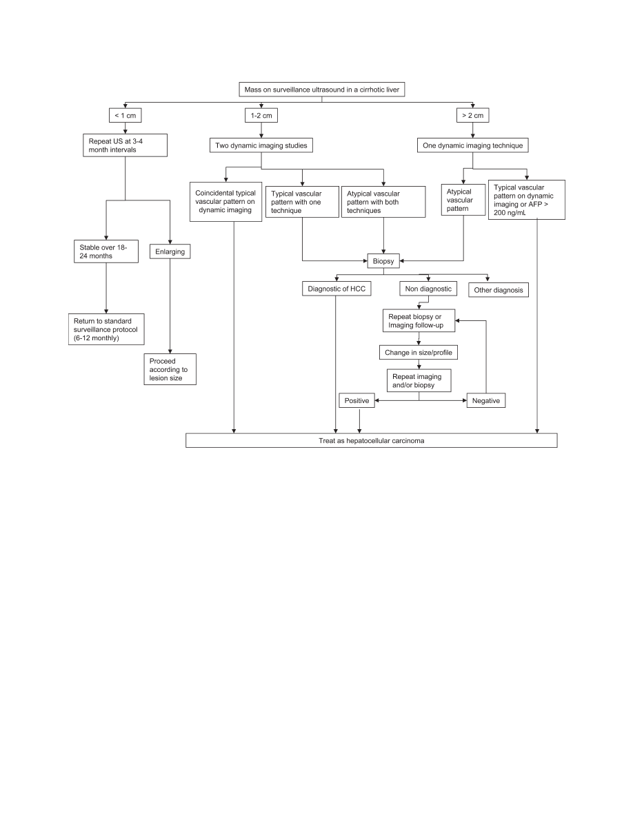
Figure 1 shows the suggested diagnostic strategy after
detection of an hepatic nodule by ultrasound.
Recommendations
7. Nodules found on ultrasound surveillance that
are smaller than 1 cm should be followed with ultra-
1216
BRUIX AND SHERMAN
HEPATOLOGY, November 2005
sound at intervals from 3-6 months (level III). If there
has been no growth over a period of up to 2 years, one
can revert to routine surveillance (level III).
8. Nodules between 1-2 cm found on ultrasound
screening of a cirrhotic liver should be investigated fur-
ther with two dynamic studies, either CT scan, contrast
ultrasound or MRI with contrast. If the appearances are
typical of HCC (i.e., hypervascular with washout in the
portal/venous phase) in two techniques the lesion should
be treated as HCC. If the findings are not characteristic
or the vascular profile is not coincidental among tech-
niques the lesion should be biopsied (level II).
9. If the nodule is larger than 2 cm at initial
diagnosis and has the typical features of HCC on a
dynamic imaging technique, biopsy is not necessary
for the diagnosis of HCC. Alternatively, if the AFP
is > 200 ng/mL biopsy is also not required. However,
if the vascular profile on imaging is not characteristic
or if the nodule is detected in a non-cirrhotic liver,
biopsy should be performed (level II).
10. Biopsies of small lesions should be evaluated by
expert pathologists. If the biopsy is negative for HCC
patients should be followed by ultrasound or CT scan-
ning at 3-6 monthly intervals until the nodule either
disappears, enlarges, or displays diagnostic characteris-
tics of HCC. If the lesion enlarges but remains atypical
for HCC a repeat biopsy is recommended (level III).
Staging Systems
The prognosis of solid tumors is generally related to
tumor stage at presentation and thus tumor stage guides
treatment decisions. However, in HCC patients the pre-
diction of prognosis is more complex because the under-
lying liver function also affects prognosis. There is no
worldwide consensus on the use of any given HCC stag-
Fig. 1.
A suggested algorithm for investigation of a nodule found on ultrasound during screening or surveillance. Note that nodules smaller than
1 cm initially which enlarge over time should be investigated using one of the other two algorithms shown depending on the size of the nodule. The
typical vascular pattern referred to means that the lesion is hypervascular in the arterial phase, and washes out in the portal/venous phase. All other
patterns are considered atypical.
HEPATOLOGY, Vol. 42, No. 5, 2005
BRUIX AND SHERMAN
1217
ing system, and which is the preferred system remains
controversial. Any staging system should classify patients
into subgroups with significantly different outcomes, and
at the same time should help to direct therapy. Histori-
cally, HCC has been classified by the TNM
172
or Okuda
staging systems.
173
The TNM system has been modified
repeatedly
174
and still does not have adequate prognostic
accuracy. In addition, its use is limited because it is based
on pathological findings and liver function is not consid-
ered. An international multicentre study developed a
score based on the evaluation of liver status (cirrhosis vs
no cirrhosis) together with tumor staging according to the
TNM classification,
175
but its validity has only been asse-
sed in patients undergoing resection. The Okuda classifi-
cation takes tumor size (on imaging/surgery) and liver
function into account. It allows the identification of end-
stage disease, but is unable to adequately stratify patients
with early or intermediate stage disease. The Child–Pugh
system
176
and the MELD score
177,178
only consider liver
function and thus, cannot be accurate. Several scoring
systems have been developed in the last few years, at-
tempting to stratify patients according to expected sur-
vival. Schemes have been proposed in Barcelona,
179
France,
180
Italy,
181
Austria,
182
China,
183
and Japan.
184,185
While these systems are able to divide patients into strata
with different prognoses, these schemes are mostly helpful
in identifying end-stage patients with a poor prognosis.
Furthermore, most classification/staging systems do not
take into account the effects of treatment, nor do they
indicate optimal forms of treatment for different disease
stages. Finally, when comparing results among systems
none have been adequately cross-validated. The lack of
reproducibility likely indicates heterogeneity among the
different patient groups and this prevents the develop-
ment of a universal staging system. Recently, Marrero et
al.
186
and Grieco et al.
187
have compared all systems avail-
able and validated the BCLC system in U.S. and Italian
patients, respectively.
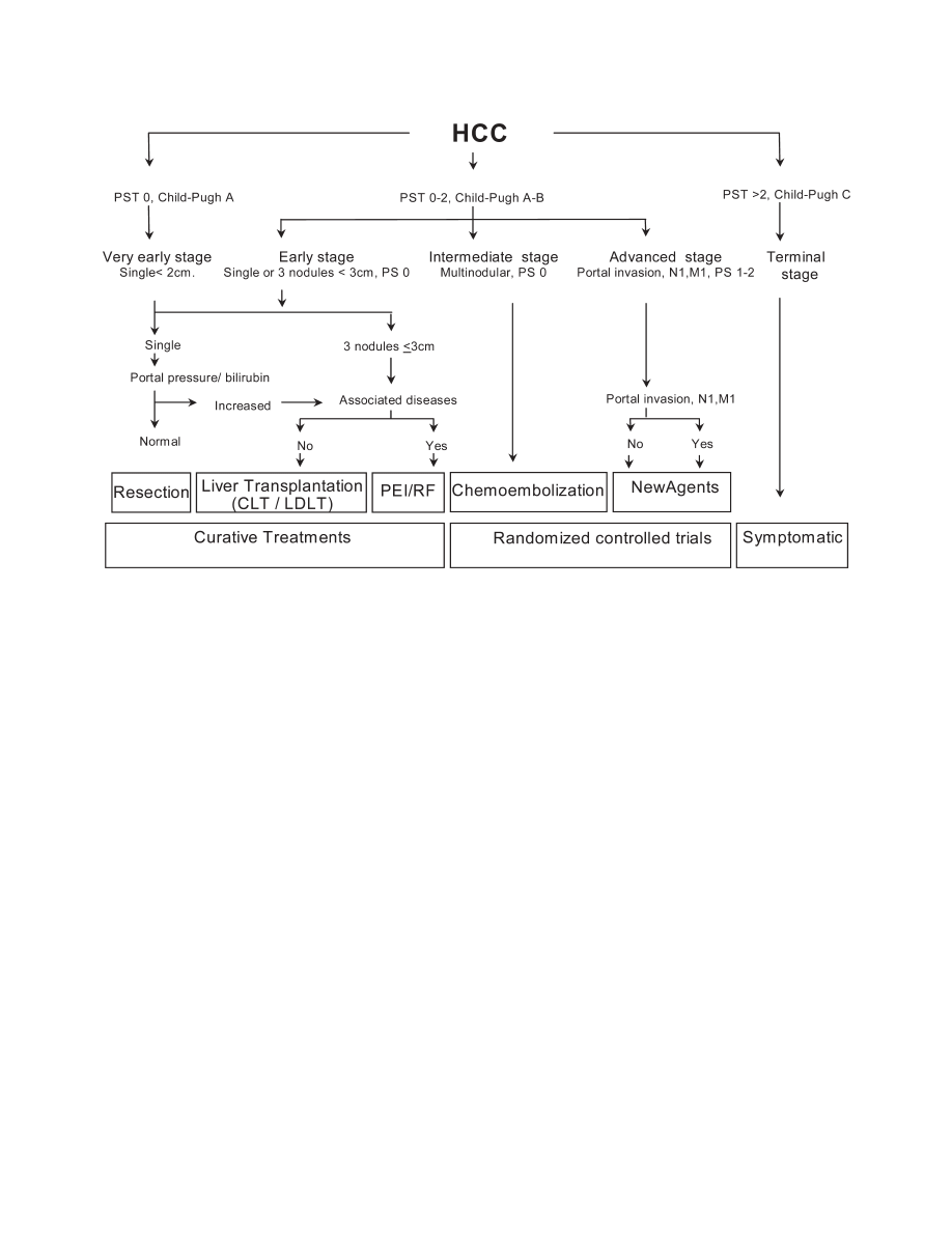
The Barcelona-Clinic- Liver-Cancer (BCLC) stag-
ing system (Fig. 2)
19,188
was developed based on the
combination of data from several independent studies
representing different disease stages and/or treatment
modalities. It includes variables related to tumor stage,
liver functional status, physical status and cancer related
symptoms.
189-191
The main advantage of the BCLC stag-
ing system is that it links staging with treatment modali-
ties and with an estimation of life expectancy that is based
on published response rates to the various treatments. It
identifies those with early HCC who may benefit from
curative therapies, those at intermediate or advanced dis-
ease stage who may benefit from palliative treatments, as
well as those at end-stage with a very poor life expectancy
Fig. 2.
Strategy for staging and treatment assignment in patients diagnosed with HCC according to the BCLC proposal.
1218
BRUIX AND SHERMAN
HEPATOLOGY, November 2005

(Fig. 2). Early stage disease includes patients with pre-
served liver function (Child–Pugh A and B) with solitary
HCC or up to 3 nodules
ⱕ3 cm in size. These patients
can be effectively treated by resection, liver transplanta-
tion or percutaneous ablation with possibility of long
term cure, with 5-year survival figures ranging from 50%
to 75%. Very early HCC is currently very difficult to
diagnose confidently prior to surgical ablation. In these
lesions the absence of microvascular invasion and dissem-
ination offers the highest likelihood of cure and thus, in
Child–Pugh A patients may theoretically achieve a 5-year
survival of almost 100%. The intermediate stage consists
of Child–Pugh A and B patients with large/multifocal
HCC who do not have cancer related symptoms and do
not have macrovascular invasion or extrahepatic spread.
Their survival at 3 years without therapy may reach 50%.
These are the optimal candidates for transarterial chemo-
embolization. Patients who present with cancer symp-
toms and/or with vascular invasion or extrahepatic spread
comprise the advanced stage. They have a shorter life
expectancy (50% survival at 1 year) and are candidates to
enter therapeutic trials with new agents. Finally, patients
with extensive tumor involvement leading to severe dete-
rioration of their physical capacity [WHO performance
status
⬎2] (Table 4)
191
] and/or major impairment of liver
function (Child–Pugh C),
173
are considered end stage.
Their median survival is less than 3 months. Ongoing
genomic and proteomic studies will characterize HCC
more accurately, such that in the future HCC patients
may be classified and treated according to their molecular
profile and not according to the rough evaluation of tu-
mor burden and conventional measures of liver function.
Recommendation
11. To best assess the prognosis of HCC patients it
is recommended that the staging system takes into
account tumor stage, liver function and physical sta-
tus. The impact of treatment should also be considered
when estimating life expectancy. Currently, the BCLC
system is the only staging system that accomplishes
these aims (level II-2).
Treatment of Hepatocellular Carcinoma
Historically, the diagnosis of HCC was almost always
made when the disease was advanced, when patients were
symptomatic and presented with a variable degree of liver
function impairment. At this late stage virtually no treat-
ment had any chance of being effective or of significantly
improving survival. In addition, the morbidity associated
with therapy (which was usually limited to surgical resec-
tion or systemic chemotherapy) was unacceptably high.
Today, many patients are diagnosed at an early stage
when liver function is preserved and there are no cancer-
related symptoms. In addition, there are several active
treatments available that will potentially have a positive
impact on survival.
17
However, to achieve the best out-
comes requires the careful selection of candidates for each
treatment option and the expert application of these treat-
ments. Given the complexity of the disease and the large
number of potentially useful therapies, patients diagnosed
with liver cancer should be referred to multidisciplinary
teams involving hepatologists, pathologists, radiologists,
surgeons and oncologists.
It is important to note that the level of evidence for
most of the therapeutic options is limited to cohort inves-
tigations with few RCT, most of which are limited to the
treatment of advanced disease.
192
There are no studies
that compare treatments considered effective for early
stage disease (surgical resection, transplantation, percuta-
neous ablation) nor are there studies comparing these
methods to no treatment. Hence, any proposed treatment
strategy has to be developed from the analysis of several
published cohorts of treated individuals. Availability of
resources also has to be considered in developing treat-
ment strategies. This is particularly relevant when consid-
ering liver transplantation, which is well established in the
United States and Europe, but in some areas of the world
transplantation is not available or has very limited appli-
cability. For patients with solitary HCC in the setting of
decompensated cirrhosis and for those with early multi-
focal disease (up to 3 lesions, none larger than 3 cm)
193
the
best option is liver transplantation,
17
but for patients with
solitary tumors in well-compensated cirrhosis the optimal
treatment strategy is still under debate.
24
It has become common to assess outcome through the
use of the disease-free survival (DFS) rate. However, al-
though this parameter is clinically informative, it can be
misleading because it is a composite index registering two
events: death and recurrence of tumor (Fig. 3). This is
especially relevant in HCC patients as they usually
present with underlying cirrhosis and thus, they are at risk
of death related either to cirrhosis itself or to tumor pro-
gression. Accordingly, different outcomes in DFS may be
Table 4. World Health Organization Performance
Status grades
Stage 0
Fully active, normal life, no symptoms.
Stage 1
Minor symptoms, able to do light activity.
Stage 2
Capable of self-care but unable to carry out work activities.
Up for more than 50% waking hours
Stage 3
Limited self care capacity. Confined to bed or chair
⬎
50% waking hours.
Stage 4
Completely disabled. Confined to bed or chair.
HEPATOLOGY, Vol. 42, No. 5, 2005
BRUIX AND SHERMAN
1219
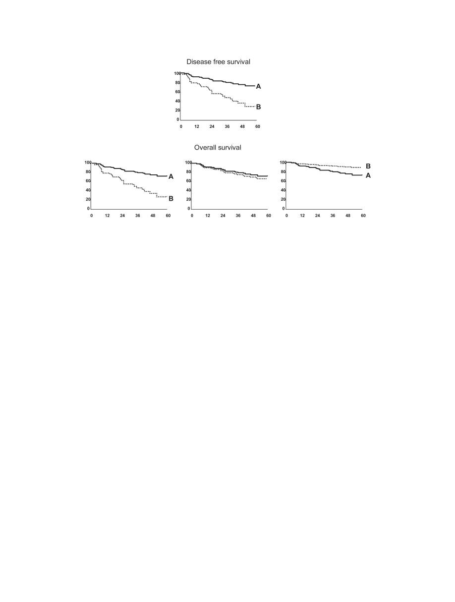
due either to differences in death rate, recurrence rate or
both. Thus, theoretically, although a treatment might be
less active against the tumor than another treatment and
thus result in a higher recurrence rate after initial treat-
ment, the overall survival might not differ or may even be
better.
Thus, the preferred parameter for primary comparison
between different therapies should be survival. These
comments are particularly relevant when discussing what
the first treatment option should be in patients with cir-
rhosis and with early HCC, surgical resection or trans-
plantation.
In the following pages we will review the outcomes that
might be achieved with the different therapeutic options
that are currently available in conventional clinical prac-
tice. We will identify the selection criteria that should be
used to offer each patient the option that provides the best
long term survival.
The therapies that are known to offer a high rate of
complete responses and thus, a potential for cure, are
surgical resection, transplantation and percutaneous abla-
tion.
17
Among non-curative therapies the only one that
has been shown to positively impact survival is transarte-
rial chemoembolization.
192
Other options such as arterial
embolization without chemotherapy
194
or internal radia-
tion do show some antitumor activity,
195
but there is no
proof of their benefit in terms of improved survival. Sys-
temic chemotherapy with several agents has marginal ac-
tivity with frequent toxicity, and is not associated with
improved survival.
196,197
Finally, agents such as tamox-
ifen,
192
anti-androgens,
198,199
or octreotide
200
are com-
pletely ineffective.
Surgical Resection
This is the treatment of choice for HCC in non-cir-
rhotic patients, who account for just 5% of the cases in
Western countries, and for about 40% in Asia. These
patients will tolerate major resections with low morbidity,
but in cirrhosis candidates for resection have to be care-
fully selected to diminish the risk of postoperative liver
failure with increased risk of death. Right hepatectomy in
cirrhotic patients has a higher risk of inducing decompensa-
tion than left hepatectomy. Two decades ago long-term sur-
vival was seldom achieved by resection. Today however, the
5-year survival after resection can exceed 50%.
27,28,159,201,202
Several major advances have increased of the long-term
survival figures. Diagnosis during the asymptomatic
phase of disease together with a more accurate staging of
the patients has allowed the identification of patients with
early stage disease. At the same time, more accurate eval-
uation of the underlying liver function has permitted the
exclusion of those in whom the resection would likely
prompt liver decompensation and death. For years the
selection of candidates for resection has been based on the
Fig. 3.
Disease free survival (DFS)(upper panel) appears to be different between option A (continuous line) and option B (dotted line). However,
the lower panel expresses outcome as overall survival and there are three different potential scenarios. Overall survival might truly be better for option
A (left lower panel). Alternatively, the improved DFS for option A may be the result of a lower recurrence rate but similar death rate; overall survival
would thus be similar to that of the control (central lower panel). Finally, the right lower panel depicts the worst possibility. In this scenario, option
A is highly effective against HCC but is associated with treatment related deaths, while option B is safe but has minimal antitumoral efficacy. Thus, the overall
survival is better with option B, and its lower DFS is due only to the lack of significant antitumoral effect with associated higher recurrence rates.
1220
BRUIX AND SHERMAN
HEPATOLOGY, November 2005

Child–Pugh classification
176
but this is known to have
inconsistent predictive value. Child–Pugh A patients may
already have significant liver functional impairment with
increased bilirubin, significant portal hypertension or
even minor fluid retention requiring diuretic therapy.
176
These features indicate advanced liver disease
203,204
and
preclude resection. Many Japanese groups rely on the In-
docyanine Green retention test. The decision whether
surgery is feasible and the extent of the resection that can
be performed is made based on the degree of retention of
the dye.
205
In contrast, in Europe and the United States,
selection of optimal candidates for resection is usually
based on the assessment of the presence of portal hyper-
tension, as assessed clinically or by hepatic vein catheter-
ization. Studies have shown that a normal bilirubin
concentration, and the absence of clinically significant
portal hypertension measured by hepatic vein catheterisa-
tion (hepatic vein pressure gradient
⬍10 mmHg) are the
best predictors of excellent outcomes after surgery, with
almost no risk for postoperative liver failure.
27,206
Such
patients will not decompensate after resection and may
achieve a 5-year survival of better than 70%.
27,206
In con-
trast, the majority of patients with significant portal hy-
pertension will develop postoperative decompensation
(mostly ascites),
206
with a 5-year survival of less than 50%.
Finally, the survival of those subjects with both adverse
predictors (portal hypertension and elevated bilirubin) is
less than 30% at 5 years, regardless of their Child–Pugh
stage.
27
Therefore, measurement of portal pressure is a
key step in the evaluation of candidates for resection. Ob-
viously, if upper endoscopy shows varices or if diuretic
treatment is needed to control ascites, portal hypertension
is already severe and catheterisation is not necessary. Clin-
ically significant portal hypertension may also be sus-
pected when the platelet count is below 100,000/mm
3
associated with significant splenomegaly.
In recent years surgeons have refined both selection
criteria and surgical techniques. Hence, blood transfusion
may be needed in fewer than 10% of the cases and treatment
related mortality should be less than 1%-3%.
27,205,207
The
use of intra-operative ultrasonography (IOUS) allows
precise localization and staging of the tumor, and also
permits anatomical resections to be performed. From an
oncological perspective anatomic resections that may in-
clude satellite lesions are more sound than limited resec-
tions without a surrounding margin. Pathological studies
in resected tumors provide support for this notion
161
but
some authors have challenged the benefits of a safety mar-
gin
208
and robust evidence is lacking.
Most groups restrict the indication for resection to pa-
tients with single tumor in a suitable location for resection
(as shown by triphasic CT scan, MRI, or other high res-
olution imaging techniques). The size of the tumor is not
a clear-cut limiting factor. As discussed previously, the
risk of vascular invasion and dissemination increases
with size,
161
but some tumors may grow as a large single
mass with no evidence of invasion. In these, surgery
may be safely performed and the risk of recurrence is
not significantly increased as compared to smaller tu-
mors.
27,209
Chemoembolization of the tumor prior to resection
offers no benefit.
210
The same is true for the general use of
portal vein embolization of the hepatic lobe hosting the
tumor
211,212
to induce compensatory liver growth and
functional capacity in the non-affected lobe prior to a
major resection. It has also been suggested that malignant
hepatocytes may also respond to the proliferative stimulus
and this could result in uncontrolled tumor progression.
In addition, portal vein obstruction may induce an acute
increase in portal pressure and result in variceal bleeding.
Clearly, large RCTs are needed to define the benefits and
risks of these procedures.
Risk of Recurrence
After resection, tumor recurrence rate exceeds 70% at 5
years,
27,209,213-216
including recurrence due to dissemina-
tion and de novo tumors.
217,218
The most powerful pre-
dictors of recurrence are the presence of microvascular
invasion and/or additional tumor sites besides the pri-
mary lesion.
27,209,213-216,219
This suggests that the majority
of recurrences are due to dissemination from the primary
tumor and not metachronous tumors developing in a liver
with cirrhosis.
217,220
Furthermore, recurrence due to dis-
semination is more likely to appear during the first 3 years
of follow-up.
218
There is no effective adjuvant therapy
that can reduce recurrence rates.
221
Preoperative chemo-
embolization or adjuvant chemotherapy are not effective
and may complicate the intervention. Internal radia-
tion
222
and adoptive immunotherapy by activated lym-
phocytes
223
may have some anti-tumor efficacy but early
promising results still have to be properly validated. This
is also the case for retinoid administration
224
and inter-
feron therapy.
225,226
As hoped in all cancers,
227-229
molecular profiling of
HCC is expected to refine risk assessment and several
studies have been published trying to correlate abnormal
gene expression with recurrence and outcome.
230-232
However, none of the proposed markers have gained wide
acceptance or become routine in clinical practice.
233
Treatment of recurrence is a poorly investigated area.
Solitary recurrence might benefit from repeat resection,
but in most patients recurrence after primary resection
HEPATOLOGY, Vol. 42, No. 5, 2005
BRUIX AND SHERMAN
1221

will be multifocal because of intra-hepatic dissemination
from the primary tumor.
29,216,234
This reflects an ad-
vanced tumor stage and there is no evidence that any
treatment provides a survival advantage. It has been sug-
gested that patients with recurrence might be candidates
for salvage transplantation.
235
Some retrospective analyses
have suggested that the majority of patients with recur-
rence might benefit from this option.
29
However, this
optimistic suggestion is not supported by an analysis of
clinical outcomes. Most of the recurrences and specially
those that appear early during follow-up are due to tumor
dissemination and have a more aggressive biological pat-
tern as compared to primary tumors.
216,218
Hence, only
those patients in whom recurrence is due to de novo on-
cogenesis can be expected to benefit from salvage trans-
plantation or repeated resection. Since the most accurate
predictors of recurrence due to dissemination (vascular
invasion, satellites) may be identified on pathology, and
since the results of transplantation in these patients is
good, some authors have proposed that this category of
patients should be listed immediately after resection.
236
This might be more effective than waiting for recurrence
to develop with excessive tumor burden possibly exclud-
ing liver transplantation. Organ allocation policies might
have to be modified to take these findings into account.
Recommendations
12. Patients who have a single lesion can be offered
surgical resection if they are non-cirrhotic or have
cirrhosis but still have well preserved liver function,
normal bilirubin and hepatic vein pressure gradient
<
10 mmHg (level II).
13. Pre or post-resection adjuvant therapy is not
recommended (level II)
Liver Transplantation
Patients with HCC were frequently part of the initial
experiences with liver transplantation because of the lack
of alternative treatment and a dismal life expectancy. This
was necessary to establish the feasibility of the interven-
tion. At the same time, the initial results provided the
rationale for the application of strict selection criteria to
candidates who might benefit from the liver transplanta-
tion.
237,238
Patients with HCC that was detected only at
surgery (incidental) because the lesion was too small to be
detected by imaging techniques had an excellent outcome
that did not differ from that of patients with non-malig-
nant disease.
238
These tumors were those that were soli-
tary and smaller than 5 cm. Later experience from
France
239
Italy,
193
Spain,
240
and Germany
30
showed that
excellent results could be achieved in patients with solitary
HCC
⬍5 cm or with up to 3 nodules smaller than 3 cm,
these criteria being known as the Milano criteria after the
seminal study by Mazzaferro et al.
193
The 5-year survival
of these early stage patients exceeds 70%. This has con-
firmed early HCC as a clear indication for liver transplan-
tation in conventional clinical practice.
The need to obtain the optimal benefit from the lim-
ited number of organs that are available has prompted the
maintainance of strict selection criteria so as to list only
those patients with early HCC who have the highest like-
lihood of survival after transplant. However, this means
that some patients with a slightly more advanced HCC in
whom transplant would offer an acceptable, but not ex-
cellent outcome, are excluded from the proce-
dure.
25,241,242
This has recently fuelled a debate about
whether and to what extent the indications for transplan-
tation as therapy for HCC can be expanded.
243
There are
very limited data to support extending the selection crite-
ria. The current more restrictive criteria were developed
when imaging techniques were not as accurate as they are
today and this has always meant a variable degree of un-
derstaging, ranging between 10% and 15%.
165,240,244
At
the same time, in most programs the waiting time for
transplant is long enough that there is a chance that the
HCC will grow beyond the listing criteria. However, for
patients with disease beyond standard listing criteria, if
progression of disease has not been extensive and there is
no macroscopic vascular invasion or extrahepatic spread,
the survival is comparable to patients transplanted for
disease within the standard listing criteria. Most groups
describe a 5-year survival of around 50% in patients trans-
planted for extended criteria
25,242,245
and this is likely the
lowest acceptable survival.
243,246
Thus, it is clear that there
is some room to expand the criteria, but at present there
are no data to define the new limits. Most of the published
studies that support an expansion of the limits are based
on an analysis of explanted livers, information that is not
available prior to surgery.
The most powerful predictor of recurrence in the ab-
sence of extrahepatic spread is macro- or microscopic vas-
cular invasion
30,247
The likelihood of this event runs in
parallel to tumor size and number
150,248
Thus expanding
the listing criteria is a very controversial issue, particularly
when considering the shortage of donors. Tumor differ-
entiation has been proposed to be a predictor for micro-
scopic vascular invasion,
249,250
but its assessment would
require biopsy. Since large tumors are known to be heter-
ogeneous, the accuracy of this strategy for clinical deci-
sion-making would be suboptimal.
The lack of sufficient liver donation is the major limi-
tation for liver transplantation. There is always a waiting
1222
BRUIX AND SHERMAN
HEPATOLOGY, November 2005

period between listing and transplantation. This varies
among programs but if long enough, the tumor will grow
and develop major contraindications (vascular invasion,
extrahepatic spread) to transplantation.
27,251
The rate of
exclusion on the waiting list may be as high as 25% if the
waiting list is longer than 12 months.
251
Obviously, if
patients with more advanced tumors are included as a
result of expanded listing criteria the dropout rate will be
higher and this will translate into poor survival figures on
an intention-to-treat analysis. Studies from Barcelona and
San Francisco have shown that if the dropout rate due to
advancing disease is 25% at 1 year this will translate into
a 60% survival rate for transplantation based on an inten-
tion-to-treat analysis of patients listed for transplant
(rather than those who actually undergo transplanta-
tion).
27,251
Data from Mount Sinai describe a 50% drop-
out rate with an even worse survival if the criteria for
transplant are expanded.
252
Furthermore, one of the most
important issues is the lack of clearly defined criteria for
removing patients from the waiting list because of exces-
sive tumor growth while waiting. If only major events
(macroscopic vascular invasion and extrahepatic spread)
are used to de-list patients this will mean that some pa-
tients will undergo transplantation who have too ad-
vanced disease. This will ultimately impair the survival
figures for transplantation for HCC and put the whole
program at risk. The listing of patients using expanded
criteria will further worsen this scenario and thus, prior to
any change in listing policy, it is essential to define the
exclusion criteria.
Priority Listing for Transplantation
Following a federal request UNOS developed a prior-
ity system to transplant those with the highest short-term
risk of mortality. The MELD score was selected as the
most clinically useful tool for this aim as it accurately
predicts early mortality in chronic liver disease of viral or
alcoholic origin.
253
However, MELD is less powerful in
predicting mortality in cholestatic liver disease and can-
not predict mortality in HCC. To give patients with
HCC equal opportunity for transplantation, HCC pa-
tients were initially given additional points aimed at
matching the risk of death in end-stage cirrhosis: 24
points for solitary HCC
⬍2cm and 29 for solitary HCC 2
to 5cm or 3 nodules each
ⱕ3 cm.
254
After implementa-
tion it was recognised that too high a priority was given to
HCC patients and this was unfair to patients without
cancer.
255
In addition, it was recognized that one fifth of
the patients listed with an HCC diagnosis and who re-
ceived priority, did not have HCC in the explanted liver.
The points for HCC patients were therefore reduced to
20 and 29, to none and 24, respectively, and finally to
none and 22, respectively.
254
In addition, a 10% point
increase is given for every three months on the waiting list.
Results of the new points allocation are unknown. The
major difficulty in setting up fair and equitable priority
policies is that there are no clear predictive data to identify
patients at higher risk of progression and thus, of dropout.
Patients with progression while waiting are clearly at
higher risk, but some may have more aggressive tumors.
Thus, if given excessive priority, the long-term results
may be less than optimal because of HCC recurrence in
the latter subset. Ongoing research should be able to clar-
ify some of these key issues and in the future it should be
possible to use clear clinical and molecular data to make
clinical decisions regarding transplantation in patients
with known HCC.
In addition to the establishment of a priority policy, most
groups treat the HCC upon listing, prior to transplantation.
Unfortunately, this area also lacks robust RCTs comparing
active intervention vs no therapy or comparing several inter-
ventions to each other. All the evidence of benefit currently
available comes from cohort assessments, usually using a per
protocol rather than an intention-to-treat approach, or from
Markov modelling using published clinical outcomes.
117
Despite some encouraging preliminary data,
248
more recent
cohort studies suggest that systemic chemotherapy is ineffec-
tive.
256-258
Most groups perform transarterial chemoemboli-
zation upon listing because it reduces tumor burden and
delays tumor progression.
259
However, it is known that in
patients with decompensated disease this treatment may in-
duce liver failure and death. Hence, it cannot be applied in all
candidates. Patients with small tumors can have ablation
either by percutaneous ethanol injection, radiofrequency or
any other technique and statistical modelling has shown that
such intervention is cost-effective if the expected waiting
time is longer than 6 months.
117
The main concern with this
approach is seeding due to tumor puncture as has been re-
ported for diagnostic biopsy. However, puncture-related
seeding is usually restricted to poorly differentiated tumors
and to peripheral tumors that cannot be approached through
a rim of non-tumoral liver.
260,261
Living Donor OLT
The most effective approach to reduce the dropout rate
on the OLT waiting list is to expand the number of avail-
able livers. Several strategies (domino transplant using
livers extracted from patients with amyloidosis, use of
viral infected livers with minimal damage, split liver trans-
plantation, non-beating heart donors) have been estab-
lished for this purpose, but the best opportunity is the
development of live donation.
262
HEPATOLOGY, Vol. 42, No. 5, 2005
BRUIX AND SHERMAN
1223

After the first successful attempt,
263
more than 3000
living donor operations have been performed worldwide
using the right hepatic lobe. Results from Asia
264-266
and a
recent survey in Japan
267
that includes all the interven-
tions performed suggests that the outcome after live do-
nor transplantation is the same as with cadaveric
donation. Interestingly, the value of the Milano criteria
are further reinforced in this study since the survival and
disease recurrence rates in patients transplanted with
HCC are significantly different according to this straifi-
cation. In any case, long-term data are eagerly awaited.
This is especially relevant for patients with hepatitis C
virus infection in whom the potential severe recurrent
liver disease is a matter of controversy.
268-271
Decision
analysis taking into account the risk of drop-out while
waiting (4% per month), the expected survival of the
recipient (70% at 5 years) and the risk for the donor
(0.3%-0.5% mortality) suggest that this is a cost-effective
approach if the waiting time exceeds 7 months.
272
How-
ever, this is a complex intervention that should only be
undertaken by expert surgeons to ensure the lowest mor-
bidity and best outcome, not only to the recipient, but
also to the donor. Complications may develop in 20% to
40% of the donors and the mortality risk for the donor is
still 0.3% to 0.5%.
262
Finally, even with liver organ do-
nation the number of donors is restricted because of blood
group incompatibility, medical contraindications or psy-
chosocial issues.
The development of living donation has further stim-
ulated the discussion about expansion of the tumor bur-
den limits for HCC patients. Since transplantation can be
done with almost no delay and staging would be recent,
several programs have proposed that living donation
might be a valid option for those patients whose tumor
stage does not allow listing for cadaveric liver transplan-
tation. Cadaveric livers would then be allocated to pa-
tients with the best potential outcome (70% at 5 years),
and living donation livers would benefit patients with a
lower expectancy, around 50% at 5 years. There are no
data to support utilizing such expanded criteria.
Posttransplant Management
There are insufficient data to support or discourage any
specific type of immunosuppression aimed at diminishing
the growth of unrecognised tumor nests disseminated
prior to the operation. Similarly, even if pathology dis-
closes vascular invasion indicating a high risk for HCC
recurrence there is no effective intervention to prevent or
diminish this unfortunate event. The sole aspect that
might be prevented by treatment is the viral reinfection of
the graft. There are several effective strategies for hepatitis
B,
273
but in patients with hepatitis C the situation is less
encouraging. The response rate in those patients who can
receive combined therapy with pegylated interferon and
ribavirin is reduced compared to the pre-transplant situ-
ation.
274
If viral replication persists, the new liver will
develop infection that will cause significant liver damage
leading to cirrhosis in enough patients and will affect both
graft and patient survival.
271,275
Thus, the goal that trans-
plantation may cure both the tumor and the underlying
liver cannot be achieved, at least in the majority of HCC
patients in Japan, the United States, and Europe, where
hepatitis C is the major cause of HCC.
Recommendations
14. Liver transplantation is an effective option for
patients with HCC corresponding to the Milan crite-
ria: solitary tumor <5 cm or up to three nodules <3
cm (level II). Living donor transplantation can be
offered for HCC if the waiting time is long enough to
allow tumor progression leading to exclusion from the
waiting list (level II)./
15. No recommendation can be made regarding
expanding the listing criteria beyond the standard
Milan Criteria (level III).
16. Preoperative therapy can be considered if the
waiting list exceeds 6 months (level II).
Percutaneous Ablation
This is the best treatment option for patients with early
stage HCC who are not suitable for resection or trans-
plantation. In some Japanese centres this is offered as the
first therapeutic option.
276
There are no RCT comparing
local ablation to resection. Destruction of tumor cells can
be achieved by the injection of chemical substances (eth-
anol, acetic acid, boiling saline) or by modifying the tem-
perature (radiofrequency, microwave, laser, cryotherapy).
The efficacy of percutaneous ablation is assessed by dy-
namic CT 1 month after therapy.
3
Although not entirely
reliable, the absence of contrast uptake within the tumor
reflects tumor necrosis, while the persistence of contrast
uptake indicates treatment failure. The recurrence rate
after ablation is as high as for resection. Some recurrences
will occur in the vicinity of the treated nodule and are due
to the presence of microscopic satellites not included in
the ablation zone.
Percutaneous ablation is usually performed under ul-
trasound guidance. Ethanol injection is the best known
and best studied approach.
277,278
It is highly effective for
small HCC and has a low rate of adverse effects. In addi-
tion, it is inexpensive. This should be the standard against
which any new therapy should be compared. Ethanol in-
1224
BRUIX AND SHERMAN
HEPATOLOGY, November 2005

jection achieves necrosis rate of 90%-100% of the HCC
smaller than 2 cm, but the necrosis rate is reduced to 70%
in tumors between 2 and 3 cm and to 50% in HCC
between 3 and 5 cm.
279-282
Long term studies indicate
that Child–Pugh A patients with successful tumor necro-
sis may achieve a 50% survival at 5 years.
28,167,278
This
compares well with the outcome of resection in those
candidates who do not fit the optimal surgical profile.
28
Ethanol injection requires repeated injections on separate
days and rarely accomplishes complete necrosis in tumors
larger than 3 cm, because the injected ethanol cannot
access the entire tumor volume. This may be due to the
presence of intra-tumoral septa. To disrupt septae and
facilitate ethanol infiltration, some authors have proposed
that ethanol injection should be preceded by arterial em-
bolization in large HCC.
283
The rate of initial response is
enhanced but development of viable intra-tumoral nests
or distant recurrence is the rule during follow-up and the
long-term outcome is no different. Thus, there have been
major efforts to develop alternative ablative techniques
that would be able to necrose larger tumors in fewer treat-
ment sessions.
Radiofrequency ablation (RFA) is the option that has
better results in that regard. The insertion of single or
multiple cooled tip electrodes or single electrodes with
J-hooked needles that deliver heat around the tip induces
a wide region of tumor necrosis. The efficacy of RFA in
tumors
⬍2 cm is similar to that of ethanol but requires
fewer treatment sessions.
284,285
The efficacy in tumors
⬎2
cm is better than with ethanol.
284-286
RCT have shown
that RFA provides better local disease control that could
result in an improved survival in RCT.
284-287
Large RCT
comparing these two options in tumors
⬍2 cm and
primarily designed to assess survival are needed.
The main drawback of radiofrequency is its higher
cost and the higher rate (up to 10%) of adverse events
(pleural effusion, peritoneal bleeding).
261,288,289
Procedure-related mortality ranges from 0% to
0.3%.
261,288,289
Subcapsular location and poor tumor
differentiation have been associated with increased risk
of peritoneal seeding
260,261
and thus, this type of tumor
should not be treated with RFA. Since the efficacy of
radiofrequency is based on heat delivery and blood
circulation inside the tumor may prevent proper heat-
ing, some authors have proposed combining radiofre-
quency with simultaneous vessel obstruction.
290
This
manoeuvre may increase the area of necrosis, but the
lack of evidence of a major benefit, together with the
more complex process has prevented its wide imple-
mentation.
Recommendations
17. Local ablation is safe and effective therapy for
patients who cannot undergo resection, or as a bridge
to transplantation (level II).
18. Alcohol injection and radiofrequency are
equally effective for tumors <2 cm. However, the
necrotic effect of radiofrequency is more predictable in
all tumor sizes and in addition, its efficacy is clearly
superior to that of alcohol injection in larger tumors
(level I).
Non-Curative Treatment
As previously discussed, the end-point of therapy is to
extend life expectancy. The only way to demonstrate this for
any therapeutic option is to perform a properly powered
RCT comparing active intervention vs. no treatment. The
systematic review of the English literature during the last 25
years showed only a limited number of RCT that properly
test the efficacy of palliative therapy
192
and the only option
for which there is adequate and positive information is
transarterial chemoembolization.
192
Systemic chemotherapy with any of the available
agents has marginal anti-tumor activity and no impact on
survival.
196,291
Despite this lack of efficacy and the associ-
ated morbidity, chemotherapy (usually doxorubicin) is
frequently administered in conventional clinical practice.
Furthermore, it is also sometimes used as a control arm in
some research studies. This policy must be discouraged,
since if a treatment is thought to be inactive and used as a
placebo, it should at least be non-toxic and easy to admin-
ister. In fact, in the absence of effective therapy, the goal of
health care providers should be to avoid unnecessary suf-
fering with impairment of quality of life. Selective intra-
arterial administration of any chemotherapy agent,
frequently suspended in lipiodol, has also negligible anti-
tumor activity and robust data supporting survival benefit
are lacking.
192,292
Selective radiation through intra-arte-
rial injection of lipiodol-I-131
195,293
or Ytrium-90 labeled
microspheres
294
has some antitumor activity but the im-
pact on survival has not been established.
There are multiple other treatment modalities such as
octreotide,
200,295
interferon,
296
external radiation,
297
tamox-
ifen,
192,298-300
or antiandrogenic therapy,
198,199,301
but none
have been shown to improve survival.
195,200,293-297,302,303
The first studies testing tamoxifen reported encouraging
results
304,305
but unfortunately, larger RCT properly de-
signed, showed unequivocal negative results.
192,298-300
The absence of effect is maintained even when given at
high doses
306
and thus, tamoxifen has no activity in pa-
tients with HCC.
HEPATOLOGY, Vol. 42, No. 5, 2005
BRUIX AND SHERMAN
1225

Some authors have suggested that HCC patients may
have mutated estrogen receptors that cannot be blocked
by tamoxifen,
307
but by megestrol.
308
Again, the small
number of patients in which this agent has been tested
prevents any firm conclusion.
Transarterial Embolization and Chemoembolization
HCC exhibits intense neo-angiogenic activity during
its progression.
150
At very early stages the tumor is not
highly vascularised and its blood supply comes from the
portal vein. As the tumor grows the blood supply becomes
progressively arterialized, so that even well differentiated
HCC is mostly dependent on the hepatic artery for blood
supply. This characteristic provides the pathologic basis
for the radiological characteristics that are used to diag-
nose the disease. It also provides the rationale to support
arterial obstruction as an effective therapeutic option.
Acute arterial obstruction induces ischemic tumor necro-
sis with a high rate of objective responses. Hepatic artery
obstruction is performed during an angiographic proce-
dure and is known as transarterial, or transcatheter arterial
embolization (TAE). When TAE is combined with the
prior injection into the hepatic artery of chemotherapeu-
tic agents, usually mixed with lipiodol, the procedure is
known as transarterial chemoembolization (TACE). He-
patic artery obstruction can be achieved by the injection
or placement of several agents. Gelfoam carefully pre-
pared as 1mm cubes is the most frequently used agent, but
polyvinyl alcohol,
309
alcohol,
310
starch microspheres,
311
metallic coils
312
or even autologous blood clots
313
have
also been used. Gelfoam powder should be not be used as
this may cause biliary damage.
314
The procedure requires the advancement of the cathe-
ter into the hepatic artery and then to lobar and segmental
branches aiming to be as selective as possible so as to
induce only minimal injury to the surrounding non-tu-
morous liver. Multifocal HCC involving both hepatic
lobes may require the obstruction of the total hepatic
artery blood flow.
Chemotherapy has to be injected prior to arterial ob-
struction. It is usual to suspend chemotherapy in lipiodol,
an oily contrast agent used for lymphographic studies.
Lipiodol is selectively retained within the tumor and this
expands the exposure of the neoplastic cells to chemother-
apy. The dose of chemotherapy to be administered has to
be distributed among the affected lobes. If the tumor
affects only one lobe, it is common policy to inject 25% of
the agent into the lobe free of tumor with the objective of
treating potentially undetected clones. Several chemo-
therapeutic agents have been used for TACE, but the
most common is to inject adriamycin or cisplatin.
315
TAE and TACE are considered for patients with non-
surgical HCC that are also ineligible for percutaneous
ablation, provided there is no extrahepatic tumor spread.
The main contraindication is the lack of portal blood flow
(because of portal vein thrombosis, portosystemic anasto-
moses or hepatofugal flow). Patients with lobar or seg-
mental portal vein thrombosis are poor candidates for
TACE, as this will cause necrosis of the tumor and of the
non-tumorous liver deprived of blood supply. This in-
creases the risk of treatment-related death due to liver
failure. Patients with advanced liver disease (Child–Pugh
class B or C) and/or clinical symptoms of end-stage cancer
should not be considered for these treatments as they have
an increased risk of liver failure and death.
The side effects of intra-arterial injection of chemo-
therapy are the same as for systemic administration: nau-
sea, vomiting, bone marrow depression, alopecia and
potentially renal failure. Hepatic artery obstruction with
acute ischemia of the HCC is associated with the so-called
post-embolization syndrome. This appears in more than
50% of the patients and consists of fever, abdominal pain
and a moderate degree of ileus. Fasting is required for 24
hours and IV hydration is mandatory. Prophylactic anti-
biotics are not routinely used
316
Fever is a reflection of
tumor necrosis, but a minority of patients may develop
severe infectious complications such as hepatic abscess or
cholecystitis. The post-embolization syndrome is usually
self-limited in less than 48 hours and the patients can be
discharged from the hospital.
Both TAE and TACE induce extensive tumor necrosis
in more than 50% of the patients.
192
Treatment response
is assessed by the decrease in the concentration of tumor
markers and the identification of large intra-tumoral ne-
crotic areas and reduction in tumor burden in dynamic
CT or MRI.
3
Immediately after arterial obstruction it is
possible to see intra-tumoral bubbles that reflect tissue
liquefaction The evaluation of the treatment response
should take into account the induction of intra-tumoral
necrotic areas in estimating the decrease in tumor load,
and not just a reduction in overall tumor size.
3
According
to conventional WHO criteria the reported rate of objec-
tive responses ranges between 16% and 60%,
192,315
there
being no differences between TAE and TACE. Fewer
than 2% of treated patients achieve a complete response.
During follow-up the residual tumor nests recover their
blood supply and the tumor continues to grow. This con-
sideration prompts treatment repetition either at regular
intervals or “a la demande” as there is no prospective
comparison to support one or other strategy.
315
The tumor progression rate is reduced after treatment
and this translates into a lower risk of vascular invasion.
1226
BRUIX AND SHERMAN
HEPATOLOGY, November 2005

Response to treatment is associated with a significant im-
provement in survival. Cumulative meta-analysis of all
published RCT indicate that patient survival is signifi-
cantly improved.
184
Until very recently, the gain in sur-
vival reported in individual trials was not statistically
significant.
194,317-319
However, studies performed in Bar-
celona
320
and Hong Kong
321
reported a significant impact
on survival have changed this negative statement. It has to
be emphasized that the available trials are heterogeneous
both in terms of patients profile, treatment schedule and
agent used. Thus, it has still to be determined which are
the best obstructing agents, the optimal chemotherapeu-
tics and the most effective re-treatment schedule.
The improvement in survival in treated patients ranges
from 20% to 60% at 2 years,
315
but it is clear that the
relevance of the improvement as compared to their out-
come if untreated, is largely dependent on the patients
baseline characteristics regarding tumor stage, liver func-
tion and general health status.
Recommendations
19. TACE is recommended as first line non-curative
therapy for non-surgical patients with large/multifo-
cal HCC who do not have vascular invasion or extra-
hepatic spread (level I).
20. Tamoxifen, antiandrogens, octreotide or he-
patic artery ligation/embolization are not recom-
mended (level I). Other options such as radio-labelled
Yttrium glass beads, radio-labelled lipiodol or immu-
notherapy cannot be recommended as standard ther-
apy for advanced HCC outside clinical trials.
21. Systemic or selective intra-arterial chemother-
apy is not recommended and should not be used as
standard of care (level II).
Treatment Algorithm
As previously stated, the establishment of an evidence-
based treatment strategy for HCC patients relies on fewer
than one hundred RCT, assessing all of the possible treat-
ment strategies. Almost all the treatment recommenda-
tions, therefore, are based on a critical reading of
observational studies. In the clinical setting patients
should be stratified by disease stage. For each stage there
should be an indicated treatment. This is the basis for the
BCLC scheme as depicted in Fig. 2.
17,19,322
The strategy
combines in a single proposal staging, indicated treatment
and estimation of prognosis, and it can be applied to the
majority of patients evaluated for HCC.
Patients diagnosed at an early HCC stage are optimal
candidates for resection, liver transplantation or percuta-
neous ablation. Resection is considered for patients with
single tumors, absence of clinically relevant portal hyper-
tension and normal bilirubin. Tumor size is not a limiting
factor, but it is uncommon to resect patients with tumors
⬎5cm. Transplantation is considered in patients with 3
nodules
⬍3 cm or with single tumors ⱕ5 cm with liver
function impairment precluding resection. If a long wait-
ing time (
⬎6 months) is expected resection or percutane-
ous treatments are recommended prior to OLT. Living
donor transplantation should also be considered. Percu-
taneous ablation is indicated in patients with small non-
surgical HCC. If these options are not feasible, patients
have to be considered for palliation.
Transarterial chemoembolization is indicated in
asymptomatic patients with multinodular tumors that
have not invaded vessels nor been disseminated outside
the liver. This type of patient is the best candidate for this
approach, particularly if they still meet the criteria for
Child–Pugh A stage. Treated patients who respond to
therapy have an improved survival and thus, this is the last
effective option in conventional clinical practice.
Patients who present with a more advanced stage be-
cause of liver failure or tumor growth with vascular in-
volvement/extrahepatic spread or physical impairment
reflected by a markedly impaired performance status
(
⬍2)
189-191
will not benefit from any treatment option,
even one with known efficacy in earlier disease. Accord-
ingly, the optimal policy for these subjects is to attempt to
enroll them in research studies testing new agents either
within phase 2 investigations or within RCT. The opti-
mal design of such studies should be the comparison of
any intervention vs. placebo or the best supportive care as
currently practiced. There is no proof that any of the
available agents has any impact on survival.
Finally, patients at a terminal stage with deeply im-
paired physical status (performance status
⬎2) and/or
massive tumor burden with heavily impaired liver func-
tion should receive symptomatic treatment to avoid un-
necessary suffering.
Future Perspectives
This practice guideline has depicted the current status
regarding the diagnosis, staging and treatment of HCC.
As discussed, there are several areas where active research
is needed, ranging from molecular pathogenesis to detec-
tion, diagnosis and treatment. The elucidation of the mo-
lecular steps that determine the transition from non-
malignant to malignant should allow the stratification of
patients according to the distinct pathways that led to
cancer and also provide for new preventive and therapeu-
tic strategies. Identification of new biomarkers to establish
the risk of cancer and/or detect its appearance at a preclin-
HEPATOLOGY, Vol. 42, No. 5, 2005
BRUIX AND SHERMAN
1227

ical stage are urgently needed. The current therapeutic
approach also needs significant improvement. Treat-
ments able to provide initial cure are hampered by a sig-
nificant rate of disease recurrence and there is also a need
for effective adjuvant therapies. Finally, the therapeutic
options for patients with advanced HCC have limited
impact and thus, development of new agents and strate-
gies for this group of patients is of major relevance. For-
tunately, the awareness of these needs by official agencies
such as the National Institutes of Health has increased the
resources allocated for sponsoring research in this area.
Hence, the action plan of the liver disease section (ww-
w.niddk.nih.gov/fund/divisions/ddn/ldrb/ldrb_action-
_plan.htm) includes specific goals in the field of liver can-
cer. Hopefully, in the years to come the management of
patients with HCC will offer a completely different perspec-
tive in which both prevention and treatment will have sig-
nificantly decreased the number of HCC related deaths.
Acknowledement: The authors thank the AASLD Prac-
tice Guidelines Committee for their help in the prepara-
tion of this practice guideline. This committee, in concert
with F. Fred Poordad, M.D., and Margaret C. Shuhart,
M.D., M.S., as the lead reviewers, provided extensive peer
review of this manuscript. The members of the Practice
Guidelines Committee include K. Rajender Reddy,
M.D., Chair, Andres Cardenas, M.D., MMSc, Robert L.
Carithers, Jr., M.D., Stanley M. Cohen, M.D., Timothy
J. Davern, M.D., Thomas W. Faust, M.D., Steven L.
Flamm, M.D., Gregory J. Gores, M.D., Steven-Huy B.
Han, M.D., Elizabeth Hespenheide, MSN, ACNP, Mi-
chael R. Lucey, M.D., David R. Nelson, M.D., F. Fred
Poordad, M.D., Margaret C. Shuhart, M.D., MS, Brent
A. Tetri, M.D., Zobair M. Younossi, M.D., MPH, and
Nizar N. Zein, M.D..
References
1. Eddy DM. Manual for Assessing Health Practices and Designing Practice
Guidelines. Philadelphia, PA: American College of Physicians, 1996.
2. American Gastroenterological Association Medical Position Statement:
guidelines for the use of enteral nutrition. Gastroenterology 1995;108:
1280-1281.
3. Bruix J, Sherman M, Llovet JM, Beaugrand M, Lencioni R, Burroughs
AK, et al. Clinical Management of Hepatocellular carcinoma: conclu-
sions of the Barcelona–2000 EASL Conference. J Hepatol 2001;35:421-
430.
4. El Serag HB, Mason AC. Rising incidence of hepatocellular carcinoma in
the United States. N Engl J Med 1999; 340:745-750.
5. Parkin DM, Bray F, Ferlay J, Pisani P. Estimating the world cancer
burden: Globocan 2000. Int J Cancer 2001;94:153-156.
6. Deuffic S, Poynard T, Buffat L, Valleron AJ. Trends in primary liver
cancer. Lancet 1998;351:214-215.
7. Stroffolini T, Andreone P, Andriulli A, Ascione A, Craxi A, Chiaramonte
M, et al. Characteristics of hepatocellular carcinoma in Italy. J Hepatol
1998;29:944-952.
8. Taylor-Robinson SD, Foster GR, Arora S, Hargreaves S, Thomas HC.
Increase in primary liver cancer in the UK, 1979-94. Lancet 1997;350:
1142-1143.
9. Bosch FX, Ribes J, Diaz M, Cleries R. Primary liver cancer: worldwide
incidence and trends. Gastroenterology 2004;127(5 Suppl 1):S5-S16.
10. Zhang BH, Yang BH, Tang ZY. Randomized controlled trial of screening
for hepatocellular carcinoma. J Cancer Res Clin Oncol 2004;130:417-
422.
11. Chen JG, Parkin DM, Chen QG, Lu JH, Shen QJ, Zhang BC, et al.
Screening for liver cancer: results of a randomised controlled trial in
Qidong, China. J Med Screen 2003;10:204-209.
12. Welch HG, Schwartz LM, Woloshin S. Are increasing 5-year survival
rates evidence of success against cancer? JAMA 2000;283:2975-2978.
13. McMahon BJ, Bulkow L, Harpster A, Snowball M, Lanier A, Sacco et al.
Screening for hepatocellular carcinoma in Alaska natives infected with
chronic hepatitis B: a 16-year population-based study. H
EPATOLOGY
2000;32(4 Pt 1):842-846.
14. Wong LL, Limm WM, Severino R, Wong LM. Improved survival with
screening for hepatocellular carcinoma. Liver Transpl 2000;6:320-325.
15. Oka H, Kurioka N, Kim K, Kanno T, Kuroki T, Mizoguchi Y, et al.
Prospective study of early detection of hepatocellular carcinoma in pa-
tients with cirrhosis. H
EPATOLOGY
1990;12(4 Pt 1):680-687.
16. Bolondi L, Sofia S, Siringo S, Gaiani S, Casali A, Zironi G, et al. Surveil-
lance programme of cirrhotic patients for early diagnosis and treatment of
hepatocellular carcinoma: a cost-effectiveness analysis. Gut 2001;48:251-
259.
17. Llovet JM, Burroughs A, Bruix J. Hepatocellular carcinoma. Lancet
2003;362:1907-1917.
18. Liu JH, Chen PW, Asch SM, Busuttil RW, Ko CY. Surgery for hepato-
cellular carcinoma: does it improve survival? Ann Surg Oncol 2004;11:
298-303.
19. Llovet JM, Bru C, Bruix J. Prognosis of hepatocellular carcinoma: the
BCLC staging classification. Semin Liver Dis 1999;19:329-338.
20. Yamamoto J, Iwatsuki S, Kosuge T, Dvorchik I, Shimada K, Marsh JW,
et al. Should hepatomas be treated with hepatic resection or transplanta-
tion? Cancer 1999;86:1151-1158.
21. Molmenti EP, Marsh JW, Dvorchik I, Oliver JH III, Madariaga J, Iwat-
suki S. Hepatobiliary malignancies. Primary hepatic malignant neo-
plasms. Surg Clin North Am 1999;79:43-57, viii.
22. Bismuth H, Majno PE, Adam R. Liver transplantation for hepatocellular
carcinoma. Semin Liver Dis 1999;19:311-322.
23. Michel J, Suc B, Montpeyroux F, Hachemanne S, Blanc P, Domergue J,
et al. Liver resection or transplantation for hepatocellular carcinoma?
Retrospective analysis of 215 patients with cirrhosis. J Hepatol 1997;26:
1274-1280.
24. Llovet JM, Bruix J, Gores GJ. Surgical resection versus transplantation for
early hepatocellular carcinoma: clues for the best strategy. H
EPATOLOGY
2000;31:1019-1021.
25. Yao FY, Ferrell L, Bass NM, Watson JJ, Bacchetti P, Venook A, et al.
Liver transplantation for hepatocellular carcinoma: expansion of the tu-
mor size limits does not adversely impact survival. H
EPATOLOGY
2001;
33:1394-1403.
26. Otto G, Heuschen U, Hofmann WJ, Krumm G, Hinz U, Herfarth C.
Survival and recurrence after liver transplantation versus liver resection
for hepatocellular carcinoma: a retrospective analysis. Ann Surg 1998;
227:424-432.
27. Llovet JM, Fuster J, Bruix J. Intention-to-Treat Analysis of Surgical
Treatment for Early Hepatocellular Carcinoma: Resection Versus Trans-
plantation. H
EPATOLOGY
1999;30:1434-1440.
28. Arii S, Yamaoka Y, Futagawa S, Inoue K, Kobayashi K, Kojiro M, et al.
Results of surgical and nonsurgical treatment for small-sized hepatocel-
lular carcinomas: A retrospective and nationwide survey in japan. H
EPA
-
TOLOGY
2000;32:1224-1229.
29. Poon RT, Fan ST, Lo CM, Liu CL, Wong J. Long-term survival and
pattern of recurrence after resection of small hepatocellular carcinoma in
1228
BRUIX AND SHERMAN
HEPATOLOGY, November 2005

patients with preserved liver function: implications for a strategy of sal-
vage transplantation. Ann Surg 2002;235:373-382.
30. Jonas S, Bechstein WO, Steinmuller T, Herrmann M, Radke C, Berg T,
et al. Vascular invasion and histopathologic grading determine outcome
after liver transplantation for hepatocellular carcinoma in cirrhosis.
H
EPATOLOGY
2001;33:1080-1086.
31. Naimark D, Naglie G, Detsky AS. The meaning of life expectancy: what
is a clinically significant gain? J Gen Intern Med 1994;9:702-707.
32. Laupacis A, Feeny D, Detsky AS, Tugwell PX. How attractive does a new
technology have to be to warrant adoption and utilization? Tentative
guidelines for using clinical and economic evaluations. CMAJ 1992;146:
473-481.
33. Sarasin FP, Giostra E, Hadengue A. Cost-effectiveness of screening for
detection of small hepatocellular carcinoma in western patients with
Child-Pugh class A cirrhosis. Am J Med 1996;101:422-434.
34. Arguedas MR, Chen VK, Eloubeidi MA, Fallon MB. Screening for hep-
atocellular carcinoma in patients with hepatitis C cirrhosis: a cost-utility
analysis. Am J Gastroenterol 2003;98679-690.
35. Lin OS, Keeffe EB, Sanders GD, Owens DK. Cost-effectiveness of
screening for hepatocellular carcinoma in patients with cirrhosis due to
chronic hepatitis C. Aliment Pharmacol Ther 2004;19:1159-1172.
36. Beasley RP, Hwang LY, Lin CC, Chien CS. Hepatocellular carcinoma
and hepatitis B virus. A prospective study of 22 707 men in Taiwan.
Lancet 1981;2:1129-1133.
37. Koike K, Tsutsumi T, Fujie H, Shintani Y, Kyoji M. Molecular mecha-
nism of viral hepatocarcinogenesis. Oncology 2002;62(Suppl 1):29-37.
38. Beasley RP. Hepatitis B virus as the etiologic agent in hepatocellular
carcinoma. H
EPATOLOGY
1982;2(Suppl):21S-26S.
39. Sakuma K, Saitoh N, Kasai M, Jitsukawa H, Yoshino I, Yamaguchi M et
al. Relative risks of death due to liver disease among Japanese male adults
having various statuses for hepatitis B s and e antigen/antibody in serum:
a prospective study. H
EPATOLOGY
1988;8:1642-1646.
40. Villeneuve JP, Desrochers M, Infante-Rivard C, Willems B, Raymond G,
Bourcier M et al. A long-term follow-up study of asymptomatic hepatitis
B surface antigen-positive carriers in Montreal. Gastroenterology 1994;
106:1000-1005.
41. McMahon BJ, Alberts SR, Wainwright RB, Bulkow L, Lanier AP. Hep-
atitis B-related sequelae. Prospective study of 1400 hepatitis B surface
antigen-positive Alaska native carriers. Arch Intern Med 1990;150:1051-
1054.
42. Sherman M, Peltekian KM, Lee C. Screening for hepatocellular carci-
noma in chronic carriers of hepatitis B virus: incidence and prevalence of
hepatocellular carcinoma in a North American urban population. H
EPA
-
TOLOGY
1995;22:432-438.
43. Fattovich G, Brollo L, Giustina G, Noventa F, Pontisso P, Alberti A, et al.
Natural history and prognostic factors for chronic hepatitis type B. Gut
1991;32:294-298.
44. Manno M, Camma C, Schepis F, Bassi F, Gelmini R, Giannini F et al.
Natural history of chronic HBV carriers in northern Italy: morbidity and
mortality after 30 years. Gastroenterology 2004;127:756-763.
45. Hsu YS, Chien RN, Yeh CT, Sheen IS, Chiou HY, Chu CM, et al.
Long-term outcome after spontaneous HBeAg seroconversion in patients
with chronic hepatitis B. H
EPATOLOGY
2002;35:1522-1527.
46. de Franchis R, Meucci G, Vecchi M, Tatarella M, Colombo M, Del
Ninno E, et al. The natural history of asymptomatic hepatitis B surface
antigen carriers. Ann Intern Med 1993;118:191-194.
47. Sanchez-Tapias JM, Costa J, Mas A, Bruguera M, Rodes J. Influence of
hepatitis B virus genotype on the long-term outcome of chronic hepatitis
B in western patients. Gastroenterology 2002;123:1848-1856.
48. Fattovich G. Natural history of hepatitis B. J Hepatol 2003;39(Suppl
1):S50-S58.
49. Yang HI, Lu SN, Liaw YF, You SL, Sun CA, Wang LY, et al. Hepatitis B
e antigen and the risk of hepatocellular carcinoma. N Engl J Med 2002;
347:168-174.
50. Evans AA, Chen G, Ross EA, Shen FM, Lin WY, London WT. Eight-
year follow-up of the 90,000-person Haimen City cohort: I. Hepatocel-
lular carcinoma mortality, risk factors, and gender differences. Cancer
Epidemiol Biomarkers Prev 2002;11:369-376.
51. Huo TI, Wu JC, Lee PC, Chau GY, Lui WY, Tsay SH, et al. Sero-
clearance of hepatitis B surface antigen in chronic carriers does not nec-
essarily imply a good prognosis. H
EPATOLOGY
1998;28:231-236.
52. Yuen MF, Wong DK, Sablon E, Tse E, Ng IO, Yuan HJ et al. HBsAg
seroclearance in chronic hepatitis B in the Chinese: virological, histolog-
ical, and clinical aspects. H
EPATOLOGY
2004;39:1694-1701.
53. Fattovich G, Giustina G, Realdi G, Corrocher R, Schalm SW. Long-term
outcome of hepatitis B e antigen-positive patients with compensated
cirrhosis treated with interferon alfa. European Concerted Action on
Viral Hepatitis (EUROHEP). H
EPATOLOGY
1997;26:1338-1342.
54. Fattovich G, Giustina G, Sanchez-Tapias J, Quero C, Mas A, Olivotto
PG et al. Delayed clearance of serum HBsAg in compensated cirrhosis B:
relation to interferon alpha therapy and disease prognosis. European
Concerted Action on Viral Hepatitis (EUROHEP) [see comments]. Am J
Gastroenterol 1998;93:896-900.
55. Yu MW, Chang HC, Liaw YF, Lin SM, Lee SD, Liu CJ, et al. Familial
risk of hepatocellular carcinoma among chronic hepatitis B carriers and
their relatives. J Natl Cancer Inst 2000;92:1159-1164.
56. Kew MC, Marcus R, Geddes EW. Some characteristics of Mozambican
Shangaans with primary hepatocellular cancer. S Afr Med J 1977;51:306-
309.
57. Kew MC, Macerollo P. Effect of age on the etiologic role of the hepatitis
B virus in hepatocellular carcinoma in blacks. Gastroenterology 1988;94:
439-442.
58. Bellentani S, Dal Morin G, Miglioli L, Croce L, Masutti F, Castiglione A.
Natural history of HBV infection: a nine year follow-up of the Dionysius
cohort. J Hepatology 2002;36:228S.
59. Fattovich G, Giustina G, Degos F, Tremolada F, Diodati G, Almasio P et
al. Morbidity and mortality in compensated cirrhosis type C: a retrospec-
tive follow-up study of 384 patients. Gastroenterology 1997;112:463-
472.
60. Niederau C, Lange S, Heintges T, Erhardt A, Buschkamp M, Hurter D,
et al. Prognosis of chronic hepatitis C: results of a large, prospective
cohort study. H
EPATOLOGY
1998;28:1687-1695.
61. Niederau C, Heintges T, Lange S, Goldmann G, Niederau CM, Mohr L
et al. Long-term follow-up of HBeAg-positive patients treated with inter-
feron alfa for chronic hepatitis B. N Engl J Med 1996;334:1422-1427.
62. Degos F, Christidis C, Ganne-Carrie N, Farmachidi JP, Degott C,
Guettier C, et al. Hepatitis C virus related cirrhosis: time to occurrence of
hepatocellular carcinoma and death. Gut 2000;47:131-136.
63. Sun CA, Wu DM, Lin CC, Lu SN, You SL, Wang LY et al. Incidence and
cofactors of hepatitis C virus-related hepatocellular carcinoma: a prospec-
tive study of 12,008 men in Taiwan. Am J Epidemiol 2003;157:674-682.
64. Yoshida H, Shiratori Y, Moriyama M, Arakawa Y, Ide T, Sata M, et al.
Interferon therapy reduces the risk for hepatocellular carcinoma: national
surveillance program of cirrhotic and noncirrhotic patients with chronic
hepatitis C in Japan. IHIT Study Group. Inhibition of Hepatocarcino-
genesis by Interferon Therapy. Ann Intern Med 1999;131:174-181.
65. Imbert-Bismut F, Ratziu V, Pieroni L, Charlotte F, Benhamou Y, Poy-
nard T. Biochemical markers of liver fibrosis in patients with hepatitis C
virus infection: a prospective study. Lancet 2001; 357:1069-1075.
66. Forns X, Ampurdanes S, Llovet JM, Aponte J, Quinto L, Martinez-Bauer
E, et al. Identification of chronic hepatitis C patients without hepatic
fibrosis by a simple predictive model. H
EPATOLOGY
2002;36(4 Pt 1):986-
992.
67. Callewaert N, Van Vlierberghe H, Van Hecke A, Laroy W, Delanghe J,
Contreras R. Noninvasive diagnosis of liver cirrhosis using DNA se-
quencer-based total serum protein glycomics. Nat Med 2004;10:429-
434.
68. Moriyama M, Matsumura H, Aoki H, Shimizu T, Nakai K, Saito T, et al.
Long-term outcome, with monitoring of platelet counts, in patients with
chronic hepatitis C and liver cirrhosis after interferon therapy. Intervirol-
ogy 2003;46:296-307.
HEPATOLOGY, Vol. 42, No. 5, 2005
BRUIX AND SHERMAN
1229

69. Velazquez RF, Rodriguez M, Navascues CA, Linares A, Perez R, Sotorrios
NG, et al. Prospective analysis of risk factors for hepatocellular carcinoma
in patients with liver cirrhosis. H
EPATOLOGY
2003;37:520-527.
70. Giannini E, Risso D, Botta F, Chiarbonello B, Fasoli A, Malfatti F, et al.
Validity and clinical utility of the aspartate aminotransferase-alanine ami-
notransferase ratio in assessing disease severity and prognosis in patients
with hepatitis C virus-related chronic liver disease. Arch Intern Med
2003;163:218-224.
71. Pineda JA, Romero-Gomez M, Diaz-Garcia F, Giron-Gonzalez JA,
Montero JL, Torre-Cisneros J, et al. HIV coinfection shortens the sur-
vival of patients with hepatitis C virus-related decompensated cirrhosis.
H
EPATOLOGY
2005;41:779-789.
72. Giordano TP, Kramer JR, Souchek J, Richardson P, El Serag HB. Cir-
rhosis and hepatocellular carcinoma in HIV-infected veterans with and
without the hepatitis C virus: a cohort study, 1992-2001. Arch Intern
Med 2004;164:2349-2354.
73. Rosenthal E, Poiree M, Pradier C, Perronne C, Salmon-Ceron D, Geffray
L, et al. Mortality due to hepatitis C-related liver disease in HIV-infected
patients in France (Mortavic 2001 study). AIDS 2003;17:1803-1809.
74. Puoti M, Bruno R, Soriano V, Donato F, Gaeta GB, Quinzan GP, et al.
Hepatocellular carcinoma in HIV-infected patients: epidemiological fea-
tures, clinical presentation and outcome. AIDS 2004;18:2285-2293.
75. Coelho-Little ME, Jeffers LJ, Bernstein DE, Goodman JJ, Reddy KR, De
Medina M, et al. Hepatitis C virus in alcoholic patients with and without
clinically apparent liver disease. Alcohol Clin Exp Res 1995;19:1173-
1176.
76. Befrits R, Hedman M, Blomquist L, Allander T, Grillner L, Kinnman N,
et al. Chronic hepatitis C in alcoholic patients: prevalence, genotypes, and
correlation to liver disease. Scand J Gastroenterol 1995;30:1113-1118.
77. Bode JC, Alscher DM, Wisser H, Bode C. Detection of hepatitis C virus
antibodies and hepatitis C virus RNA in patients with alcoholic liver
disease. Alcohol Alcohol 1995;30:97-103.
78. Hassan MM, Hwang LY, Hatten CJ, Swaim M, Li D, Abbruzzese JL et al.
Risk factors for hepatocellular carcinoma: synergism of alcohol with viral
hepatitis and diabetes mellitus. H
EPATOLOGY
2002;36:1206-1213.
79. Schoniger-Hekele M, Muller C, Kutilek M, Oesterreicher C, Ferenci P,
Gangl A. Hepatocellular carcinoma in Austria: aetiological and clinical
characteristics at presentation. Eur J Gastroenterol Hepatol 2000;12:
941-948.
80. El Serag HB, Mason AC. Risk factors for the rising rates of primary liver
cancer in the United States. Arch Intern Med 2000;160:3227-3230.
81. Adami HO, Chow WH, Nyren O, Berne C, Linet MS, Ekbom A, et al.
Excess risk of primary liver cancer in patients with diabetes mellitus.
J Natl Cancer Inst 1996;88:1472-1477.
82. Bugianesi E, Leone N, Vanni E, Marchesini G, Brunello F, Carucci P et
al. Expanding the natural history of nonalcoholic steatohepatitis: From
cryptogenic cirrhosis to hepatocellular carcinoma. Gastroenterology
2002;123:134-140.
83. Shimada M, Hashimoto E, Taniai M, Hasegawa K, Okuda H, Hayashi
N, et al. Hepatocellular carcinoma in patients with non-alcoholic steato-
hepatitis. J Hepatol 2002;37:154-160.
84. Elmberg M, Hultcrantz R, Ekbom A, Brandt L, Olsson S, Olsson R, et al.
Cancer risk in patients with hereditary hemochromatosis and in their
first-degree relatives. Gastroenterology 2003;125:1733-1741.
85. Hsing AW, McLaughlin JK, Olsen JH, Mellemkjar L, Wacholder S,
Fraumeni JF Jr. Cancer risk following primary hemochromatosis: a pop-
ulation-based cohort study in Denmark. Int J Cancer 1995;60:160-162.
86. Fracanzani AL, Conte D, Fraquelli M, Taioli E, Mattioli M, Losco A, et
al. Increased cancer risk in a cohort of 230 patients with hereditary hemo-
chromatosis in comparison to matched control patients with non-iron-
related chronic liver disease. H
EPATOLOGY
2001;33:647-651.
87. Caballeria L, Pares A, Castells A, Gines A, Bru C, Rodes J. Hepatocellular
carcinoma in primary biliary cirrhosis: similar incidence to that in hepa-
titis C virus-related cirrhosis. Am J Gastroenterol 2001;96:1160-1163.
88. Elzouki AN, Eriksson S. Risk of hepatobiliary disease in adults with severe
alpha 1-antitrypsin deficiency (PiZZ): is chronic viral hepatitis B or C an
additional risk factor for cirrhosis and hepatocellular carcinoma? Eur J
Gastroenterol Hepatol 1996;8:989-994.
89. Eriksson S, Carlson J, Velez R. Risk of cirrhosis and primary liver cancer
in alpha 1-antitrypsin deficiency. N Engl J Med 1986;314:736-739.
90. Fattovich G, Giustina G, Degos F, Diodati G, Tremolada F, Nevens F et
al. Effectiveness of interferon alfa on incidence of hepatocellular carci-
noma and decompensation in cirrhosis type C. European Concerted Ac-
tion on Viral Hepatitis (EUROHEP). J Hepatol 1997;27:201-205.
91. Benvegnu L, Chemello L, Noventa F, Fattovich G, Pontisso P, Alberti A.
Retrospective analysis of the effect of interferon therapy on the clinical
outcome of patients with viral cirrhosis. Cancer 1998;83:901-909.
92. Lin SM, Sheen IS, Chien RN, Chu CM, Liaw YF. Long-term beneficial
effect of interferon therapy in patients with chronic hepatitis B virus
infection. H
EPATOLOGY
1999;29:971-975.
93. Yuen MF, Hui CK, Cheng CC, Wu CH, Lai YP, Lai CL. Long-term
follow-up of interferon alfa treatment in Chinese patients with chronic
hepatitis B infection: The effect on hepatitis B e antigen seroconversion
and the development of cirrhosis-related complications. H
EPATOLOGY
2001;34:139-145.
94. Liaw YF, Sung JJ, Chow WC, Farrell G, Lee CZ, Yuen H, et al. Lami-
vudine for patients with chronic hepatitis B and advanced liver disease.
N Engl J Med 2004;351:1521-1531.
95. Nishiguchi S, Shiomi S, Nakatani S, Takeda T, Fukuda K, Tamori A, et
al. Prevention of hepatocellular carcinoma in patients with chronic active
hepatitis C and cirrhosis. Lancet 2001;357:196-197.
96. Valla DC, Chevallier M, Marcellin P, Payen JL, Trepo C, Fonck M, et al.
Treatment of hepatitis C virus-related cirrhosis: a randomized, controlled
trial of interferon alfa-2b versus no treatment. H
EPATOLOGY
1999;29:
1870-1875.
97. Camma C, Giunta M, Andreone P, Craxi A. Interferon and prevention of
hepatocellular carcinoma in viral cirrhosis: an evidence-based approach.
J Hepatol 2001;34:593-602.
98. HCC-Int.Interferon-alpha StudyGroup. Effect of interferon-alpha on
progression of cirrhosis to hepatocellular carcinoma: a retrospective co-
hort study. International Interferon-alpha Hepatocellular Carcinoma
Study Group. Lancet 1998;351:1535-1539.
99. Hayashi K, Kumada T, Nakano S, Takeda I, Kiriyama S, Sone Y, et al.
Incidence of hepatocellular carcinoma in chronic hepatitis C after inter-
feron therapy. Hepatogastroenterology 2002;49:508-512.
100. Ikeda K, Saitoh S, Kobayashi M, Suzuki Y, Suzuki F, Tsubota A, et al.
Long-term interferon therapy for 1 year or longer reduces the hepatocel-
lular carcinogenesis rate in patients with liver cirrhosis caused by hepatitis
C virus: a pilot study. J Gastroenterol Hepatol 2001;16:406-415.
101. Tanaka H, Tsukuma H, Kasahara A, Hayashi N, Yoshihara H, Masuzawa
M, et al. Effect of interferon therapy on the incidence of hepatocellular
carcinoma and mortality of patients with chronic hepatitis C: a retrospec-
tive cohort study of 738 patients. Int J Cancer 2000;87:741-749.
102. Okanoue T, Itoh Y, Minami M, Sakamoto S, Yasui K, Sakamoto M, et al.
Interferon therapy lowers the rate of progression to hepatocellular carci-
noma in chronic hepatitis C but not significantly in an advanced stage: a
retrospective study in 1148 patients. Viral Hepatitis Therapy Study
Group. J Hepatol 1999;30:653-659.
103. Toyoda H, Kumada T, Nakano S, Takeda I, Sugiyama K, Kiriyama S, et
al. Effect of the dose and duration of interferon-alpha therapy on the
incidence of hepatocellular carcinoma in noncirrhotic patients with a
nonsustained response to interferon for chronic hepatitis C. Oncology
2001;61:134-142.
104. Oka H, Tamori A, Kuroki T, Kobayashi K, Yamamoto S. Prospective
study of alpha-fetoprotein in cirrhotic patients monitored for develop-
ment of hepatocellular carcinoma. H
EPATOLOGY
1994;19:61-66.
105. Zhang JY, Wang X, Han SG, Zhuang H. A case-control study of risk
factors for hepatocellular carcinoma in Henan, China. Am J Trop Med
Hyg 1998;59:947-951.
106. Colombo M, de Franchis R, Del Ninno E, Sangiovanni A, De Fazio C,
Tommasini M, et al. Hepatocellular carcinoma in Italian patients with
cirrhosis. N Engl J Med 1991; 325:675-680.
1230
BRUIX AND SHERMAN
HEPATOLOGY, November 2005

107. Hytiroglou P, Theise ND, Schwartz M, Mor E, Miller C, Thung SN.
Macroregenerative nodules in a series of adult cirrhotic liver explants:
issues of classification and nomenclature. H
EPATOLOGY
1995;21:703-
708.
108. Ganne-Carrie N, Chastang C, Chapel F, Munz C, Pateron D, Sibony M,
et al. Predictive score for the development of hepatocellular carcinoma
and additional value of liver large cell dysplasia in Western patients with
cirrhosis. H
EPATOLOGY
1996;23:1112-1118.
109. Lee RG, Tsamandas AC, Demetris AJ. Large cell change (liver cell dys-
plasia) and hepatocellular carcinoma in cirrhosis: matched case-control
study, pathological analysis, and pathogenetic hypothesis. H
EPATOLOGY
1997;26:1415-1422.
110. Shibata M, Morizane T, Uchida T, Yamagami T, Onozuka Y, Nakano
M, et al. Irregular regeneration of hepatocytes and risk of hepatocellular
carcinoma in chronic hepatitis and cirrhosis with hepatitis-C-virus infec-
tion. Lancet 1998;351:1773-1777.
111. Tiniakos DG, Brunt EM. Proliferating cell nuclear antigen and Ki-67
labeling in hepatocellular nodules: a comparative study. Liver 1999;19:
58-68.
112. Ballardini G, Groff P, Zoli M, Bianchi G, Giostra F, Francesconi R, et al.
Increased risk of hepatocellular carcinoma development in patients with
cirrhosis and with high hepatocellular proliferation. J Hepatol 1994;20:
218-222.
113. Dutta U, Kench J, Byth K, Khan MH, Lin R, Liddle C, et al. Hepato-
cellular proliferation and development of hepatocellular carcinoma: a
case-control study in chronic hepatitis C. Hum Pathol 1998;29:1279-
1284.
114. Borzio M, Trere D, Borzio F, Ferrari AR, Bruno S, Roncalli M, et al.
Hepatocyte proliferation rate is a powerful parameter for predicting hep-
atocellular carcinoma development in liver cirrhosis. Mol Pathol 1998;
51:96-101.
115. Donato MF, Arosio E, Del Ninno E, Ronchi G, Lampertico P, Morabito
A, et al. High rates of hepatocellular carcinoma in cirrhotic patients with
high liver cell proliferative activity. H
EPATOLOGY
2001;34:523-528.
116. Saab S, Ly D, Nieto J, Kanwal F, Lu D, Raman S, et al. Hepatocellular
carcinoma screening in patients waiting for liver transplantation: a deci-
sion analytic model. Liver Transpl 2003;9:672-681.
117. Llovet JM, Mas X, Aponte JJ, Fuster J, Navasa M, Christensen E, et al.
Cost effectiveness of adjuvant therapy for hepatocellular carcinoma dur-
ing the waiting list for liver transplantation. Gut 2002;50:123-128.
118. Trevisani F, D’Intino PE, Morselli-Labate AM, Mazzella G, Accogli E,
Caraceni P, et al. Serum alpha-fetoprotein for diagnosis of hepatocellular
carcinoma in patients with chronic liver disease: influence of HBsAg and
anti-HCV status. J Hepatol 2001;34:570-575.
119. Pateron D, Ganne N, Trinchet JC, Aurousseau MH, Mal F, Meicler C et
al. Prospective study of screening for hepatocellular carcinoma in Cauca-
sian patients with cirrhosis [see comments]. J Hepatol 1994;20:65-71.
120. Zoli M, Magalotti D, Bianchi G, Gueli C, Marchesini G, Pisi E. Efficacy
of a surveillance program for early detection of hepatocellular carcinoma.
Cancer 1996;78:977-985.
121. Izuno K, Fujiyama S, Yamasaki K, Sato M, Sato T. Early detection of
hepatocellular carcinoma associated with cirrhosis by combined assay of
des-gamma-carboxy prothrombin and alpha-fetoprotein: a prospective
study. Hepatogastroenterology 1995;42:387-393.
122. Sherman M. Alphafetoprotein: An obituary. J Hepatol 2001;34:603-
605.
123. Grazi GL, Mazziotti A, Legnani C, Jovine E, Miniero R, Gallucci A, et al.
The role of tumor markers in the diagnosis of hepatocellular carcinoma,
with special reference to the des-gamma-carboxy prothrombin. Liver
Transpl Surg 1995;1:249-255.
124. Tsai SL, Huang GT, Yang PM, Sheu JC, Sung JL, Chen DS. Plasma
des-gamma-carboxyprothrombin in the early stage of hepatocellular car-
cinoma. H
EPATOLOGY
1990;11:481-488.
125. Suehiro T, Sugimachi K, Matsumata T, Itasaka H, Taketomi A, Maeda
T. Protein induced by vitamin K absence or antagonist II as a prognostic
marker in hepatocellular carcinoma. Comparison with alpha-fetoprotein.
Cancer 1994;73:2464-2471.
126. Marrero JA, Su GL, Wei W, Emick D, Conjeevaram HS, Fontana RJ et
al. Des-gamma carboxyprothrombin can differentiate hepatocellular car-
cinoma from nonmalignant chronic liver disease in American patients.
H
EPATOLOGY
2003;37:1114-1121.
127. Koike Y, Shiratori Y, Sato S, Obi S, Teratani T, Imamura M, et al. Des-
gamma-carboxy prothrombin as a useful predisposing factor for the develop-
ment of portal venous invasion in patients with hepatocellular carcinoma: a
prospective analysis of 227 patients. Cancer 2001;91:561-569.
128. Yamashita F, Tanaka M, Satomura S, Tanikawa K. Monitoring of lectin-
reactive alpha-fetoproteins in patients with hepatocellular carcinoma
treated using transcatheter arterial embolization. Eur J Gastroenterol
Hepatol 1995;7:627-633.
129. Hayashi K, Kumada T, Nakano S, Takeda I, Sugiyama K, Kiriyama S et
al. Usefulness of measurement of Lens culinaris agglutinin-reactive frac-
tion of alpha-fetoprotein as a marker of prognosis and recurrence of small
hepatocellular carcinoma. Am J Gastroenterol 1999;94:3028-3033.
130. Okuda K, Tanaka M, Kanazawa N, Nagashima J, Satomura S, Kinoshita
et al. Evaluation of curability and prediction of prognosis after surgical
treatment for hepatocellular carcinoma by lens culinaris agglutinin-reac-
tive alpha-fetoprotein. Int J Oncol 1999;14:265-271.
131. Kumada T, Nakano S, Takeda I, Kiriyama S, Sone Y, Hayashi et al.
Clinical utility of Lens culinaris agglutinin-reactive alpha- fetoprotein in
small hepatocellular carcinoma: special reference to imaging diagnosis.
J Hepatol 1999;30:125-130.
132. Sato Y, Nakata K, Kato Y, Shima M, Ishii N, Koji T, et al. Early recog-
nition of hepatocellular carcinoma based on altered profiles of alpha-
fetoprotein. N Engl J Med 1993;328:1802-1806.
133. Shiraki K, Takase K, Tameda Y, Hamada M, Kosaka Y, Nakano T. A
clinical study of lectin-reactive alpha-fetoprotein as an early indicator of
hepatocellular carcinoma in the follow-up of cirrhotic patients. H
EPATOL
-
OGY
1995;22:802-807.
134. Taketa K, Endo Y, Sekiya C, Tanikawa K, Koji T, Taga H, et al. A
collaborative study for the evaluation of lectin-reactive alpha- fetopro-
teins in early detection of hepatocellular carcinoma. Cancer Res 1993;53:
5419-5423.
135. Ishizuka H, Nakayama T, Matsuoka S, Gotoh I, Ogawa M, Suzuki K et
al. Prediction of the development of hepato-cellular-carcinoma in pa-
tients with liver cirrhosis by the serial determinations of serum alpha-L-
fucosidase activity. Intern Med 1999;38:927-931.
136. Giardina MG, Matarazzo M, Morante R, Lucariello A, Varriale A, Guar-
dasole V, et al. Serum alpha-L-fucosidase activity and early detection of
hepatocellular carcinoma: a prospective study of patients with cirrhosis.
Cancer 1998;83:2468-2474.
137. Capurro M, Wanless IR, Sherman M, Deboer G, Shi W, Miyoshi E, et al.
Glypican-3: a novel serum and histochemical marker for hepatocellular
carcinoma. Gastroenterology 2003;125:89-97.
138. Nakatsura T, Yoshitake Y, Senju S, Monji M, Komori H, Motomura Y, et al.
Glypican-3, overexpressed specifically in human hepatocellular carcinoma, is
a novel tumor marker. Biochem Biophys Res Commun 2003;306:16-25.
139. Paradis V, Degos F, Dargere D, Pham N, Belghiti J, Degott C, et al.
Identification of a new marker of hepatocellular carcinoma by serum
protein profiling of patients with chronic liver diseases. H
EPATOLOGY
2005;41:40-47.
140. Sherman M. Screening for hepatocellular carcinoma. Baillieres Best Pract
Res Clin Gastroenterol 1999;13:623-635.
141. Chen TH, Chen CJ, Yen MF, Lu SN, Sun CA, Huang GT, et al. Ultra-
sound screening and risk factors for death from hepatocellular carcinoma
in a high risk group in Taiwan. Int J Cancer 2002;98:257-261.
142. Larcos G, Sorokopud H, Berry G, Farrell GC. Sonographic screening for
hepatocellular carcinoma in patients with chronic hepatitis or cirrhosis:
an evaluation. AJR Am J Roentgenol 1998;171:433-435.
143. Bartolozzi C, Lencioni R. In: Bartolozzi C, Lencioni R, eds. Liver Malig-
nancies. Diagnostic and Interventional Radiology. Berlin: Springer-Ver-
lag, 1999.
HEPATOLOGY, Vol. 42, No. 5, 2005
BRUIX AND SHERMAN
1231

144. Zhang B, Yang B. Combined alpha fetoprotein testing and ultrasonography
as a screening test for primary liver cancer. J Med Screen 1999;6:108-110.
145. Kobayashi K, Sugimoto T, Makino H, Kumagai M, Unoura M, Tanaka
N, et al. Screening methods for early detection of hepatocellular carci-
noma. H
EPATOLOGY
1985;5:1100-1105.
146. Takayasu K, Moriyama N, Muramatsu Y, Makuuchi M, Hasegawa H,
Okazaki N, et al. The diagnosis of small hepatocellular carcinomas: effi-
cacy of various imaging procedures in 100 patients. AJR Am J Roentgenol
1990;155:49-54.
147. Miller WJ, Baron RL, Dodd GD, III, Federle MP. Malignancies in pa-
tients with cirrhosis: CT sensitivity and specificity in 200 consecutive
transplant patients. Radiology 1994;193:645-650.
148. Trevisani F, De NS, Rapaccini G, Farinati F, Benvegnu L, Zoli M, et al.
Semiannual and annual surveillance of cirrhotic patients for hepatocellu-
lar carcinoma: effects on cancer stage and patient survival (Italian experi-
ence). Am J Gastroenterol 2002;97:734-744.
149. Santagostino E, Colombo M, Rivi M, Rumi MG, Rocino A, Linari S et al. A
6-month versus a 12-month surveillance for hepatocellular carcinoma in 559
hemophiliacs infected with the hepatitis C virus. Blood 2003;102:78-82.
150. Nakashima T, Kojiro M. Hepatocellular Carcinoma. Tokyo: Springer
Verlag, 1987.
151. Torzilli G, Minagawa M, Takayama T, Inoue K, Hui AM, Kubota K, et
al. Accurate preoperative evaluation of liver mass lesions without fine-
needle biopsy. H
EPATOLOGY
1999;30:889-893.
152. Levy I, Greig PD, Gallinger S, Langer B, Sherman M. Resection of
hepatocellular carcinoma without preoperative tumor biopsy. Ann Surg
2001;234:206-209.
153. Quaia E, Calliada F, Bertolotto M, Rossi S, Garioni L, Rosa L, et al.
Characterization of focal liver lesions with contrast-specific US modes
and a sulfur hexafluoride-filled microbubble contrast agent: diagnostic
performance and confidence. Radiology 2004;232:420-430.
154. Nicolau C, Catala V, Vilana R, Gilabert R, Bianchi L, Sole M, et al.
Evaluation of hepatocellular carcinoma using SonoVue, a second gener-
ation ultrasound contrast agent: correlation with cellular differentiation.
Eur Radiol 2004;14:1092-1099.
155. Gaiani S, Celli N, Piscaglia F, Cecilioni L, Losinno F, Giangregorio F, et
al. Usefulness of contrast-enhanced perfusional sonography in the assess-
ment of hepatocellular carcinoma hypervascular at spiral computed to-
mography. J Hepatol 2004;41:421-426.
156. Bizollon T, Rode A, Bancel B, Gueripel V, Ducerf C, Baulieux J, et al.
Diagnostic value and tolerance of Lipiodol-computed tomography for
the detection of small hepatocellular carcinoma: correlation with patho-
logic examination of explanted livers. J Hepatol 1998;28:491-496.
157. Kojiro M. Focus on dysplastic nodules and early hepatocellular carci-
noma: an Eastern point of view. Liver Transpl 2004;10(2 Suppl 1):S3-S8.
158. Caturelli E, Solmi L, Anti M, Fusilli S, Roselli P, Andriulli A, et al. Ultra-
sound guided fine needle biopsy of early hepatocellular carcinoma complicat-
ing liver cirrhosis: a multicentre study. Gut 2004;53:1356-1362.
159. Takayama T, Makuuchi M, Hirohashi S, Sakamoto M, Yamamoto J,
Shimada K, et al. Early hepatocellular carcinoma as an entity with a high
rate of surgical cure. H
EPATOLOGY
1998;28:1241-1246.
160. Sakamoto M, Hirohashi S. Natural history and prognosis of adenomatous
hyperplasia and early hepatocellular carcinoma: multi-institutional analysis of
53 nodules followed up for more than 6 months and 141 patients with single
early hepatocellular carcinoma treated by surgical resection or percutaneous
ethanol injection. Jpn J Clin Oncol 1998;28:604-608.
161. Nakashima Y, Nakashima O, Tanaka M, Okuda K, Nakashima M,
Kojiro M. Portal vein invasion and intrahepatic micrometastasis in small
hepatocellular carcinoma by gross type. Hepatol Res 2003;26:142-147.
162. Mueller GC, Hussain HK, Carlos RC, Nghiem HV, Francis IR. Effec-
tiveness of MR imaging in characterizing small hepatic lesions: routine
versus expert interpretation. AJR Am J Roentgenol 2003;180:673-680.
163. Martin J, Puig J, Darnell A, Donoso L. Magnetic resonance of focal liver
lesions in hepatic cirrhosis and chronic hepatitis. Semin Ultrasound CT
MR 2002;23:62-78.
164. Yu JS, Kim KW, Kim EK, Lee JT, Yoo HS. Contrast enhancement of
small hepatocellular carcinoma: usefulness of three successive early image
acquisitions during multiphase dynamic MR imaging. AJR Am J Roent-
genol 1999;173:597-604.
165. Burrel M, Llovet JM, Ayuso C, Iglesias C, Sala M, Miquel R, et al. MRI
angiography is superior to helical CT for detection of HCC prior to liver
transplantation: An explant correlation. H
EPATOLOGY
2003;38:1034-1042.
166. Shimizu A, Ito K, Koike S, Fujita T, Shimizu K, Matsunaga N. Cirrhosis
or chronic hepatitis: evaluation of small (
⬍or⫽2-cm) early-enhancing
hepatic lesions with serial contrast-enhanced dynamic MR imaging. Ra-
diology 2003;226:550-555.
167. Sala M, Llovet JM, Vilana R, Bianchi L, Sole M, Ayuso C, et al. Initial
response to percutaneous ablation predicts survival in patients with hep-
atocellular carcinoma. H
EPATOLOGY
2004;40:1352-1360.
168. Iwasaki M, Furuse J, Yoshino M, Ryu M, Moriyama N, Mukai K. Sono-
graphic appearances of small hepatic nodules without tumor stain on
contrast-enhanced computed tomography and angiography. J Clin Ul-
trasound 1998;26:303-307.
169. Jeong YY, Mitchell DG, Kamishima T. Small (
⬍20 mm) enhancing
hepatic nodules seen on arterial phase MR imaging of the cirrhotic liver:
clinical implications. AJR Am J Roentgenol 2002;178:1327-1334.
170. Fracanzani AL, Burdick L, Borzio M, Roncalli M, Bonelli N, Borzio F, et
al. Contrast-enhanced doppler ultrasonography in the diagnosis of hep-
atocellular carcinoma and premalignant lesions in patients with cirrhosis.
H
EPATOLOGY
2001;34:1109-1112.
171. Takayama T, Makuuchi M, Hirohashi S, Sakamoto M, Okazaki N,
Takayasu K, et al. Malignant transformation of adenomatous hyperplasia
to hepatocellular carcinoma. Lancet 1990;336:1150-1153.
172. Fleming ID. AJCC/TNM cancer staging, present and future. J Surg
Oncol 2001;77:233-236.
173. Okuda K, Ohtsuki T, Obata H, Tomimatsu M, Okazaki N, Hasegawa
H, et al. Natural history of hepatocellular carcinoma and prognosis in
relation to treatment. Study of 850 patients. Cancer 1985;56:918-928.
174. Greene FL, Page DL, Fleming ID, Fritz AG, Balck CM, Haller DG,
Morrow M, eds. AJCC. Cancer Staging Handbook. New York: Springer
Verlag, 2002.
175. Vauthey JN, Lauwers GY, Esnaola NF, Do KA, Belghiti J, Mirza N, et al.
Simplified staging for hepatocellular carcinoma. J Clin Oncol 2002;20:
1527-1536.
176. Pugh RN, Murray-Lyon IM, Dawson JL, Pietroni MC, Williams R.
Transection of the oesophagus for bleeding oesophageal varices. Br J Surg
1973;60:646-649.
177. Kamath PS, Wiesner RH, Malinchoc M, Kremers W, Therneau TM,
Kosberg CL, et al. A model to predict survival in patients with end-stage
liver disease. H
EPATOLOGY
2001;33:464-470.
178. Christensen E. Prognostic models including the Child-Pugh, MELD and
Mayo risk scores—where are we and where should we go? J Hepatol
2004;41:344-350.
179. Calvet X, Bruix J, Gines P, Bru C, Sole M, Vilana R, et al. Prognostic
factors of hepatocellular carcinoma in the west: a multivariate analysis in
206 patients. H
EPATOLOGY
1990;12(4 Pt 1):753-760.
180. Chevret S, Trinchet JC, Mathieu D, Rached AA, Beaugrand M, Chastang
C. A new prognostic classification for predicting survival in patients with
hepatocellular carcinoma. Groupe d’Etude et de Traitement du Carci-
nome Hepatocellulaire. J Hepatol 1999;31:133-141.
181. The Cancer of the Liver Italian Program (CLIP) investigators. A new
prognostic system for hepatocellular carcinoma: a retrospective study of
435 patients. H
EPATOLOGY
1998;28:751-755.
182. Schoniger-Hekele M, Muller C, Kutilek M, Oesterreicher C, Ferenci P,
Gangl A. Hepatocellular carcinoma in Central Europe: prognostic fea-
tures and survival. Gut 2001;48:103-109.
183. Leung TW, Tang AM, Zee B, Lau WY, Lai PB, Leung KL, et al. Con-
struction of the Chinese University Prognostic Index for hepatocellular
carcinoma and comparison with the TNM staging system, the Okuda
staging system, and the Cancer of the Liver Italian Program staging sys-
tem: a study based on 926 patients. Cancer 2002;94:1760-1769.
1232
BRUIX AND SHERMAN
HEPATOLOGY, November 2005

184. Kudo M, Chung H, Osaki Y. Prognostic staging system for hepatocellular
carcinoma (CLIP score): its value and limitations, and a proposal for a
new staging system, the Japan Integrated Staging Score (JIS score). J
Gastroenterol 2003;38:207-215.
185. Omagari K, Honda S, Kadokawa Y, Isomoto H, Takeshima F, Hayashida
K, et al. Preliminary analysis of a newly proposed prognostic scoring
system (SLiDe score) for hepatocellular carcinoma. J Gastroenterol
Hepatol 2004;19:805-811.
186. Marrero JA, Fontana RJ, Barrat A, Askari F, Conjeevaram HS, Su GL, et
al. Prognosis of hepatocellular carcinoma: comparison of 7 staging sys-
tems in an American cohort. H
EPATOLOGY
2005;41:707-716.
187. Grieco A, Pompili M, Caminiti G, Miele L, Covino M, Alfei B, et al.
Prognostic factors for survival in patients with early-intermediate hepa-
tocellular carcinoma undergoing non-surgical therapy: comparison of
Okuda, CLIP, and BCLC staging systems in a single Italian centre. Gut
2005;54:411-418.
188. Bruix J, Boix L, Sala M, Llovet JM. Focus on hepatocellular carcinoma.
Cancer Cell 2004;5:215-219.
189. Conill C, Verger E, Salamero M. Performance status assessment in cancer
patients. Cancer 1990;65:1864-1866.
190. Verger E, Salamero M, Conill C. Can Karnofsky performance status be
transformed to the Eastern Cooperative Oncology Group scoring scale
and vice versa? Eur J Cancer 1992;28A:1328-1330.
191. Sorensen JB, Klee M, Palshof T, Hansen HH. Performance status assess-
ment in cancer patients. An inter-observer variability study. Br J Cancer
1993;67:773-775.
192. Llovet JM, Bruix J. Systematic review of randomized trials for unresect-
able hepatocellular carcinoma: Chemoembolization improves survival.
H
EPATOLOGY
2003;37:429-442.
193. Mazzaferro V, Regalia E, Doci R, Andreola S, Pulvirenti A, Bozzetti F, et
al. Liver transplantation for the treatment of small hepatocellular carci-
nomas in patients with cirrhosis. N Engl J Med 1996;334:693-699.
194. Bruix J, Llovet JM, Castells A, Montana X, Bru C, Ayuso MC, et al.
Transarterial embolization versus symptomatic treatment in patients with
advanced hepatocellular carcinoma: results of a randomized, controlled
trial in a single institution. H
EPATOLOGY
1998;27:1578-1583.
195. Raoul JL, Guyader D, Bretagne JF, Heautot JF, Duvauferrier R, Bour-
guet P, et al. Prospective randomized trial of chemoembolization versus
intra-arterial injection of 131I-labeled-iodized oil in the treatment of
hepatocellular carcinoma. H
EPATOLOGY
1997;26:1156-1161.
196. Nerenstone SR, Ihde DC, Friedman MA. Clinical trials in primary hep-
atocellular carcinoma: current status and future directions. Cancer Treat
Rev 1988;15:1-31.
197. Okada S, Okazaki N, Nose H, Yoshimori M, Aoki K. Prognostic factors
in patients with hepatocellular carcinoma receiving systemic chemother-
apy. H
EPATOLOGY
1992;16:112-117.
198. Grimaldi C, Bleiberg H, Gay F, Messner M, Rougier P, Kok TC, et al.
Evaluation of antiandrogen therapy in unresectable hepatocellular carci-
noma: results of a European Organization for Research and Treatment of
Cancer multicentric double-blind trial. J Clin Oncol 1998;16:411-417.
199. Randomized trial of leuprorelin and flutamide in male patients with
hepatocellular carcinoma treated with tamoxifen. H
EPATOLOGY
2004;40:
1361-1369.
200. Yuen MF, Poon RT, Lai CL, Fan ST, Lo CM, Wong KW, et al. A random-
ized placebo-controlled study of long-acting octreotide for the treatment of
advanced hepatocellular carcinoma. H
EPATOLOGY
2002;36:687-691.
201. Fong Y, Sun RL, Jarnagin W, Blumgart LH. An analysis of 412 cases of
hepatocellular carcinoma at a Western center. Ann Surg 1999;229:790-799.
202. Grazi GL, Ercolani G, Pierangeli F, Del Gaudio M, Cescon M, Cavallari
A, et al. Improved results of liver resection for hepatocellular carcinoma
on cirrhosis give the procedure added value. Ann Surg 2001;234:71-78.
203. D’Amico G, Morabito A, Pagliaro L, Marubini E. Survival and prognos-
tic indicators in compensated and decompensated cirrhosis. Dig Dis Sci
1986;31:468-475.
204. Gines P, Quintero E, Arroyo V, Teres J, Bruguera M, Rimola A, et al.
Compensated cirrhosis: natural history and prognostic factors. H
EPATOL
-
OGY
1987;7:122-128.
205. Torzilli G, Makuuchi M, Inoue K, Takayama T, Sakamoto Y, Sugawara
Y, et al. No-mortality liver resection for hepatocellular carcinoma in
cirrhotic and noncirrhotic patients: is there a way? A prospective analysis
of our approach. Arch Surg 1999;134:984-992.
206. Bruix J, Castells A, Bosch J, Feu F, Fuster J, Garcia-Pagan JC, et al.
Surgical resection of hepatocellular carcinoma in cirrhotic patients: prog-
nostic value of preoperative portal pressure. Gastroenterology 1996;111:
1018-1022.
207. Rees M, Plant G, Wells J, Bygrave S. One hundred and fifty hepatic
resections: evolution of technique towards bloodless surgery. Br J Surg
1996;83:1526-1529.
208. Poon RT, Fan ST, Ng IO, Wong J. Significance of resection margin in
hepatectomy for hepatocellular carcinoma: A critical reappraisal. Ann
Surg 2000;231:544-551.
209. Okada S, Shimada K, Yamamoto J, Takayama T, Kosuge T, Yamasaki S,
et al. Predictive factors for postoperative recurrence of hepatocellular
carcinoma. Gastroenterology 1994;106:1618-1624.
210. Yamasaki S, Hasegawa H, Kinoshita H, Furukawa M, Imaoka S,
Takasaki K, et al. A prospective randomized trial of the preventive effect
of pre- operative transcatheter arterial embolization against recurrence of
hepatocellular carcinoma. Jpn J Cancer Res 1996;87:206-211.
211. Tanaka H, Hirohashi K, Kubo S, Shuto T, Higaki I, Kinoshita H. Pre-
operative portal vein embolization improves prognosis after right hepa-
tectomy for hepatocellular carcinoma in patients with impaired hepatic
function. Br J Surg 2000;87:879-882.
212. Farges O, Belghiti J, Kianmanesh R, Regimbeau JM, Santoro R, Vilgrain
V, et al. Portal vein embolization before right hepatectomy: prospective
clinical trial. Ann Surg 2003;237:208-217.
213. Shirabe K, Kanematsu T, Matsumata T, Adachi E, Akazawa K, Sugima-
chi K. Factors linked to early recurrence of small hepatocellular carci-
noma after hepatectomy: univariate and multivariate analyses.
H
EPATOLOGY
1991;14:802-805.
214. Adachi E, Maeda T, Matsumata T, Shirabe K, Kinukawa N, Sugimachi
K, et al. Risk factors for intrahepatic recurrence in human small hepato-
cellular carcinoma. Gastroenterology 1995;108:768-775.
215. Poon RT, Fan ST, Lo CM, Liu CL, Wong J. Intrahepatic recurrence after
curative resection of hepatocellular carcinoma: long-term results of treat-
ment and prognostic factors. Ann Surg 1999;229:216-222.
216. Minagawa M, Makuuchi M, Takayama T, Kokudo N. Selection criteria
for repeat hepatectomy in patients with recurrent hepatocellular carci-
noma. Ann Surg 2003;238:703-710.
217. Chen YJ, Yeh SH, Chen JT, Wu CC, Hsu MT, Tsai SF, et al. Chromo-
somal changes and clonality relationship between primary and recurrent
hepatocellular carcinoma. Gastroenterology 2000;119:431-440.
218. Imamura H, Matsuyama Y, Tanaka E, Ohkubo T, Hasegawa K, Miya-
gawa S, et al. Risk factors contributing to early and late phase intrahepatic
recurrence of hepatocellular carcinoma after hepatectomy. J Hepatol
2003;38:200-207.
219. Nagasue N, Uchida M, Makino Y, Takemoto Y, Yamanoi A, Hayashi T,
et al. Incidence and factors associated with intrahepatic recurrence fol-
lowing resection of hepatocellular carcinoma. Gastroenterology 1993;
105:488-494.
220. Morimoto O, Nagano H, Sakon M, Fujiwara Y, Yamada T, Nakagawa
H, et al. Diagnosis of intrahepatic metastasis and multicentric carcino-
genesis by microsatellite loss of heterozygosity in patients with multiple
and recurrent hepatocellular carcinomas. J Hepatol 2003;39:215-221.
221. Schwartz JD, Schwartz M, Mandeli J, Sung M. Neoadjuvant and adju-
vant therapy for resectable hepatocellular carcinoma: review of the ran-
domised clinical trials. Lancet Oncol 2002;3:593-603.
222. Lau WY, Leung TW, Ho SK, Chan M, Machin D, Lau J, et al. Adjuvant
intra-arterial iodine-131-labelled lipiodol for resectable hepatocellular
carcinoma: a prospective randomised trial. Lancet 1999;353:797-801.
HEPATOLOGY, Vol. 42, No. 5, 2005
BRUIX AND SHERMAN
1233

223. Takayama T, Sekine T, Makuuchi M, Yamasaki S, Kosuge T, Yamamoto
J, et al. Adoptive immunotherapy to lower postsurgical recurrence rates of
hepatocellular carcinoma: a randomised trial. Lancet 2000;356:802-807.
224. Muto Y, Moriwaki H, Ninomiya M, Adachi S, Saito A, Takasaki KT.
Prevention of second primary tumors by an acyclic retinoid, polyprenoic
acid, in patients with hepatocellular carcinoma. N Engl J Med 1996;334:
1561-1567.
225. Ikeda K, Arase Y, Saitoh S, Kobayashi M, Suzuki Y, Suzuki F, et al.
Interferon beta prevents recurrence of hepatocellular carcinoma after
complete resection or ablation of the primary tumor-A prospective ran-
domized study of hepatitis C virus-related liver cancer. H
EPATOLOGY
2000;32:228-232.
226. Kubo S, Nishiguchi S, Hirohashi K, Tanaka H, Shuto T, Kinoshita H.
Randomized clinical trial of long-term outcome after resection of hepa-
titis C virus-related hepatocellular carcinoma by postoperative interferon
therapy. Br J Surg 2002;89:418-422.
227. Golub TR, Slonim DK, Tamayo P, Huard C, Gaasenbeek M, Mesirov
JP, et al. Molecular classification of cancer: class discovery and class pre-
diction by gene expression monitoring. Science 1999;286:531-537.
228. Khan J, Wei JS, Ringner M, Saal LH, Ladanyi M, Westermann F, et al.
Classification and diagnostic prediction of cancers using gene expression
profiling and artificial neural networks. Nat Med 2001;7:673-679.
229. Rosenblatt KP, Bryant-Greenwood P, Killian JK, Mehta A, Geho D,
Espina V, et al. Serum proteomics in cancer diagnosis and management.
Annu Rev Med 2004;55:97-112.
230. Iizuka N, Oka M, Yamada-Okabe H, Nishida M, Maeda Y, Mori N, et al.
Oligonucleotide microarray for prediction of early intrahepatic recur-
rence of hepatocellular carcinoma after curative resection. Lancet 2003;
361:923-929.
231. Ye QH, Qin LX, Forgues M, He P, Kim JW, Peng AC, et al. Predicting
hepatitis B virus-positive metastatic hepatocellular carcinomas using gene
expression profiling and supervised machine learning. Nat Med 2003;9:
416-423.
232. Smith MW, Yue ZN, Geiss GK, Sadovnikova NY, Carter VS, Boix L, et
al. Identification of novel tumor markers in hepatitis C virus-associated
hepatocellular carcinoma. Cancer Res 2003;63:859-864.
233. Llovet JM, Wurmbach E. Gene expression profiles in hepatocellular car-
cinoma: not yet there. J Hepatol 2004;41:336-339.
234. Takayasu K, Muramatsu Y, Moriyama N, Hasegawa H, Makuuchi M,
Okazaki N, et al. Clinical and radiologic assessments of the results of
hepatectomy for small hepatocellular carcinoma and therapeutic arterial
embolization for postoperative recurrence. Cancer 1989;64:1848-1852.
235. Majno PE, Sarasin FP, Mentha G, Hadengue A. Primary liver resection
and salvage transplantation or primary liver transplantation in patients
with single, small hepatocellular carcinoma and preserved liver function:
An outcome-oriented decision analysis. H
EPATOLOGY
2000;31:899-906.
236. Sala M, Fuster J, Llovet JM, Navasa M, Sole M, Varela M, et al. High
pathological risk of recurrence after surgical resection for hepatocellular
carcinoma: an indication for salvage liver transplantation. Liver Transpl
2004;10:1294-1300.
237. Ringe B, Pichlmayr R, Wittekind C, Tusch G. Surgical treatment of
hepatocellular carcinoma: experience with liver resection and transplan-
tation in 198 patients. World J Surg 1991;15:270-285.
238. Iwatsuki S, Gordon RD, Shaw BW Jr, Starzl TE. Role of liver transplan-
tation in cancer therapy. Ann Surg 1985;202:401-407.
239. Bismuth H, Chiche L, Adam R, Castaing D. Surgical treatment of hep-
atocellular carcinoma in cirrhosis: liver resection or transplantation?
Transplant Proc 1993;25(1 Pt 2):1066-1067.
240. Llovet JM, Bruix J, Fuster J, Castells A, Garcia-Valdecasas JC, Grande L,
et al. Liver transplantation for treatment of small hepatocellular carci-
noma: the tumor-node-metastasis classification does not have prognostic
power. H
EPATOLOGY
1998;27:1572-1577.
241. Marsh JW, Dvorchik I, Bonham CA, Iwatsuki S. Is the pathologic TNM
staging system for patients with hepatoma predictive of outcome? Cancer
2000;88:538-543.
242. Marsh JW, Dvorchick I. Liver organ allocation for hepatocellular carci-
noma: are we sure? Liver Transpl 2003;9:693-696.
243. Bruix J, Fuster J, Llovet JM. Liver transplantation for hepatocellular
carcinoma: Foucault pendulum versus evidence-based decision. Liver
Transpl 2003;9:700-702.
244. Libbrecht L, Bielen D, Verslype C, Vanbeckevoort D, Pirenne J, Nevens
F, et al. Focal lesions in cirrhotic explant livers: pathological evaluation
and accuracy of pretransplantation imaging examinations. Liver Transpl
2002;8:749-761.
245. Roayaie S, Frischer JS, Emre SH, Fishbein TM, Sheiner PA, Sung M, et
al. Long-term results with multimodal adjuvant therapy and liver trans-
plantation for the treatment of hepatocellular carcinomas larger than 5
centimeters. Ann Surg 2002;235:533-539.
246. Neuberger J. Developments in liver transplantation. Gut 2004;53:759-768.
247. Plessier A, Codes L, Consigny Y, Sommacale D, Dondero F, Cortes A, et
al. Underestimation of the influence of satellite nodules as a risk factor for
post-transplantation recurrence in patients with small hepatocellular car-
cinoma. Liver Transpl 2004;10(Suppl 2):S86-S90.
248. Hsu HC, Wu TT, Wu MZ, Sheu JC, Lee CS, Chen DS. Tumor inva-
siveness and prognosis in resected hepatocellular carcinoma. Clinical and
pathogenetic implications. Cancer 1988;61:2095-2099.
249. Wayne JD, Lauwers GY, Ikai I, Doherty DA, Belghiti J, Yamaoka Y, et al.
Preoperative predictors of survival after resection of small hepatocellular
carcinomas. Ann Surg 2002;235:722-730.
250. Cillo U, Vitale A, Bassanello M, Boccagni P, Brolese A, Zanus G, et al.
Liver transplantation for the treatment of moderately or well-differenti-
ated hepatocellular carcinoma. Ann Surg 2004;239:150-159.
251. Yao FY, Bass NM, Nikolai B, Davern TJ, Kerlan R, Wu V, et al. Liver
transplantation for hepatocellular carcinoma: Analysis of survival accord-
ing to the intention-to-treat principle and dropout from the waiting list.
Liver Transpl 2002;8:873-883.
252. Roayaie S, Haim MB, Emre S, Fishbein TM, Sheiner PA, Miller CM, et
al. Comparison of surgical outcomes for hepatocellular carcinoma in
patients with hepatitis B versus hepatitis C: a western experience. Ann
Surg Oncol 2000;7:764-770.
253. Freeman RB, Jr., Wiesner RH, Harper A, McDiarmid SV, Lake J, Ed-
wards E, et al. The new liver allocation system: moving toward evidence-
based transplantation policy. Liver Transpl 2002;8:851-858.
254. Freeman RB, Wiesner RH, Edwards E, Harper A, Merion R, Wolfe R.
Results of the first year of the new liver allocation plan. Liver Transpl
2004;10:7-15.
255. Sharma P, Balan V, Hernandez JL, Harper AM, Edwards EB, Rodriguez-
Luna H, et al. Liver transplantation for hepatocellular carcinoma: the
MELD impact. Liver Transpl 2004;10:36-41.
256. Stone MJ, Klintmalm G, Polter D, Husberg B, Egorin MJ. Neoadjuvant
chemotherapy and orthotopic liver transplantation for hepatocellular car-
cinoma. Transplantation 1989;48:344-347.
257. Holman M, Harrison D, Stewart A, Stone M, Goldstein R, Husberg B, et
al. Neoadjuvant chemotherapy and orthotopic liver transplantation for
hepatocellular carcinoma. N J Med 1995;92:519-522.
258. Pokorny H, Gnant M, Rasoul-Rockenschaub S, Gollackner B, Steiner B,
Steger G, et al. Does additional doxorubicin chemotherapy improve out-
come in patients with hepatocellular carcinoma treated by liver transplan-
tation? Am J Transplant 2005;5(4 Pt 1):788-794.
259. Majno PE, Adam R, Bismuth H, Castaing D, Ariche A, Krissat J, et al.
Influence of preoperative transarterial lipiodol chemoembolization on
resection and transplantation for hepatocellular carcinoma in patients
with cirrhosis. Ann Surg 1997;226:688-701.
260. Llovet JM, Vilana R, Bru C, Bianchi L, Salmeron JM, Boix L, et al. Increased
risk of tumor seeding after percutaneous radiofrequency ablation for single
hepatocellular carcinoma. H
EPATOLOGY
2001;33:1124-1129.
261. Livraghi T, Solbiati L, Meloni MF, Gazelle GS, Halpern EF, Goldberg
SN. Treatment of focal liver tumors with percutaneous radio-frequency
ablation: complications encountered in a multicenter study. Radiology
2003;226:441-451.
1234
BRUIX AND SHERMAN
HEPATOLOGY, November 2005

262. Trotter JF, Wachs M, Everson GT, Kam I. Adult-to-adult transplanta-
tion of the right hepatic lobe from a living donor. N Engl J Med 2002;
346:1074-1082.
263. Strong RW, Lynch SV, Ong TH, Matsunami H, Koido Y, Balderson GA.
Successful liver transplantation from a living donor to her son. N Engl
J Med 1990;322:1505-1507.
264. Kawasaki S. Living-donor liver transplantation for hepatocellular carci-
noma. Hepatogastroenterology 2002;49:53-55.
265. Steinmuller T, Pascher A, Sauer I, Theruvath T, Muller A, Settmacher U,
et al. Living-donation liver transplantation for hepatocellular carcinoma:
time to drop the limitations? Transplant Proc 2002;34:2263-2264.
266. Gondolesi GE, Roayaie S, Munoz L, Kim-Schluger L, Schiano T, Fish-
bein TM et al. Adult living donor liver transplantation for patients with
hepatocellular carcinoma: extending UNOS priority criteria. Ann Surg
2004; 239(2):142-149.
267. Todo S, Furukawa H. Living donor liver transplantation for adult pa-
tients with hepatocellular carcinoma: experience in Japan. Ann Surg
2004; 240(3):451-459.
268. Gaglio PJ, Malireddy S, Levitt BS, Lapointe-Rudow D, Lefkowitch J,
Kinkhabwala M et al. Increased risk of cholestatic hepatitis C in recipients
of grafts from living versus cadaveric liver donors. Liver Transpl 2003;
9(10):1028-1035.
269. Forman LM, Trotter JF, Emond J. Living donor liver transplantation and
hepatitis C. Liver Transpl 2004; 10(3):347-348.
270. Garcia-Retortillo M, Forns X, Llovet JM, Navasa M, Feliu A, Massaguer
A et al. Hepatitis C recurrence is more severe after living donor compared
to cadaveric liver transplantation. H
EPATOLOGY
2004; 40(3):699-707.
271. Forman LM, Lewis JD, Berlin JA, Feldman HI, Lucey MR. The associ-
ation between hepatitis C infection and survival after orthotopic liver
transplantation. Gastroenterology 2002; 122(4):889-896.
272. Sarasin FP, Majno PE, Llovet JM, Bruix J, Mentha G, Hadengue A.
Living donor liver transplantation for early hepatocellular carcinoma: A
life-expectancy and cost-effectiveness perspective. H
EPATOLOGY
2001;
33(5):1073-1079.
273. Roche B, Samuel D. Liver transplantation for hepatitis B virus-related
liver disease: indications, prevention of recurrence and results. J Hepatol
2003; 39 Suppl 1:S181-S189.
274. Forns X, Garcia-Retortillo M, Serrano T, Feliu A, Suarez F, de la MM et
al. Antiviral therapy of patients with decompensated cirrhosis to prevent
recurrence of hepatitis C after liver transplantation. J Hepatol 2003;
39(3):389-396.
275. Berenguer M, Lopez-Labrador FX, Wright TL. Hepatitis C and liver
transplantation. J Hepatol 2001; 35(5):666-678.
276. Sato S, Shiratori Y, Imamura M, Teratani T, Obi S, Koike Y et al. Power
Doppler signals after percutaneous ethanol injection therapy for hepato-
cellular carcinoma predict local recurrence of tumors: a prospective study
using 199 consecutive patients. J Hepatol 2001;35:225-234.
277. Shiina S, Tagawa K, Unuma T, Terano A. Percutaneous ethanol injection
therapy for the treatment of hepatocellular carcinoma. AJR Am J Roent-
genol 1990;154:947-951.
278. Livraghi T, Giorgio A, Marin G, Salmi A, de Sio I, Bolondi L, et al.
Hepatocellular carcinoma and cirrhosis in 746 patients: long-term results
of percutaneous ethanol injection. Radiology 1995;197:101-108.
279. Livraghi T, Bolondi L, Lazzaroni S, Marin G, Morabito A, Rapaccini GL, et
al. Percutaneous ethanol injection in the treatment of hepatocellular carci-
noma in cirrhosis. A study on 207 patients. Cancer 1992;69:925-929.
280. Vilana R, Bruix J, Bru C, Ayuso C, Sole M, Rodes J. Tumor size deter-
mines the efficacy of percutaneous ethanol injection for the treatment of
small hepatocellular carcinoma. H
EPATOLOGY
1992;16:353-357.
281. Ishii H, Okada S, Nose H, Okusaka T, Yoshimori M, Takayama T, et al.
Local recurrence of hepatocellular carcinoma after percutaneous ethanol
injection. Cancer 1996; 77(9):1792-1796.
282. Okada S. Local ablation therapy for hepatocellular carcinoma. Semin
Liver Dis 1999; 19(3):323-328.
283. Lencioni R, Vignali C, Caramella D, Cioni R, Mazzeo S, Bartolozzi C.
Transcatheter arterial embolization followed by percutaneous ethanol
injection in the treatment of hepatocellular carcinoma. Cardiovasc Inter-
vent Radiol 1994; 17(2):70-75.
284. Livraghi T, Goldberg SN, Lazzaroni S, Meloni F, Solbiati L, Gazelle GS.
Small hepatocellular carcinoma: treatment with radio-frequency ablation
versus ethanol injection. Radiology 1999; 210(3):655-661.
285. Lencioni RA, Allgaier HP, Cioni D, Olschewski M, Deibert P, Crocetti L
et al. Small hepatocellular carcinoma in cirrhosis: randomized compari-
son of radio-frequency thermal ablation versus percutaneous ethanol in-
jection. Radiology 2003; 228(1):235-240.
286. Lin SM, Lin CJ, Lin CC, Hsu CW, Chen YC. Radiofrequency ablation
improves prognosis compared with ethanol injection for hepatocellular
carcinoma
⬍/⫽4 cm. Gastroenterology 2004; 127(6):1714-1723.
287. Shiina S, Teratani T, Obi S, Sato S, Tateishi R, Fujishima T et al. A
randomized controlled trial of radiofrequency ablation with ethanol in-
jection for small hepatocellular carcinoma. Gastroenterology 2005;
129(1):122-130.
288. Giorgio A, Tarantino L, de Stefano G, Coppola C, Ferraioli G. Compli-
cations after percutaneous saline-enhanced radiofrequency ablation of
liver tumors: 3-year experience with 336 patients at a single center. AJR
Am J Roentgenol 2005; 184(1):207-211.
289. Tateishi R, Shiina S, Teratani T, Obi S, Sato S, Koike Y et al. Percuta-
neous radiofrequency ablation for hepatocellular carcinoma. An analysis
of 1000 cases. Cancer 2005;103:1201-1209.
290. Yamasaki T, Kurokawa F, Shirahashi H, Kusano N, Hironaka K, Okita
K. Percutaneous radiofrequency ablation therapy for patients with hepa-
tocellular carcinoma during occlusion of hepatic blood flow. Comparison
with standard percutaneous radiofrequency ablation therapy. Cancer
2002;95:2353-2360.
291. Okada S. Chemotherapy in hepatocellular carcinoma. Hepatogastroen-
terology 1998;45(Suppl 3):1259-1263.
292. Kawai S, Tani M, Okamura J, Ogawa M, Ohashi Y, Monden M, et al.
Prospective and randomized trial of lipiodol-transcatheter arterial chemo-
embolization for treatment of hepatocellular carcinoma: a comparison of
epirubicin and doxorubicin (second cooperative study). The Cooperative
Study Group for Liver Cancer Treatment of Japan. Semin Oncol 1997;
24(2 Suppl 6):S6.
293. Raoul JL, Guyader D, Bretagne JF, Duvauferrier R, Bourguet P, Bekhe-
chi D, et al. Randomized controlled trial for hepatocellular carcinoma
with portal vein thrombosis: intra-arterial iodine-131-iodized oil versus
medical support. J Nucl Med 1994;35:1782-1787.
294. Carr BI. Hepatic arterial 90Yttrium glass microspheres (Therasphere) for
unresectable hepatocellular carcinoma: interim safety and survival data on
65 patients. Liver Transpl 2004;10(2 Suppl 1):S107-S110.
295. Kouroumalis E, Skordilis P, Thermos K, Vasilaki A, Moschandrea J,
Manousos ON. Treatment of hepatocellular carcinoma with octreotide: a
randomised controlled study. Gut 1998;42:442-447.
296. Llovet JM, Sala M, Castells L, Suarez Y, Vilana R, Bianchi L, et al.
Randomized Controlled Trial of Interferon Treatment for Advanced
Hepatocellular Carcinoma. H
EPATOLOGY
2000;31:1-5.
297. Seong J, Park HC, Han KH, Chon CY. Clinical results and prognostic
factors in radiotherapy for unresectable hepatocellular carcinoma: a ret-
rospective study of 158 patients. Int J Radiat Oncol Biol Phys 2003;55:
329-336.
298. Castells A, Bruix J, Bru C, Ayuso C, Roca M, Boix L, et al. Treatment of
hepatocellular carcinoma with tamoxifen: a double-blind placebo-con-
trolled trial in 120 patients. Gastroenterology 1995;109:917-922.
299. CLIP Group. Tamoxifen in treatment of hepatocellular carcinoma: a
randomised controlled trial. Lancet 1998;352:17-20.
300. Riestra S, Rodriguez M, Delgado M, Suarez A, Gonzalez N, de la Mata
M, et al. Tamoxifen does not improve survival of patients with advanced
hepatocellular carcinoma. J Clin Gastroenterol 1998;26:200-203.
301. Manesis EK, Giannoulis G, Zoumboulis P, Vafiadou I, Hadziyannis SJ.
Treatment of hepatocellular carcinoma with combined suppression and
inhibition of sex hormones: a randomized, controlled trial. H
EPATOLOGY
1995;21:1535-1542.
HEPATOLOGY, Vol. 42, No. 5, 2005
BRUIX AND SHERMAN
1235

302. Nagasue N, Ito A, Yukaya H, Ogawa Y. Estrogen receptors in hepatocel-
lular carcinoma. Cancer 1986;57:87-91.
303. Boix L, Bruix J, Castells A, Fuster J, Bru C, Visa J, et al. Sex hormone
receptors in hepatocellular carcinoma. Is there a rationale for hormonal
treatment? J Hepatol 1993;17:187-191.
304. Farinati F, Salvagnini M, De Maria N, Fornasiero A, Chiaramonte M,
Rossaro L, et al. Unresectable hepatocellular carcinoma: a prospective
controlled trial with tamoxifen. J Hepatol 1990;11:297-301.
305. Martinez Cerezo FJ, Tomas A, Donoso L, Enriquez J, Guarner C, Bal-
anzo J, et al. Controlled trial of tamoxifen in patients with advanced
hepatocellular carcinoma. J Hepatol 1994;20:702-706.
306. Chow PK, Tai BC, Tan CK, Machin D, Win KM, Johnson PJ, et al.
High-dose tamoxifen in the treatment of inoperable hepatocellular carci-
noma: A multicenter randomized controlled trial. H
EPATOLOGY
2002;36:
1221-1226.
307. Villa E, Camellini L, Dugani A, Zucchi F, Grottola A, Merighi A, et al.
Variant estrogen receptor messenger RNA species detected in human
primary hepatocellular carcinoma. Cancer Res 1995;55:498-500.
308. Villa E, Dugani A, Fantoni E, Camellini L, Buttafoco P, Grottola A, et al.
Type of estrogen receptor determines response to antiestrogen therapy.
Cancer Res 1996;56:3883-3885.
309. Chuang VP, Wallace S, Soo CS, Charnsangavej C, Bowers T. Therapeu-
tic Ivalon embolization of hepatic tumors. AJR 1982;138:289-294.
310. Ito K, Kusunoki H, Okamoto E, Ozawa M, Ishikawa A, Matsuura M, et
al. Intra-arterial alcoholization of advanced hepatocellular carcinoma.
Cancer Chemother Pharmacol 1994;33(Suppl):42-47.
311. Carr BI, Zajko A, Bron K, Orons P, Sammon J, Baron R. Phase II study
of Spherex (degradable starch microspheres) injected into the hepatic
artery in conjunction with doxorubicin and cisplatin in the treatment of
advanced-stage hepatocellular carcinoma: interim analysis. Semin Oncol
1997;24(2 Suppl 6):S6.
312. Bruix J, Castells A, Montanya X, Calvet X, Bru C, Ayuso C, et al. Phase II
study of transarterial embolization in European patients with hepatocellular
carcinoma: need for controlled trials. H
EPATOLOGY
1994;20:643-650.
313. Gunji T, Kawauchi N, Ohnishi S, Ishikawa T, Nakagama H, Kaneko
T, et al. Treatment of hepatocellular carcinoma associated with ad-
vanced cirrhosis by transcatheter arterial chemoembolization using
autologous blood clot: a preliminary report. H
EPATOLOGY
1992;15:
252-257.
314. Makuuchi M, Sukigara M, Mori T, Kobayashi J, Yamazaki S, Hasegawa
H, et al. Bile duct necrosis: complication of transcatheter hepatic arterial
embolization. Radiology 1985;156:331-334.
315. Bruix J, Sala M, Llovet JM. Chemoembolization for hepatocellular car-
cinoma. Gastroenterology 2004; 127(5 Suppl 1):S179-S188.
316. Castells A, Bruix J, Ayuso C, Bru C, Montanya X, Boix L, et al. Transarterial
embolization for hepatocellular carcinoma. Antibiotic prophylaxis and clin-
ical meaning of postembolization fever. J Hepatol 1995;22:410-415.
317. Lin DY, Liaw YF, Lee TY, Lai CM. Hepatic arterial embolization in
patients with unresectable hepatocellular carcinoma—a randomized con-
trolled trial. Gastroenterology 1988;94:453-456.
318. GRETCH. A comparison of lipiodol chemoembolization and conserva-
tive treatment for unresectable hepatocellular carcinoma. Groupe
d’Etude et de Traitement du Carcinome Hepatocellulaire. N Engl J Med
1995;332:1256-1261.
319. Pelletier G, Ducreux M, Gay F, Luboinski M, Hagege H, Dao T, et al.
Treatment of unresectable hepatocellular carcinoma with lipiodol che-
moembolization: a multicenter randomized trial. J Hepatol 1998;29:
129-134.
320. Llovet JM, Real MI, Montanya X, Planas R, Coll S, Aponte AJ, et al.
Arterial embolization, chemoembolization versus symptomatic treatment
in patients with unresectable hepatocellular carcinoma: a randomized
controlled trial. Lancet 2002;359:1734-1739.
321. Lo CM, Ngan H, Tso WK, Liu CL, Lam CM, Poon RT, et al. Random-
ized controlled trial of transarterial lipiodol chemoembolization for un-
resectable hepatocellular carcinoma. H
EPATOLOGY
2002;35:1164-1171.
322. Bruix J, Llovet JM. Prognostic prediction and treatment strategy in
HCC. H
EPATOLOGY
2002;35:519-524.
1236
BRUIX AND SHERMAN
HEPATOLOGY, November 2005
Wyszukiwarka
Podobne podstrony:
Diagnosis and Management of Hemochromatosis
Long term Management of chronic hepatitis B
Management of severe psoriasis
Guidelines for the Management of Aneurysmal Subarachnoid Hemorrhage
Optimal Management of HBV
Management of Adult Patients With Ascites Due to ascites
Introduction Blocking stock in warehouse management and the management of ATP
Diagnosis and Management of hepatitis
Practical Evaluation and Management of Atrophic Acne Scars
Management of gastro oesophageal reflux disease in general practice
Professional Management of Housekeeping Operations
Skill 21[1] Management of Gastrointestinal Suction
Management of infectionous diarrhoea
Combination Therapy in the Management of Atrophic Acne Scars
Management of infections resistant on antibiotics
4 Probiotics in the Management of Atopic Eczema
więcej podobnych podstron