
J
OURNAL OF
C
LINICAL
M
ICROBIOLOGY
, Dec. 2002, p. 4738–4740
Vol. 40, No. 12
0095-1137/02/$04.00
⫹0 DOI: 10.1128/JCM.40.12.4738–4740.2002
Copyright © 2002, American Society for Microbiology. All Rights Reserved.
Detection of Mycobacterial DNA in Andean Mummies
Nami Konomi,
1
Eve Lebwohl,
1
Ken Mowbray,
2
Ian Tattersall,
2
and David Zhang
1
*
Department of Pathology, Mount Sinai School of Medicine, New York University,
1
and
American Museum of Natural History,
2
New York, New York
Received 20 June 2002/Returned for modification 18 July 2002/Accepted 3 September 2002
The identification of genetic material from pathogenic organisms in ancient tissues provides a powerful tool
for the study of certain infectious diseases in historic populations. We have obtained tissue samples from the
genital areas of 12 mummies in the American Museum of Natural History collection in New York, N.Y. The
mummies were excavated in the Andes Mountain region of South America, and radiocarbon dating estimates
that the mummies date from
A
.
D
. 140 to 1200. DNAs were successfully extracted from all tissues and were
suitable for PCR analysis. PCRs were carried out to detect Mycobacterium tuberculosis complex and mycobac-
teria other than M. tuberculosis (MOTB). M. tuberculosis complex was detected in 2 out of 12 samples, and
MOTB were detected in 7 samples. This study confirmed the adequate preservation of genetic material in
mummified tissues and the existence of mycobacteria, including M. tuberculosis, in historic populations in
South America.
The identification of genetic material from pathogenic or-
ganisms in ancient tissues provides a powerful tool for the
study of certain infectious diseases in historic populations.
Specifically, the analysis of ancient bacterial DNA may help
clarify the uncertainty of macromorphologic analyses. More-
over, the identification of bacterial DNA provides direct evi-
dence of the occurrence and frequency of infectious diseases in
historic populations and may provide information about the
evolution of microorganisms and their associated diseases (5).
Environmental conditions have ensured the preservation of a
wealth of evidence from antiquity that provides information
about diseases of the time.
Molecular studies performed to date on bacteria and other
microorganisms in ancient human remains have mainly ad-
dressed typing and characterization of pathogen DNA. Several
recent reports describe the isolation of Mycobacterium tuber-
culosis in ancient human skeletal remains and soft tissue re-
mains (1, 2, 6, 8, 10–12). We used PCR analysis of genital tissue
samples from 12 ancient mummies from South America in
order to detect the presence of mycobacteria in this popula-
tion.
Twelve dried tissue samples were obtained from mummies
in the collection of the American Museum of Natural History
in New York, N.Y. Archaeological findings and radiocarbon
dating estimate that the mummies date to before
A
.
D
. 1220.
The tissue samples, taken from histologically confirmed skin
samples in the pelvic region, were in dried form. Specifically,
skin samples were taken from preserved genitalia when iden-
tifiable or from adjacent skin. Positive controls of M. tubercu-
losis specimens were obtained from a clinical laboratory.
Mummy tissue samples were cut into small fragments (5
mm
3
), placed in 1.5-ml microcentrifuge tubes, homogenized in
50 to 100
l of phosphate-buffered saline (PBS) (Sigma, St.
Louis, Mo.) with a homogenizer, and further diluted with 1,000
l of PBS. After centrifugation, the supernatants were aspi-
rated and the pellets were washed with PBS three times. The
pellets were lysed in a 5 M guanidinium thiocyanate (GTC)
buffer containing 5 M GTC (Sigma), 0.5% bovine serum albu-
min (Sigma), 80 mM EDTA, 400 mM Tris HCl (pH 7.5), and
0.5% sodium-N-lauroylsarcosine (Sigma) at 60°C for 1 h and
then at 37°C overnight (7). DNA was extracted twice with
phenol-chloroform (Sigma) at a 1:1 ratio, followed by chloro-
form once, and then precipitated by the addition of a 1/10
volume of 3 M sodium acetate (pH 5.2) and 2.5 volumes of
absolute ethanol. The pellets were washed with 70% ethanol
and air dried. They were dissolved in Tris-EDTA (10 mM
Tris-HCl [pH 8.0], 1 mM EDTA) buffer (Sigma) and stored at
⫺20°C for later use.
PCR was carried out in 50
l of a reaction mixture com-
posed of 1.5 mM MgCl
2
, 200
M (each) deoxynucleoside
triphosphate, various concentrations of each primer, 2 U of
AmpliTaq Gold DNA polymerase (Roche, Indianapolis, Ind.),
50 mM KCl, and 10 mM Tris-HCl (pH 8.3). The PCR was
initiated by preheating the mixture at 95°C for 10 min, followed
by temperature cycles (for GAPDH [glyceraldehyde-3-phos-
phate dehydrogenase], 94°C for 1 min, 55°C for 30 s, and 72°C
for 1 min for 40 cycles; for M. tuberculosis, 94°C for 1 min, 64°C
for 30 s, and 72°C for 1 min for 40 cycles; for MOTB, 94°C for
1 min, 60°C for 30 s, and 72°C for 1 min for 45 cycles), in a
thermal cycler (Perkin-Elmer model 9600). These temperature
cycles were followed by a final extension step at 72°C for 5 min.
PCR primers were used to detect the GAPDH gene (TCACT
GCCACCCAGAAGACT and TTCTAGACGGCAGGTCAG
GT) (15), M. tuberculosis (Tb-A, CTCGTCCAGCGCCGCTT
CGG; Tb-B, CCTGCGAGCGTAGGCGTCGG) (4, 10), and
MOTB (Tb11, ACCAACGATGGTGTGTCCAT; Tb12, CTT
GTCGAACCGCATACCCT) (9, 13). To increase the detec-
tion sensitivity, a second PCR was carried out for M. tubercu-
losis. Two microliters of the first PCR product was transferred
to a tube containing the second set of PCR primers (Tb-C,
GCTTCGGACCACCAGCACCT; Tb-D, GCGTCGGTGAC
* Corresponding author. Mailing address: Molecular Pathology Lab-
oratory, Mount Sinai School of Medicine, Box 1122, One Gustave Levy
Pl., New York, NY 10021. Phone: (212) 659-8173. Fax: (212) 427-2082.
E-mail: David.Zhang@Msnyuhealth.org.
4738
AAAGGCCAC) and amplified with the appropriate tempera-
ture cycles (94°C for 1 min, 50°C for 30 s, and 72°C for 1 min
for 40 cycles). The PCR products were examined by electro-
phoresis and visualized under UV light after being stained with
ethidium bromide.
Restriction fragment length polymorphism analyses were
used to ensure that the PCR products were specific for the M.
tuberculosis complex (4, 10) and to specify MOTB. For M.
tuberculosis, the second group of PCR products was digested
with 10 U of SalI (GIBCO BRL, Grand Island, N.Y.) in a 20-
l
reaction mixture in the presence of the appropriate buffer at
37°C for 3 h. For MOTB (9, 13), the PCR products were
analyzed by digestion with 7 U of BstEII (GIBCO BRL) for 1 h
at 60°C or with 10 U of HaeIII (GIBCO BRL) for 1 h at 37°C
in a 20-
l reaction mixture with the appropriate buffer.
To confirm that the DNA in mummy tissues was still intact
after hundreds of years and that the DNA could be detected in
and extracted from the dried samples, the GAPDH gene was
used as a marker DNA for PCR. Our results showed that
DNAs were efficiently isolated from mummy tissues by the 5 M
GTC method and that they were adequate for PCR amplifi-
cation. Human genomic DNA was detected in all samples by
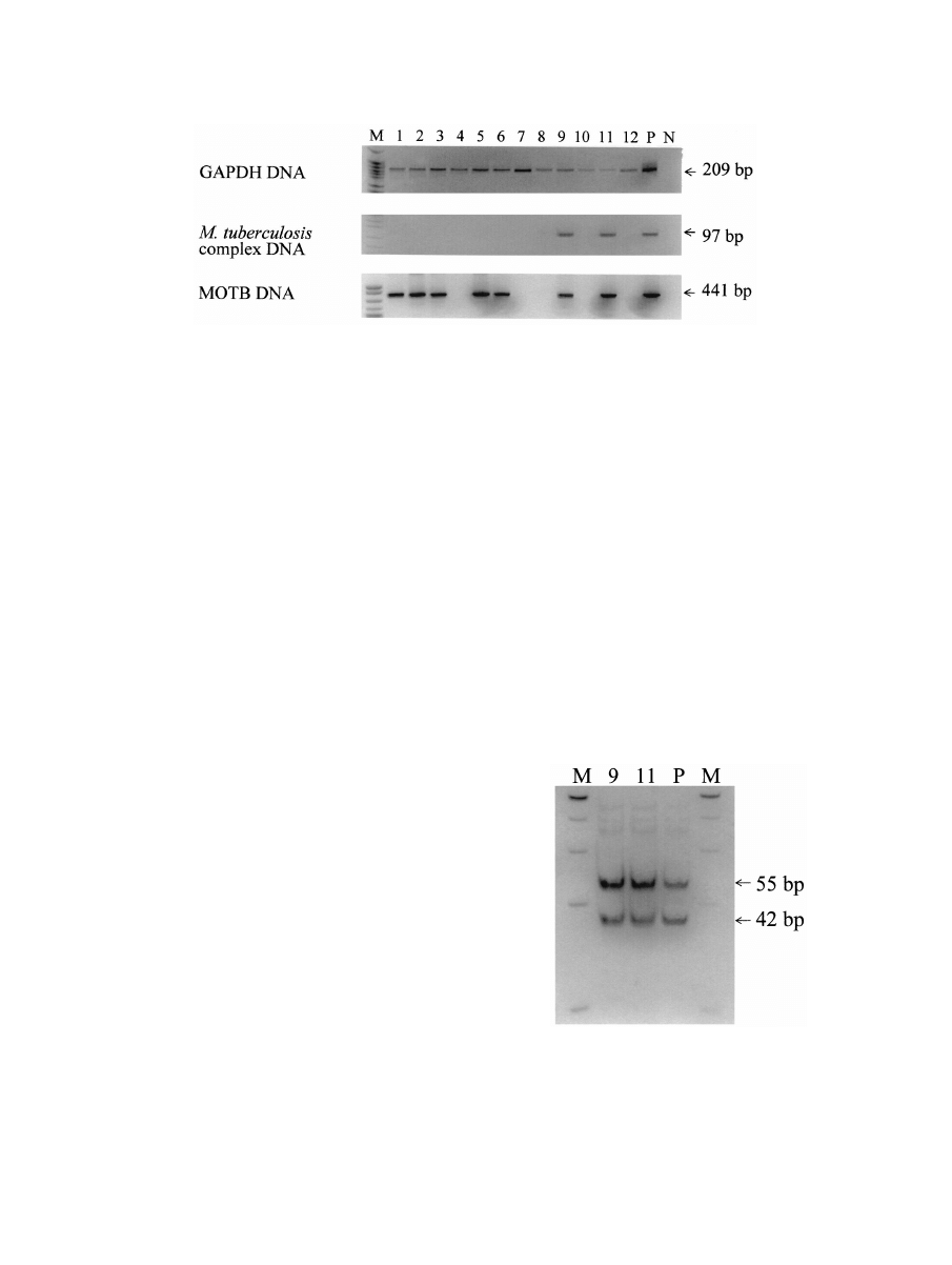
PCR with the GAPDH gene (Fig. 1).
In two samples (no. 9 and 11), a 97-bp DNA fragment was
detected by PCR (Fig. 1) with primers specific for insertion
sequence IS6110, which is unique to the M. tuberculosis com-
plex (14). This fragment was further digested with SalI and
gave rise to two expected fragments of 42 and 55 bp (Fig. 2),
confirming the presence of M. tuberculosis complex in these
two samples. The IS6110 element, which was initially identified
from a clinical isolate of M. tuberculosis, is specific for the M.
tuberculosis complex (M. tuberculosis, Mycobacterium bovis, and
Mycobacterium simiae). Generally, the IS6110 element is
present in high copy numbers in most strains of M. tuberculosis
and low copy numbers in M. bovis strains (3). The specificity of
detecting M. tuberculosis complex by PCR with the primers for
IS6110 was confirmed by Eisenach et al. (4). The primers
detected strains of M. tuberculosis, M. bovis, and M. simiae. The
IS6110 sequence was also detected in mummified tissues (10),
and it was found to be identical to that of contemporary M.
tuberculosis as reported by Eisenach et al. (4). In this study, we
applied nested PCR to detect the IS6110 sequence in ancient
DNA in order to increase sensitivity and specificity. Our results
confirmed the presence of M. tuberculosis complex in 2 of 12
mummy samples, although we could not determine the species
in the M. tuberculosis complex.
A 441-bp DNA fragment was amplified in seven samples
(no. 1, 2, 3, 5, 6, 9, and 11) (Fig. 1) by using primers specific for
the 65-kDa heat shock protein present in all MOTB (9, 13).
The DNA fragments were further digested with BstEII and
HaeIII to determine the species of MOTB according to the
algorithm reported by Telenti et al. (13) and Rastogi et al. (9).
Six samples could not be digested by BstEII but were able to be
digested with HaeIII and gave rise to a 140-bp band (Fig. 3).
Based on band size and patterns, they were considered to be
Mycobacterium flavescens I. In one sample (no. 1), two DNA
fragments were observed after digestion with BstEII and three
FIG. 1. Detection of GAPDH and mycobacterial DNA in mummified tissues. DNAs were extracted from the genital tissues of 12 mummies.
The PCR products were examined on a 10% polyacrylamide gel. GAPDH DNA was detected in all mummified tissues, indicating that the DNA
was adequate for PCR analysis. DNAs of M. tuberculosis complex were detected in 2 of the 12 samples, and MOTB DNAs were detected in 7 of
the 12 samples. Lane M, DNA size markers (PBR322 DNA digested with MspI); lanes 1 to 12, mummy samples; lane P, positive control; lane N,
negative control.
FIG. 2. Confirmation of M. tuberculosis complex by SalI digestion.
PCR products from two positive samples and one positive control were
further digested with SalI and separated on a 10% polyacrylamide gel.
We observed two bands (55 and 42 bp), which are identical to those of
the positive control (M. tuberculosis). These results confirmed the
presence of M. tuberculosis complex in these two samples. Lane M,
DNA size markers (PBR322 DNA digested with MspI); lanes 9 and 11,
mummy samples 9 and 11, respectively; lane P, positive control (M.
tuberculosis).
V
OL
. 40, 2002
NOTES
4739
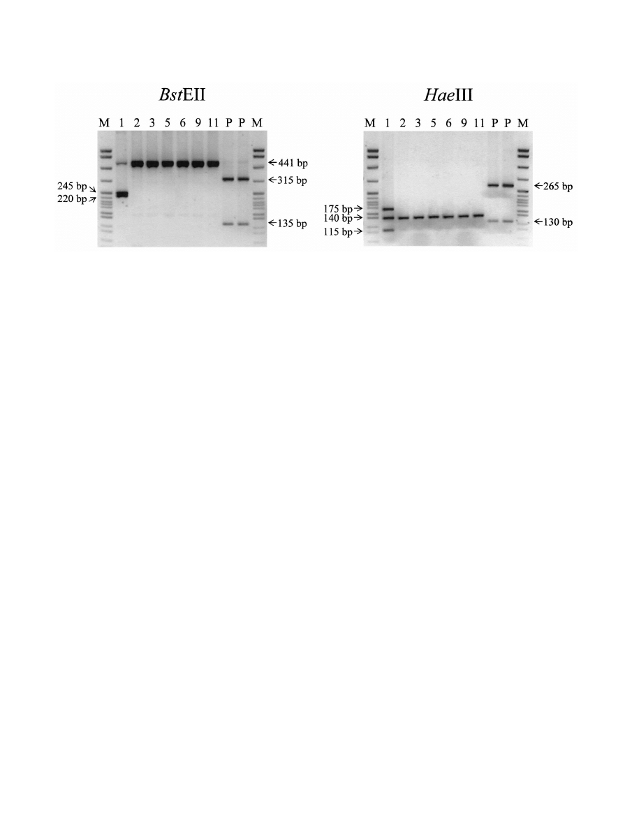
fragments were observed after digestion with HaeIII (Fig. 3).
With these patterns, the fragments could not be assigned to a
specific species. However, we believe that there may be two
species present in this sample, i.e., M. flavescens I and another
MOTB not included in the algorithm. Since these mycobacte-
ria are present in soil and water, we do not believe that their
identification indicates the presence of clinical disease; this is
in contrast to M. tuberculosis, which must be considered patho-
genic.
We also tested samples for other infectious agents, including
Mycobacterium leprae, Treponema pallidum, Leishmania spp.,
herpes simplex virus, human papillomavirus, and human T-cell
lymphotropic virus type 1. No PCR products were observed.
REFERENCES
1. Arriaza, B. T., W. Salo, A. C. Aufderheide, and T. A. Holcomb. 1995. Pre-
Columbian tuberculosis in northern Chile: molecular and skeletal evidence.
Am. J. Phys. Anthropol. 98:37–45.
2. Baron, H., S. Hummel, and B. Herrmann. 1996. Mycobacterium tuberculosis
complex DNA in ancient human bones. J. Archaeol. Sci. 23:667–671.
3. Cave, M. D., K. D. Eisenach, P. F. McDermott, J. H. Bates, and J. T.
Crawford.
1991. IS6110: conservation of sequence in the Mycobacterium
tuberculosis complex and its utilization in DNA fingerprinting. Mol. Cell.
Probes 5:73–80.
4. Eisenach, K. D., M. D. Cave, J. H. Bates, and J. T. Crawford. 1990. Poly-
merase chain reaction amplification of a repetitive DNA sequence specific
for Mycobacterium tuberculosis. J. Infect. Dis. 161:977–981.
5. Haas, C. J., A. Zink, G. Palfi, U. Szeimies, and A. G. Nerlich. 2000. Detection
of leprosy in ancient human skeletal remains by molecular identification of
Mycobacterium leprae. Am. J. Clin. Pathol. 114:428–436.
6. Nerlich, A. G., C. J. Haas, A. Zink, U. Szeimies, and H. G. Hagedorn. 1997.
Molecular evidence for tuberculosis in an ancient Egyptian mummy. Lancet
350:
1404.
7. Park, Y. N., K. Abe, H. Li, T. Hsuih, S. N. Thung, and D. Y. Zhang. 1996.
Detection of hepatitis C virus RNA using ligation-dependent polymerase
chain reaction in formalin-fixed, paraffin-embedded liver tissues. Am. J.
Pathol. 149:1485–1491.
8. Rafi, A., M. Spigelman, J. Stanford, E. Lemma, H. Donoghue, and J. Zias.
1994. Mycobacterium leprae DNA from ancient bone detected by PCR. Lan-
cet 343:1360–1361.
9. Rastogi, N., K. S. Goh, and M. Berchel. 1999. Species-specific identification
of Mycobacterium leprae by PCR-restriction fragment length polymorphism
analysis of the hsp65 gene. J. Clin. Microbiol. 37:2016–2019.
10. Salo, W. L., A. C. Aufderheide, J. Buikstra, and T. A. Holcomb. 1994.
Identification of Mycobacterium tuberculosis DNA in a pre-Columbian Pe-
ruvian mummy. Proc. Natl. Acad. Sci. USA 91:2091–2094.
11. Spigelman, M., and E. Lemma. 1993. The use of the polymerase chain
reaction (PCR) to detect Mycobacterium tuberculosis in ancient skeletons.
Int. J. Osteoarchaeol. 3:137–143.
12. Taylor, G. M., M. Grossey, J. Saldanha, and T. Waldron. 1996. DNA from
Mycobacterium tuberculosis identified in mediaeval human skeletal remains
using polymerase chain reaction. J. Archaeol. Sci. 23:789–798.
13. Telenti, A., F. Marchesi, M. Balz, F. Bally, E. C. Bottger, and T. Bodmer.
1993. Rapid identification of mycobacteria to the species level by polymerase
chain reaction and restriction enzyme analysis. J. Clin. Microbiol. 31:175–
178.
14. Thierry, D., M. D. Cave, K. D. Eisenach, J. T. Crawford, J. H. Bates, B.
Gicquel, and J. L. Guesdon.
1990. IS6110, an IS-like element of Mycobac-
terium tuberculosis complex. Nucleic Acids Res. 18:188.
15. Tokunaga, K., Y. Nakamura, K. Sakata, K. Fujimori, M. Ohkubo, K.
Sawada, and S. Sakiyama.
1987. Enhanced expression of a glyceraldehyde-
3-phosphate dehydrogenase gene in human lung cancers. Cancer Res. 47:
5616–5619.
FIG. 3. Identification of MOTB by BstEII and HaeIII. PCR products from seven positive samples were further digested with BstEII and HaeIII
and separated on a 2.5% agarose gel. A 441-bp band was seen in all seven samples after digestion with BstEII. However, sample 1 gave rise to two
bands of 245 and 220 bp. After digestion with HaeIII, a 140-bp band was seen in all samples; however, two bands of 175 and l15 bp were observed
in sample 1. The results suggest that M. flavescens I was present in all samples and that there was an additional species present in sample 1. Lane
P, positive control (M. leprae); lanes M, DNA size markers; lanes 1, 2, 3, 5, 6, 9, and 11, mummy samples 1, 2, 3, 5, 6, 9, and 11, respectively.
4740
NOTES
J. C
LIN
. M
ICROBIOL
.
Wyszukiwarka
Podobne podstrony:
[42]Oxidative breakage of cellular DNA by plant polyphenols A putative mechanism for anticancer prop
Wójcik, Katarzyna A Polymorphism of the DNA Base Excision Repair Genes in Keratoconus (2014)
Farren, Mick DNA Cowboys 01 The Quest of the DNA Cowboys 1 0b
The Quest of the DNA Cowboys Mick Farren
Dental DNA fingerprinting in identification of human remains
The positive and negative?fects of dna profiling
Patterns of damage in genomic DNA sequences from a Neandertal
Detection and Molecular Characterization of 9000 Year Old Mycobacterium tuberculosis from a Neolithi
4 10 DNA Profiling of Other Species
The role of BRCA1 in DNA damage response
4 8 Early Uses of DNA Profiling v1
Awakening the Power of a Modern God Unlock the Mystery and Healing of Your Spiritual DNA by Gregg Br
więcej podobnych podstron